SYMBIOTIC INTERACTION and GALL FORMATIONby M. Halit UMAR |
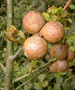
Plant galls on a young oak tree. D. Walker. |
SYMBIOTIC INTERACTION and GALL FORMATIONby M. Halit UMAR |
Plant galls on a young oak tree. D. Walker. |
One of the main characteristics of LIFE is the inevitable interaction between organisms. Several forms of such interactions are described with a word: Symbiosis, meaning living together. In my previous presentations, different forms of symbiosis were illustrated: An animal parasite called Demodex folliculorum lives in the sebaceous glands and hair follicles of men. The parasites usually cause no harm to the hosts. Various kinds of bacteria and other organisms live on the surfaces of human, animal and plant bodies. Also many types of microbes are inseparably associated with internal structures like the gut in men and animals. 'Inseparable' because their absence is often a severe condition, if not a disease, in men and animals alike. You could say that we live together with many kinds of micro-organisms in harmony. Both symbiont and host find one or more benefits from such an association. We also see a nice example of a very useful relationship between growing plant roots and some species of soil fungi. These are generally called mycorrhizas.
You may already be asking whether symbiosis is always beneficial for both parties? Rightly, the answer is NO. Not all associations are without physiological or anatomical hazards for the hosts. Therefore, they may react differently in some forms of symbiosis to demarcate the symbionts, because they do not really want to be associated. Obviously, hosts may have different mechanisms of sensing and examining for any infiltrating organisms.
Despite very powerful sensing systems, some organisms come into close association with other living organisms; they may even invade them to some extent. In this case we are now speaking about parasites instead of symbionts. They parasitize the hosts and the interaction results mainly in an observable structural change of the parasitized organ or tissues. This is a process called parasitosis which frequently leads to a disease condition.
Let me give you a simple example: If you drink water, which is not obtained under hygienic conditions, you may then unwillingly drink amoebae that may subsequently parasitize your gut and cause severe and dangerous diseases. Humans react to parasitizing amoeba by diarrhea, and by doing this, elimination of parasites can naturally be attempted (but mostly without success). So, as we see in this example, Nature also provides some defence mechanisms to both parties in a symbiotic process.
In this article, I aim to illustrate a symbiotic interaction between plant leaves and the larvae of a parasite. The neat result is the formation of so-called galls in plants. Galls are reactive, tumor-like structures against parasites, and are sometimes also associated with fungal hyphae and bacteria. Galls demarcate the larvae within a newly formed cavity where they grow and reach near adult form and size. Then, galls disintegrate (e.g. when the leaf falls), and parasites may begin a new life cycle when the gall cavity has broken down. On the other hand, galls demarcate the parasite into a limited and relatively small area of the plants which live further without harmful consequences of such leaf tumours.
But the symbiotic interaction sometimes results in a fundamental change and sequelae in the host. An example of such an extreme process can probably be the subject of another article.
In practice, you can encounter galls when you take a walk along forest trails. Look carefully at the fallen leaves and try to discriminate normal leaves from a leaf with tumorous structures. Grossly, the galls are between a few millimetres to a few centimetres in diameter. They are mostly spherical structures and their colour changes from yellow to red to dark brown. When opened, galls frequently contain barely moving parasites/larvae, mostly transparent. You can put them on a glass slide and directly observe through your microscope.
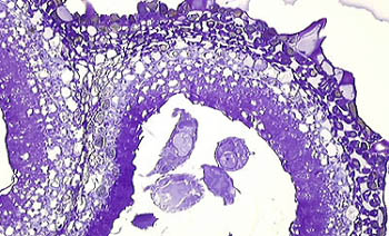
Finally, the wall of the galls is a kind of reactive, secondarily formed plant tissue. This is why some plant pathologists think about galls as a tumour or, at least, as tumour-like tissues. The thick wall consists usually of proliferating large plant cells. The following microscopic images show a leaf with small (2-3 mm across), multiple galls containing numerous parasites/larvae confined within the cavity. The parasite causing these galls is not particularly determined for the taxonomy, the name of genus and species.
.
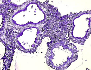
Multiple, small galls with a large cavity
in which small parasites can be seen..
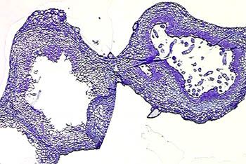
One of these galls, (on the right),
contains numerous parasites..
Note that the inner wall of the cavity is formed by densely stained plant cells. The outer wall is made of large and vacuolated plant cells.
A step higher magnification.
.
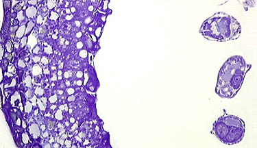
Note that only a portion of the wall
and three
of the parasites are present in this
field.
.

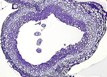
The cytoplasm of these cells contain many cell organelles and is rarely vacuolated. Their nuclei are also larger than other plant cells in the outer part of the wall. This fact may be an indication for their continuous proliferation. They probably secrete some kinds of substances which work as a humoral defence mechanism of the host against the presence and dissemination of a parasite.This image shows densely structured plant cells in the inner zone of the wall. In the upper part of the image we see a cut surface of a parasite around which a few bacteria are also present. Approximately 1000x..
Technical Notes: These microscopic images are obtained from tissue samples which were embedded in a plastic matrix and cut by a glass knife to 2 micrometre thick. Sections were stained with aqueous crystal violet (0.5%).
For a very interesting link about plant
galls with excellent illustrations on the Internet:
Just
click here.
.
Comments to the author M. Halit Umar are welcomed.
Please report any Web problems or offer general
comments to the Micscape
Editor,
via the contact on current Micscape Index.
Micscape is the on-line monthly magazine of the
Microscopy UK web
site at Microscopy-UK
WIDTH=1