|
New Old Slides: Part 2 by Richard L. Howey, Wyoming, USA |
In this part, we will be looking at plant sections from the same box that we talked about in Part 1 only this time more images and less rambling commentary (except when necessary, or course, or when I happen to feel like it).
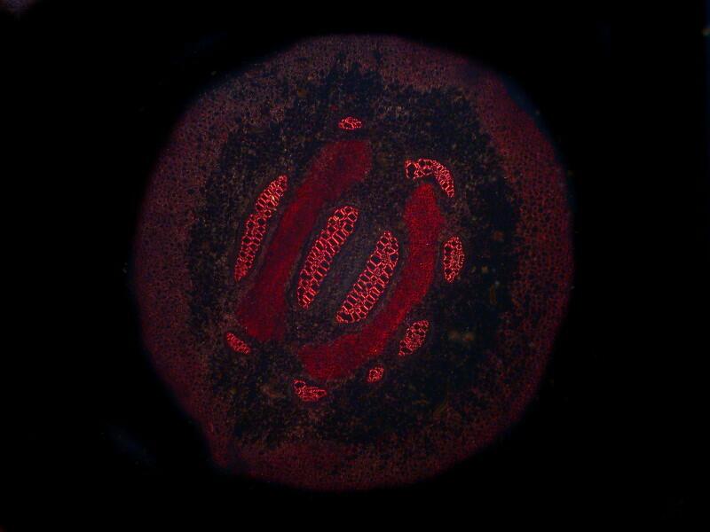
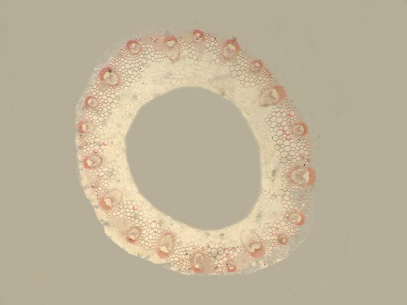
Let’s start right off with a couple of examples that I find quite striking. The first is a section of a stem from Typha latifolia which is better known as a Cattail or Bulrush. Marsh plants, such as, cattails, which, under ideal conditions, have access to large quantities of water rich in organic and mineral materials often develop, as a consequence, relatively large vesicles to transport fluids. I remind you; brightfield section first, then the same section polarized.
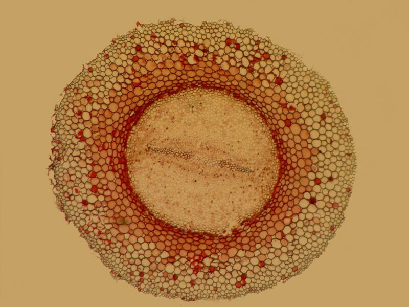
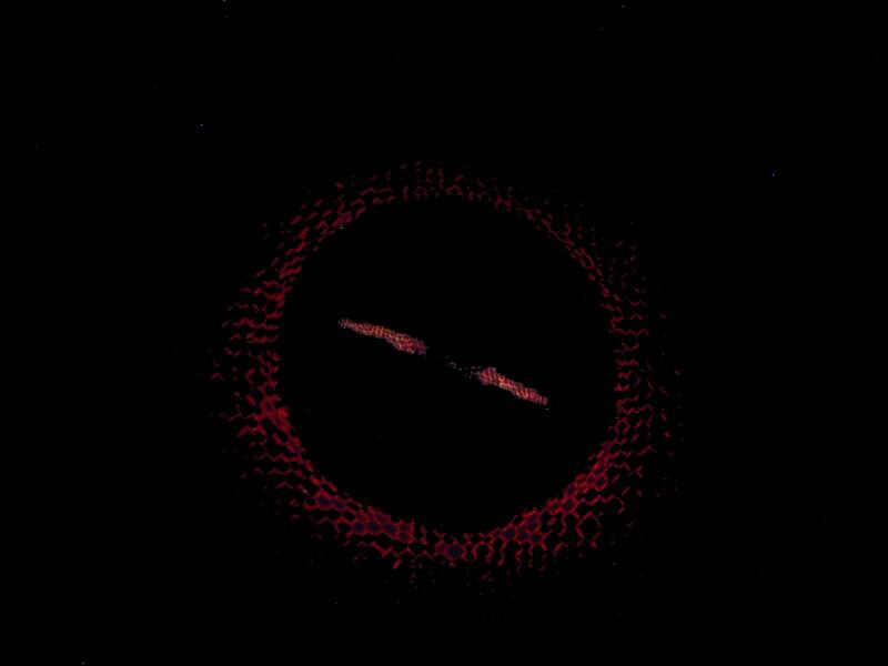
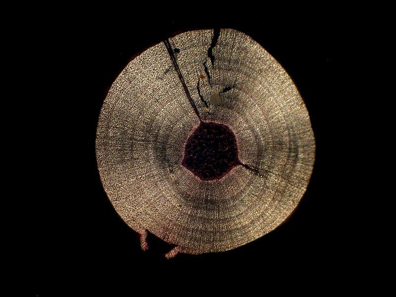
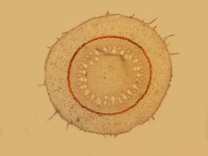
The next example is a cross section of Botrychium, a moonwort
sometimes known as a Rattlesnake fern because the habitats in which it grows are frequently populated by these reptiles. I quite like the juxtaposition of these 2 images because in the brightfield view, there is a great deal of cellular detail in the outer ring, but not much distinctive in the center until we look at the polarized image and then we have 2 narrow central bands that jump out at us giving us the impression that we are looking at some distant stellar entity.
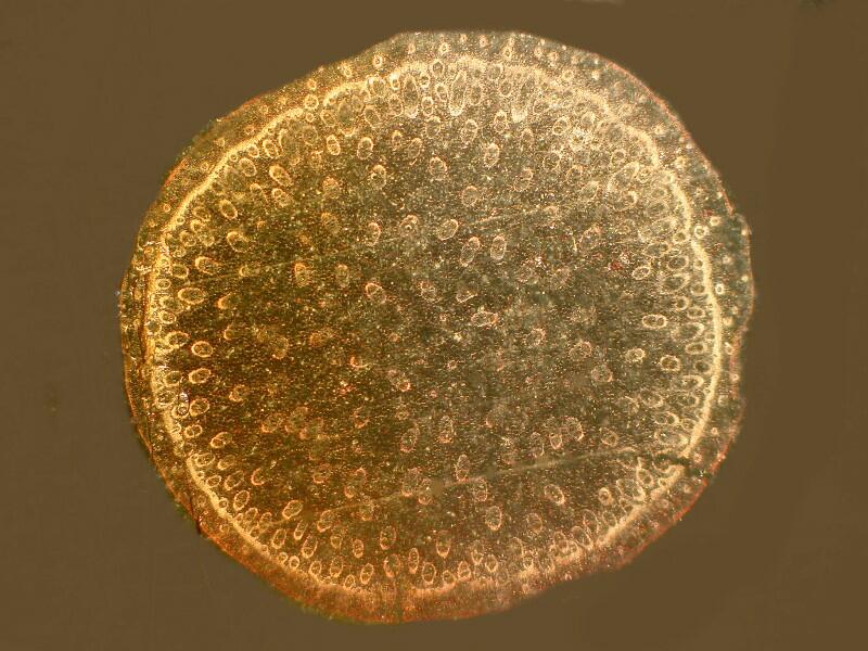
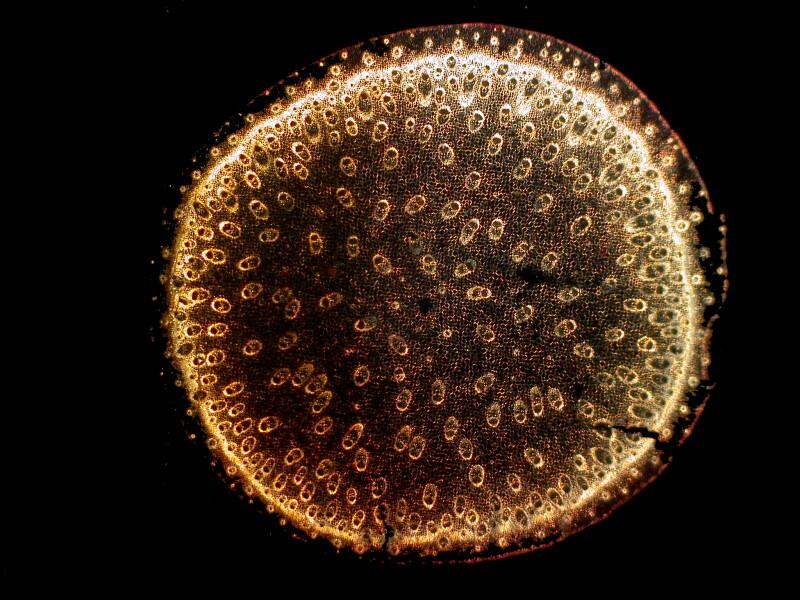
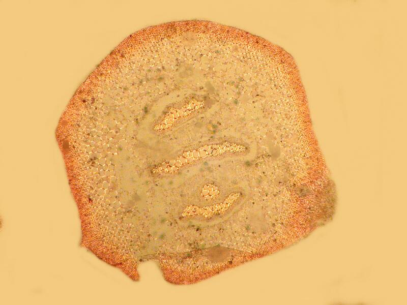
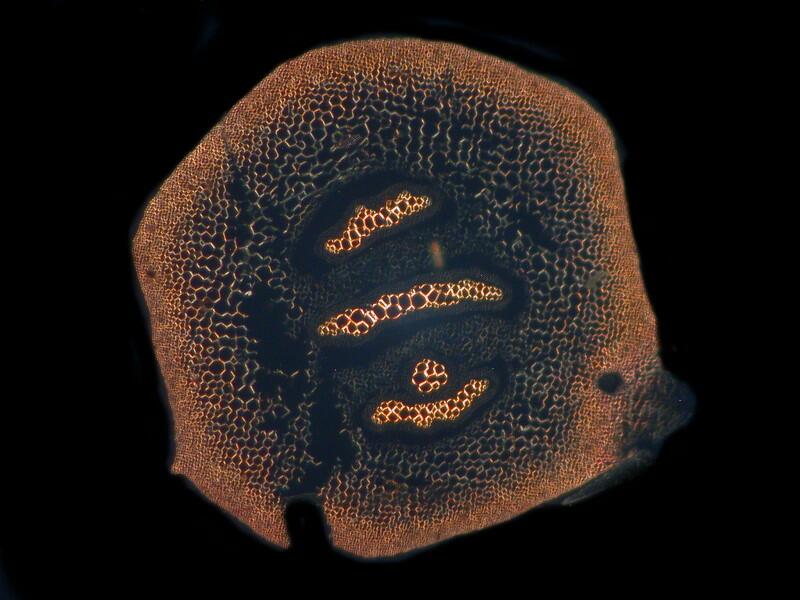
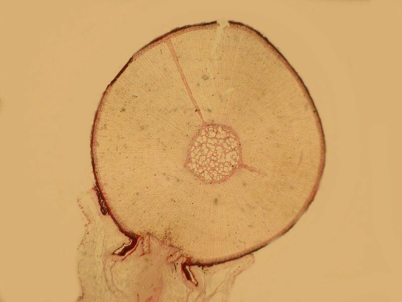
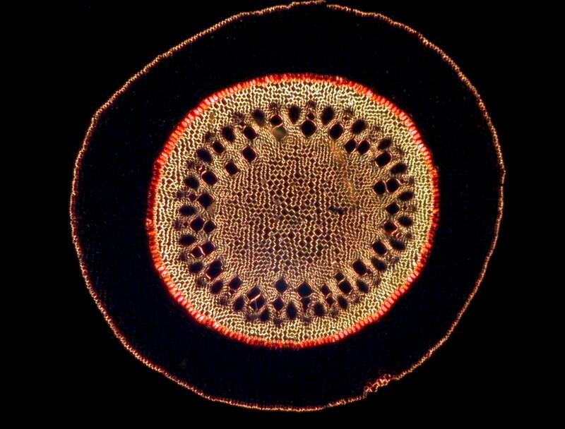
Next, we’ll look at a section of remarkable club moss, Selaginella, also know as the Resurrection Plant. Various craft and novelty shops carry these in a dried state in plastic or cellophane sacks. They are in a dormant state but, when placed in water will green up and begin to grow. Then, according to the propaganda on the package, you can let it dry up, repackage it and later revive it–apparently numerous times. I have a package of one of these but, I haven’t yet had the opportunity to try it out. If one of you have had success with this, I’d appreciate knowing and then I’ll make a goal of becoming a Resurrection Plant in my next reincarnation. These images are of a cross section of a stem.
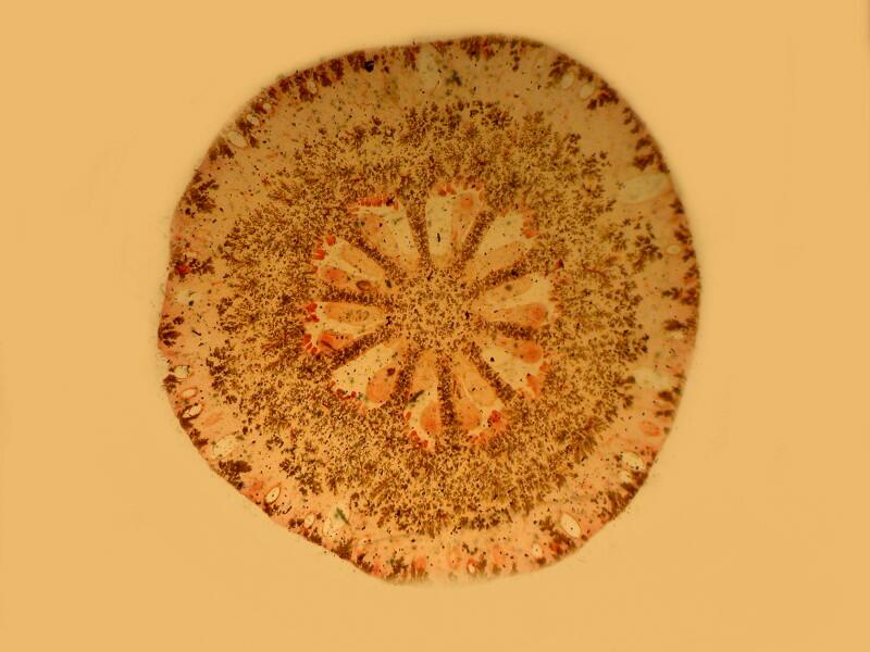
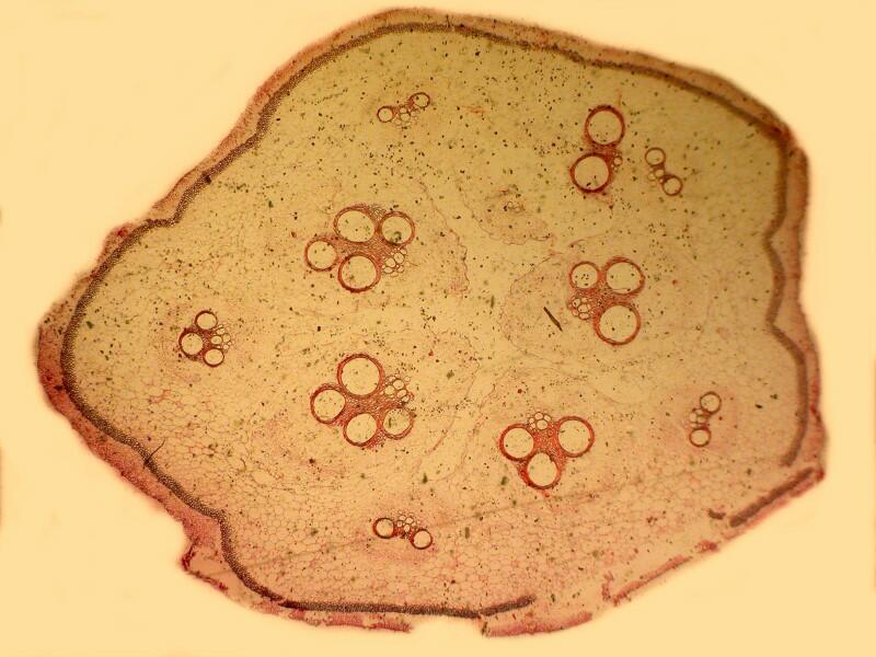
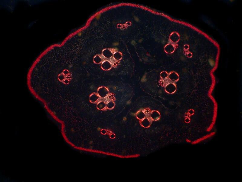
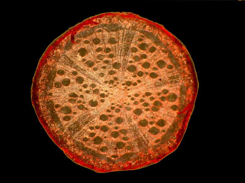
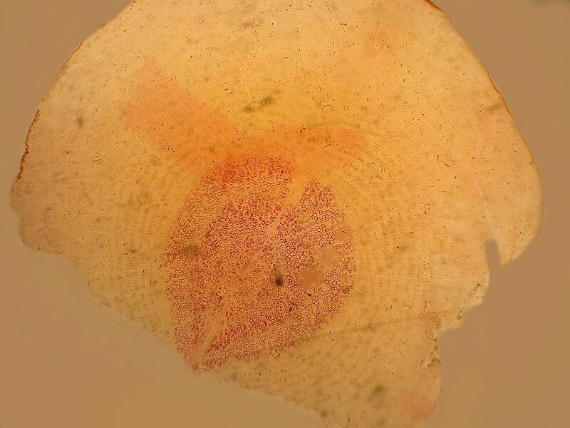
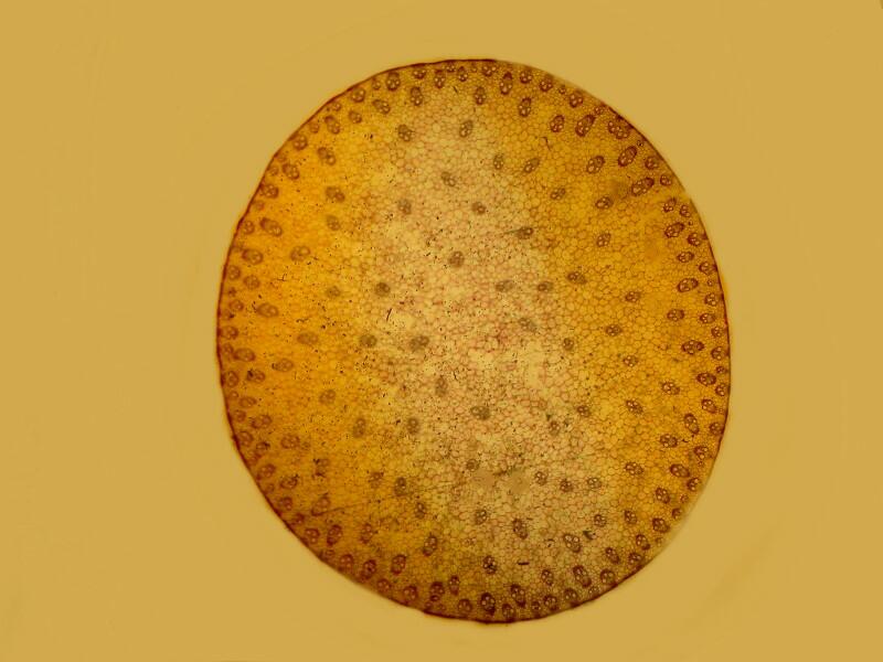
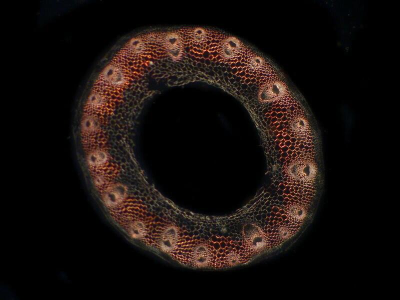
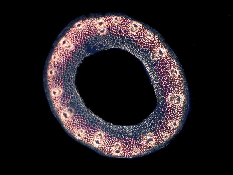
Nature is constantly experimenting with trying out new solutions and patterns. A wonderful exploratory investigation of some of these patterns can be found in the magnum opus of the Scottish biologist D’Arcy Wentworth Thompson’s On Growth and Form. To illustrate one aspect of his theory about patterns I want to show you 2 quite disparate comparisons. I have long been enchanted with the beautiful radial symmetry of the cross sections of sea urchin spines and I have been fortunate enough over the years to acquire a few 19th Century slides of good quality sections. First, I want to show you a botanical section of a citron (Citrus medica). This is a transverse section of a young fruit showing the oil glands.

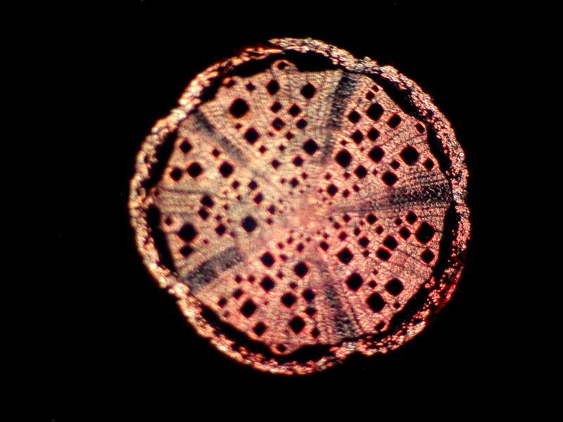
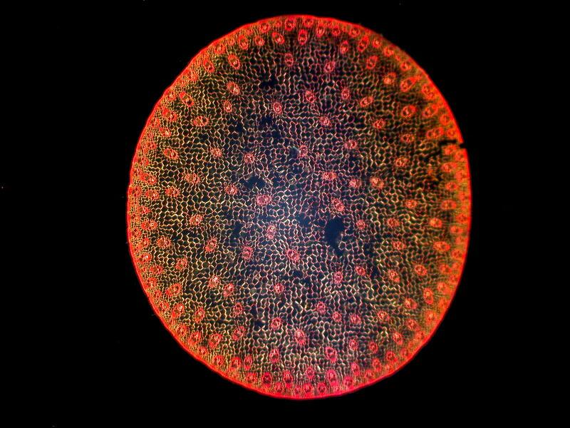
Next, 3 transverse sections of spines of Echinus. (Note: The genus here may well not correspond with modern taxonomy, since it was the habit of 19th Century slide makers to identify a wide variety of different specimens as Echinus.)
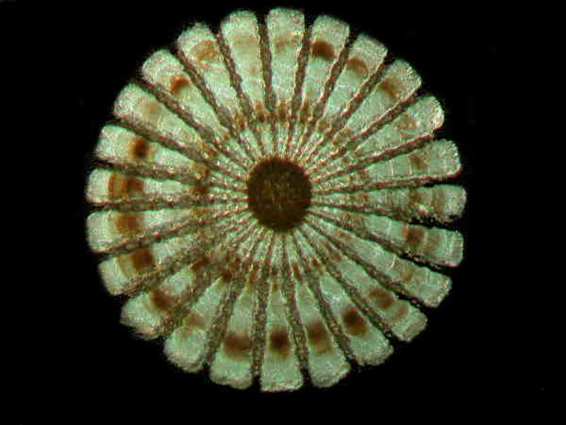
Many echinoderms (the classic conception of the starfish, for example) exhibit pentamerous symmetry (5 or multiples thereof) and it is interesting that the citron section shows 10 spokes and indeed, in many superficial respects, resembles the echinoid sections. so, here we have an intriguing pattern comparison between the plant world and the animal world.
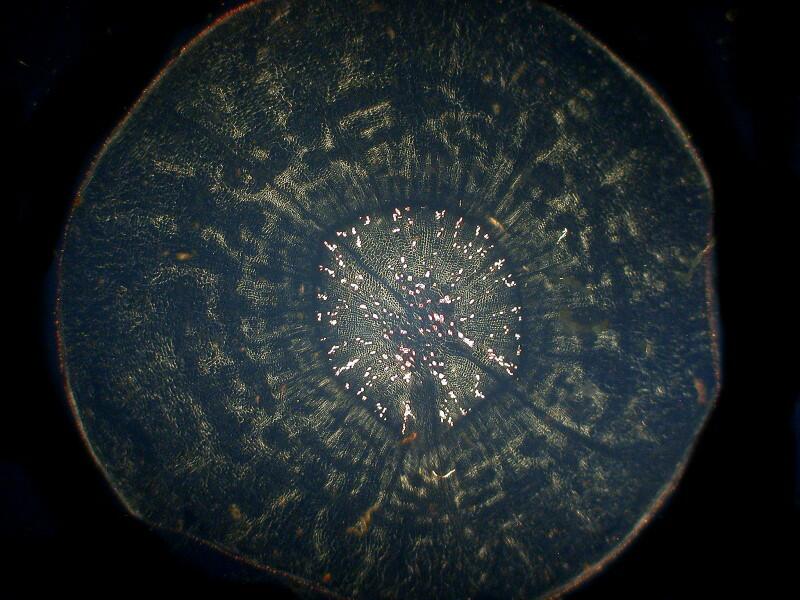
The second comparison involves equally remote realms. First, let’s take a look at a section of a very young Northern White Cedar stem, Thuja occidentalis. Here I’ll show you just one image which was taken using polarized illumination.
This time the comparison is with a crystalline substance, Vitamin C, which although it is now synthesized, was, of course, originally derived from plants. So, for comparison here are some Vitamin C images.
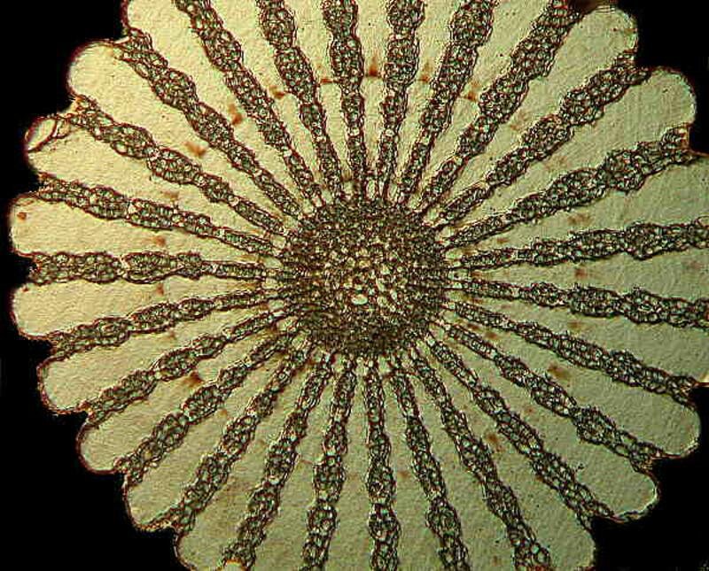
These disks can be roughly grouped into what I call the “shield” pattern which show up with surprising frequency as micro-structures of plant, animals, and crystals. I want to give you 2 more examples, the first of which involves 2 plants and the one we’re primarily interested in is Comandra livida, also known as false toadflax and here it is engaged in warfare against an Andromeda which is a Bog Rosemary; in other words, it is a parasite which occurs in peaty bogs. In the brightfield image, you can see its tissue extending out beyond is circular perimeter.
However, with the polarized image, we get a “shield”, a rather vulnerable one if that central black area is simply a hole, although it doesn’t appear to be empty from the brightfield image so, perhaps, were it not so plain, a micro-Achilles might have used it!
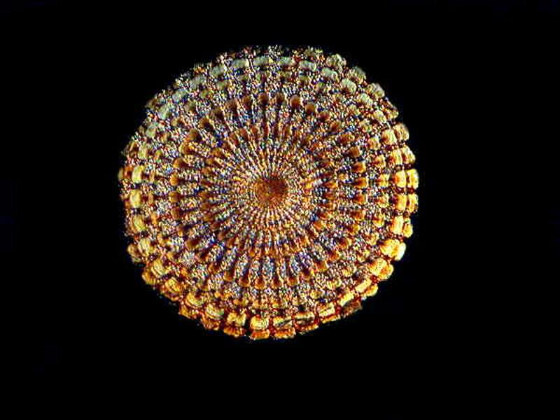
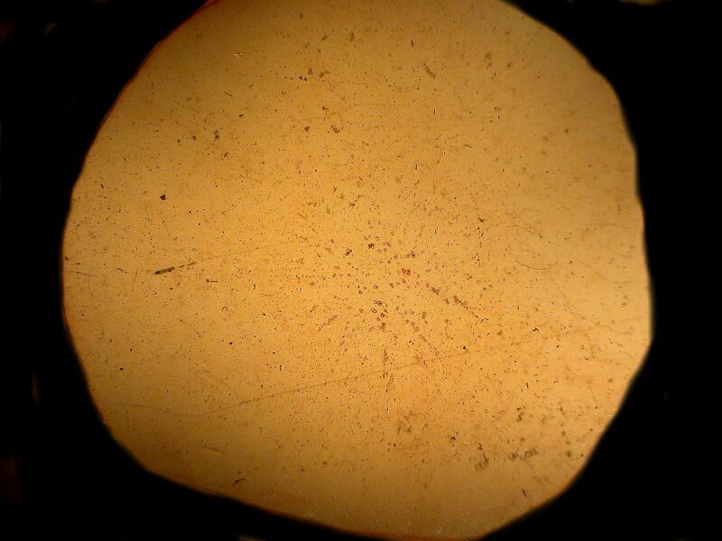
The second example is intriguing for 2 reasons: 1) it is a cross section of a stem of Queen Anne’s Lace. There are a number of legends regarding the origin of the name; one of the most popular in this country is that Anne of Denmark (1574-1619), queen consort of King James I, was an expert at making fine lace. 2) The brightfield image shows virtually no structural detail. By the way, this is Daucus carota.
However, when we shift to polarized illumination, we get a shield worthy of Atlas. Suddenly, where there was nothing before, we have a clear indication of structure including a central disk within the larger disk.
This underscores the argument that I present ad nauseam, namely, that you should use as many techniques as you have available when you are trying to understand the intricacies of the structure of a given specimen.
Sometimes structures that are readily visible in stained, brightfield sections take on a quite different character under polarized light. An excellent example is Cucurbita pepo which is a cross section of pumpkin stem. In this section , the vesicles show up marvelously in both brightfield and polarized light, but with a radically different shift in emphasis.
Sometimes this relatively simple technique seems to me like an act of conjuring, a bit of magic but, with a reversal–“Now you don’t see it; now you do.”
I’ll show you 2 instances. The first is a cross section of a stem of a bracken fern, Pteris aquilina. In the brightfield image the 2 central curved rods are readily visible.
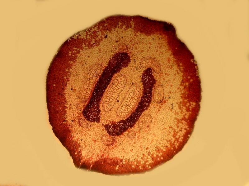
Now, notice what happens when we cross the polars; a series of smaller, more brightly-colored rods suddenly appear.
One of these days, when I’m not feeling so lazy, I want to take a look at this section using Nomarski DIC to see how much of this detail I can see using this technique.
The second example is a stem section of Smilax herbacea, the Smooth Carrion Flower, the extract of which Haight-Ashbury Hippies used as perfume, along with Patchouli. It’s name does, of course, derive from the fact that it has a foul odor and a variety of plants employ such a device in order to attract flies and other vermin in order to cross pollinate. The brightfield image is interesting in its own way as you can see and you will notice that there are tiny hairs projecting from around the edge. (These are probably involved in producing the stench.)
However, when we cross the polars, we get another example of micro-prestidigitation. Once again, I am reminded of a cross section of a sea urchin spine and one of intricate beauty and complexity.
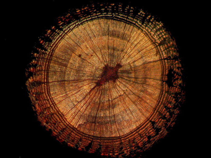
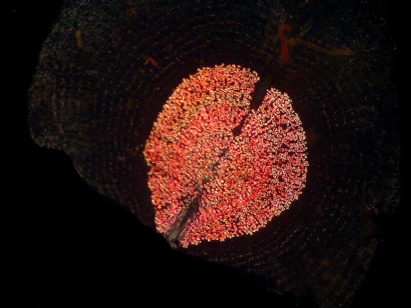
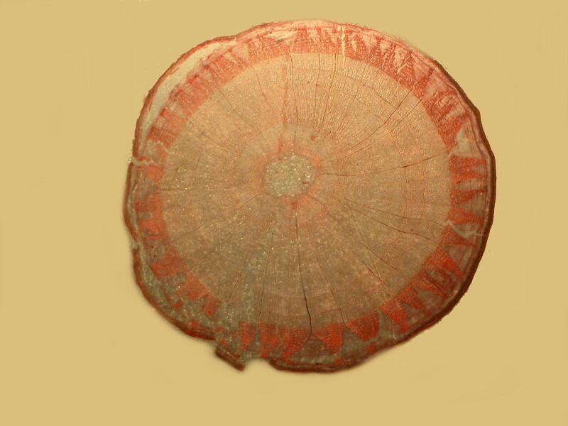
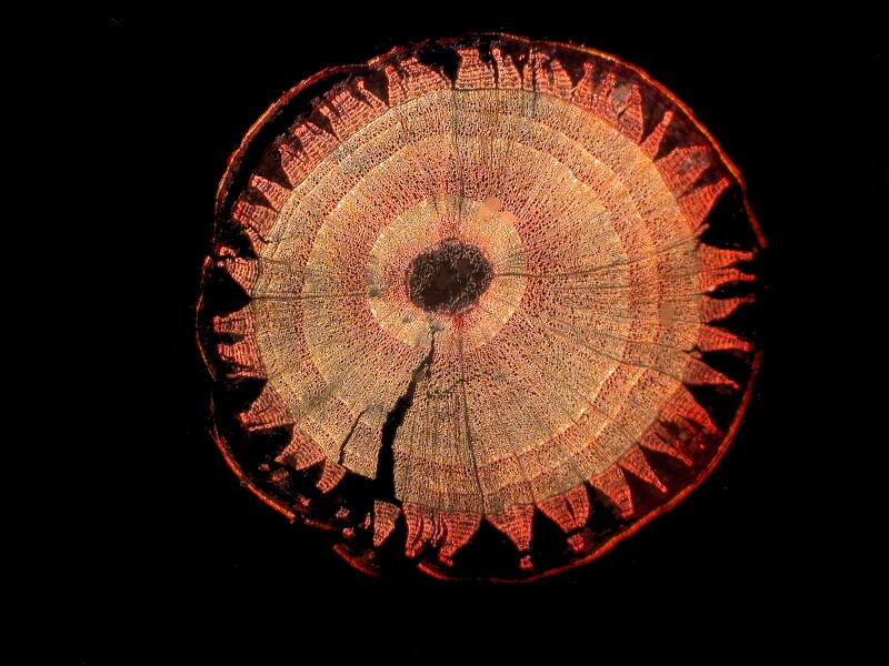
Since somehow, no doubt through the divine inspiration of Queztalcoatl, we have returned to one of my favorite subjects, namely yet another stem section which reminds me of a section of sea urchin spine. Interestingly, this is a section of a stem of a tree, a red oak, Quereus rubra. This is a tricky one to photograph so the first image is what one might describe as semi-polarized, that is, it was taken with the polarizer in place, but with the analyzer at 45 degrees. The black background was, in this case, added using graphics software.
However, when the polars are crossed the section is much more
dramatic and certainly more closely patterns a sea urchin spine section.
One of the great delights in using a variety of techniques is that a particular approach, in our case polarization, can reveal aspects of a specimen that we have only the vaguest notion of from the brightfield image. The section we are looking at here was taken from that plant that most gardeners hate, Taraxacum officinale, the notorious dandelion. The label on this slide is downright chatty and we learn that the section is from the fleshy root, was fixed in Carnoy’s fluid, and stained with Hematoxylin and Safranin O.
Certainly, in the central area, we can see something lurking but, it is quite indistinct. However, when we cross the polars a heart-shaped structure leaps out at us and we have quite a different perspective to investigate. Mother Nature didn’t realize how sneaky we can be in trying to wrest secrets from Her using such underhanded techniques as chemical warfare, stains, and special illumination.
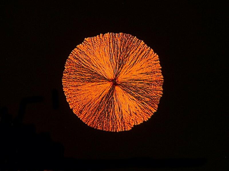
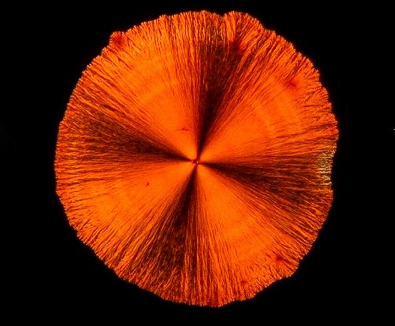
There is a wood which certain craftsmen prize for its properties; Its scientific name is Tilia americanum, more commonly known as Basswood or Linden. The section under brightfield is interesting, but not compelling.
However, simply by crossing the polars, we get an unexpected “sunburst”.
Sometimes, in our passion to seek out the exotic and unusual, we overlook the extraordinary in the ordinary things around us. Almost everyone likes sweet corn (Zea mays) and most farmers wouldn’t mind letting you take a small cutting to make a section of a stem. Again the contrast between brightfield and a stained polarized image is quite remarkable.
Finally, I’ll show you 3 images of one of my favorites, Ranunculus acris, the tall buttercup. This brilliantly yellow little flower is something to gladden the heart and I find it quite wonderful that a section of its stem reveals a very different kind of symmetry and pattern. The first image is brightfield and while suggestive remains rather pale and hidden.
The second image is more provocative and was taken with the polars only partially crossed.
The third image with the polars fully crossed reveals what one might describe as a micro-ring nebula.
I hope you have enjoyed this brief tour and will find the occasion to investigate some plant sections for yourself.
In Part 3, I shall move on to the second box of antique/vintage slides which I recently acquired.
All comments to the author Richard Howey are welcomed.
Editor's note: Visit Richard Howey's new website at http://rhowey.googlepages.com/home where he plans to share aspects of his wide interests.
Microscopy UK Front
Page
Micscape
Magazine
Article
Library
Published in the May 2012 edition of Micscape Magazine.
Please report any Web problems or offer general comments to the Micscape Editor .
Micscape is the on-line monthly magazine of the Microscopy UK website at Microscopy-UK .