|
Photographing Cannabis Under The Microscope
By Ted Kinsman emkpph@rit.edu Rochester Institute of Technology Rochester, NY |
Over the course of the past year I have been working on creating images for a recently released book. Cannabis Under The Microscope: A Visual Exploration of Medicinal Sativa and C. Indica by Ford McCann. The book is a digital release on Kindle. The images cover numerous techniques from macro photography to scanning electron microscopy. I hope these images will be of interest to Micscape readers.
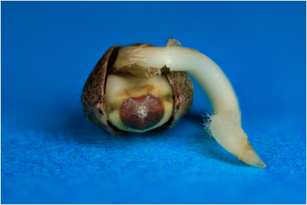
A cannabis seed at three days old. The seeds were incubated at (21ºC) on a damp paper towel and kept in the dark. The image was taken at 3x with a Canon macro lens and was focus stacked from 20 individual images. The images were collected with a stack-shot manufactured by Cognisys and combined using Zerene stacker software. Many of the following optical images used similar techniques.
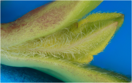
Young sprout at 5 days, Note the concentration of trichome structures to deter bugs from eating the new leaves. The fresh leaves are the target for insects, so the plant increases the concentration of toxins at these locations to detour attack. The trichomes (spherical or needle like structures) are the location on the plant with the highest concentration of THC. THC is the active chemical that is responsible for the principal psychoactive properties of the cannabis plant. The plant uses this toxin to repel insect attack. This image is focus stacked from 20 images and was collected at 5x with a Canon 68mm macro lens. Images were focus stacked with Zerene software.
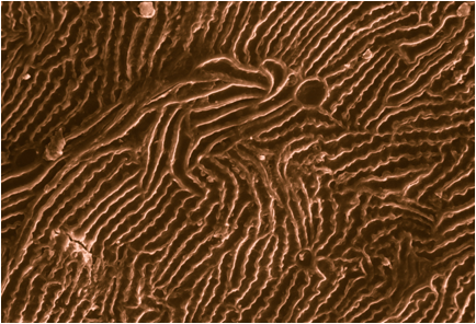
The surface of a cannabis seed seen under a scanning electron microscope. Magnification of the image shows a section of the seed coat approximately 0.2 mm across. The surface of the seed is a grooved structure that serves two different purposes: to absorb water and the strange shape is postulated to make the growth of bacteria difficult. Image taken with a Cambridge S200 scanning electron microscope. [Editor's note: Ted runs the SEM from his house, see this Micscape article.]
A scanning electron microscope image SEM image of a nice elongated trichome structure on the top of a cannabis leaf. These cell structures act as thorns on the leaf top. The image was false colorized in Photoshop and represents a section of the leaf 0.2 mm wide.
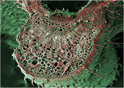
An SEM image of the cross section of the major leaf vein. The bottom of the image is the bottom of the leaf. The structure is for support as well as holding the pith cells that help transport nutrients throughout the leaf. The image represents a section of the leaf approximately 3 mm across.
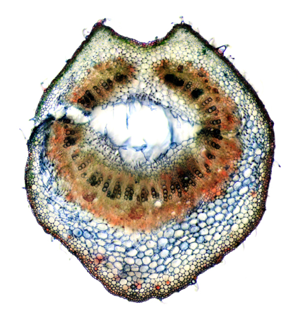
An optical image of the cross section of a leaf support (petiole). The notch in the structure points up and is believed to be associated with water flow over the leaf structure. The center of the structure is where the pith cells are located, in a mature leaf the center section would be completely filled with pith cells. This section has been dyed with neutral red and brilliant crystal blue. This picture of the petiole shows a 3 mm wide field of view. The image is a panoramic made from 18 individual images and combined with the panoramic feature in Photoshop.
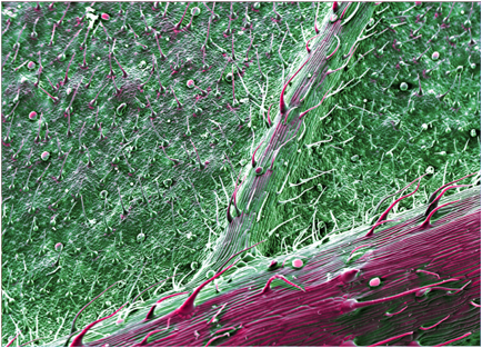
A sample of the bottom of a cannabis leaf about 3 mm wide. Two types of defensive cell structures are clearly visible: the tall needlelike trichomes and the short ball like glandular trichomes that are the source of the highest levels of THC. The high density of trichomes on modern cannabis leaves should make them a good source of THC. This is a false color SEM image with false color looking from the central rib on the bottom of a leaf towards the edge of the blade.
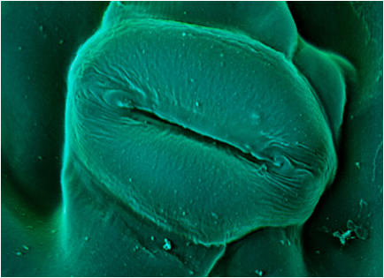
This is a highly magnification SEM image of the cell structure that allows the carbon dioxide to enter inside the leaf. This is the leaf stoma. The structure is made of two cells called the guard cells that open and close the vent depending on the time of day.
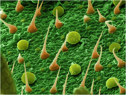
All three shots are SEM images of the bottom of the leaf. Just look at all those trichomes! Tall needlelike defensive trichomes, and the short spherical glandular trichomes which produce high levels of THC. The chemical THC is produced by the plant to ward off insect and herbivore predators. It just so happens that humans find this chemical of medical interest, and have crossbred the plant to increase the THC levels. THC is shorthand for Tetrahydrocannabinol also known as ((6aR,10aR)-delta-9-tetrahydrocannabinol), it is the psychoactive chemical in the cannabis plant. THC was first isolated in 1964 by Israeli scientists Raphael Mechoulam and Yechiel Gaoni. If you look closely at the surface you can see the stoma structures scattered around the leaf surface that allow the leaf to breathe.
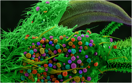
Scanning electron microscope images of the bud of a cannabis plant. This image shows the diverse forms the trichomes exhibit at the plant bud. The bud of the female plant is also the location of the highest concentration of the medicine THC. Recent measurements have shown that the concentration of THC has been found to be above 8% of the weight of the bud. These measurements are highly variable, depending on water content of the bud and techniques used to measure. The concentration of THC continues to increase due to active breading research.
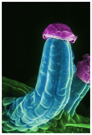
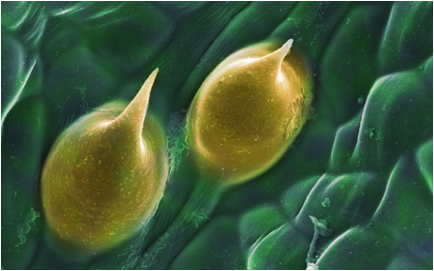
An individual glandular trichome. Approximately 0.05 mm high, the glandular trichome is the source of the highest concentration of THC in cannabis most often found on the bottom of the leaf and in the plant bud.
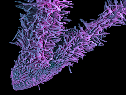
A 4 mm wide SEM view of the female flower pistil. These are very difficult structures to prepare for viewing in the SEM vacuum. The sign of a well prepared sample is that the majority of the glandular structures are full and not deflated. Samples like this were prepared with a 2.5% solution of glutaraldehyde within minutes of being collected from a living plant in California. The specimen then when through a series of increasing concentration of ethanol before removal of any liquids with a critical point dryer. All of the SEM samples were taken through a similar process and gold plated in a sputter coater before imaging. The gold coating insures that the sample is electrically conductive, a requirement for achieving good images in a scanning electron microscope.
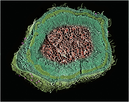
A cross section of a mature stem. The image is 6 mm wide. The different layers of the stem have been given different colors. The layer that is several cells below the skin is the section of thick-walled fiber cells. This highlights one potential uses of cannabis: an excellent source of fibers for paper, rope, and fabric. The legalizing of cannabis in several states in the United States, opens research in this very profitable aspect of the plant.
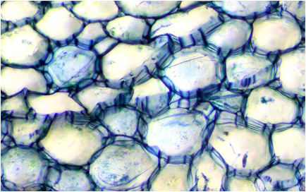
An optical microscope shows the pith cells and the cell wall structures. Dyed with brilliant crystal blue stain to show the cell walls. The pith cells are located in the center of the stem and are responsible for storing and transporting nutrients throughout the plant.
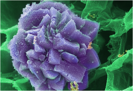
A false color SEM image of a calcium oxalate crystal. Even a small dose of calcium oxalate is enough to cause intense sensations of burning in the mouth and throat. Commonly found in popular houseplants, such as Dumbcane, the crystals effects or symptoms may last for a week or more, making raphides a non-desirable ingredient in medicinal cannabis. It is surprising that calcium oxalate is rarely discussed in literature about medical marijuana (cannabis).
If you would like to see the rest of the images, the full book is available at Amazon.com here.
Microscopy UK Front Page
Micscape Magazine
Article Library
© Microscopy UK or their contributors.
Published in the
May 2014 edition of Micscape.
Please report any Web
problems or offer general comments to the Micscape Editor.
Micscape is the on-line monthly
magazine of the Microscopy UK web
site at Microscopy-UK
© Onview.net Ltd, Microscopy-UK, and all contributors 1995 onwards. All rights reserved. Main site is at www.microscopy-uk.org.uk