|
Mysteries of Dried Stuff: Chiridota Part 4 by Richard L. Howey, Wyoming, USA |
Previous parts in the 'dried' series
Yes, I have become preoccupied; no, obsessed; nay, deranged; as mad as King Lear. My poor battered, Philippine sea cucumber continues to pose puzzles for me and yet, at the same time, I feel I have just barely begun to investigate this intriguing creature–-ordinary enough in one sense, but like an alien being from some other world in another sense.
Because of the condition of the specimen, it’s impossible to determine whether or not the wheel spicules occur in clumps covered by a membrane which is typical in other members of this genus or whether this is an exception where those spicules are widely distributed throughout the dermis and may, in some instances, actually develop right on the surface of the epidermis. My inclination at this moment is toward the latter view that they are widely distributed.
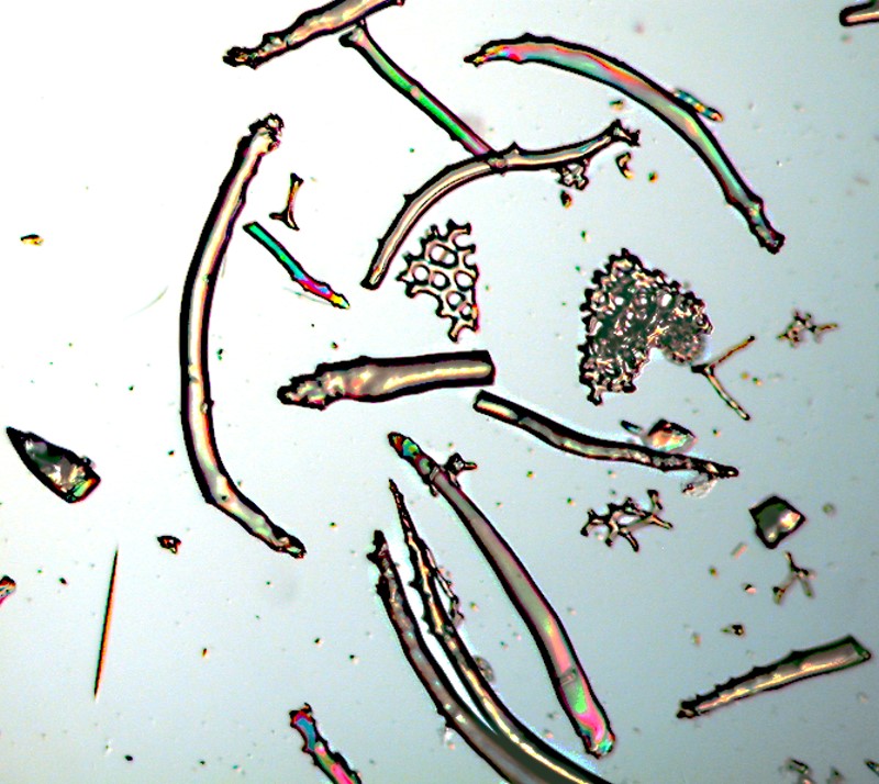
I have succeeded in finding some tube feet and have observed something rather peculiar. Around the tip of the tube foot are a large number of curved calcareous rods and they appear to form a sort of circular ring or enclosure around the tip.
Immediately below this area, there are, on the surface or just below the surface, a significant number of very small wheel spicules and these are present down to about a halfway point on the tube foot where they are then replaced by the large wheel spicules which seem especially abundant around the base of the foot.
To get the image below of the curved rods, I excised one of the tube feet, treated it with household bleach to dissolve the surrounding tissue and got this impressive array of these spicules.
I took 3 other samples from the surface tissue and subjected them to bleach as well. With very small samples, it requires 24 to 48 hours to get good results and thereafter comes the rather tedious and repetitive process of rinsing to remove all traces of the bleach. While I was waiting for the chemical and the distilled water to do their respective jobs, I went back to the specimen itself which I had preserved in 70% isopropyl alcohol. As I scanned the surface, I would look for small areas that appeared to be particularly suitable for sampling and subsequent bleach treatment. However, before I got to that stage I was finding some unexpected anomalies spicule-wise.
Some of the forms which Dr. Lambert suggested might be aberrations resulting from an acidic preservative began showing up in tissue which also had “intact” wheels, which made me begin to consider that perhaps Hamblin was right about the developmental stage but, since he only had either tiny, very early stages or adults, he likely had an incomplete picture of a species that produces spicules in a fashion different from the one I am dealing with. See part 3 of this series on Micscape.
From the sample which I treated, I began finding a number of atypical forms and I’ll give you a brief visual survey of them here.
This image above was a surprise for me because it showed a “wheel” spicule much smaller than the ones I customarily observed and a second spicule which appeared not to be fully formed. To my eye, there is not convincing evidence of fracture.
The next spicule is also anomalous and quite distinctive. I found a number of this type scattered around throughout this specimen of Chirodota. If one imagines a calcareous ring connecting the outer points, then it would begin to take on the appearance of a typical “wheel” spicule. As it is, it has more of the appearance of sharp-toothed “gear” than a wheel.
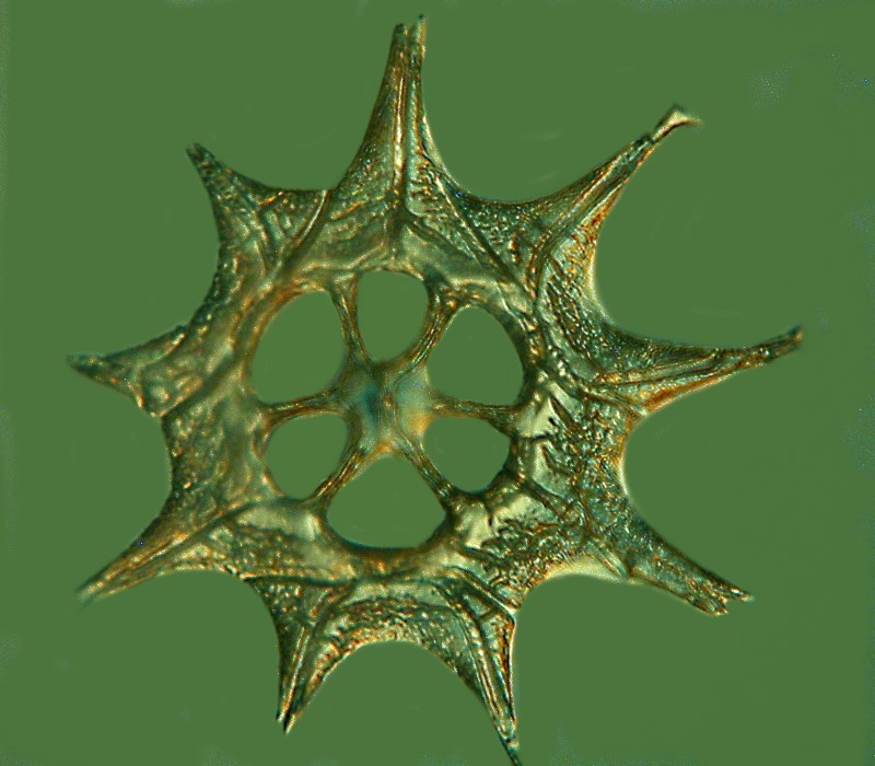
Actually, we don’t have to work very hard to imagine it, since the next image comes very close to showing what it would look like.
With the next example, we come much closer to the typical wheel, but again, as yet, not fully formed and again I see no good evidence of fracture.
The next is very similar, but illuminated differently and shows a central triangle quite clearly; one is visible in the previous image as well, but it is not as evident. Also note that there appears to be a protrusion of some sort at the very center. Again, such can be seen in other examples as well.
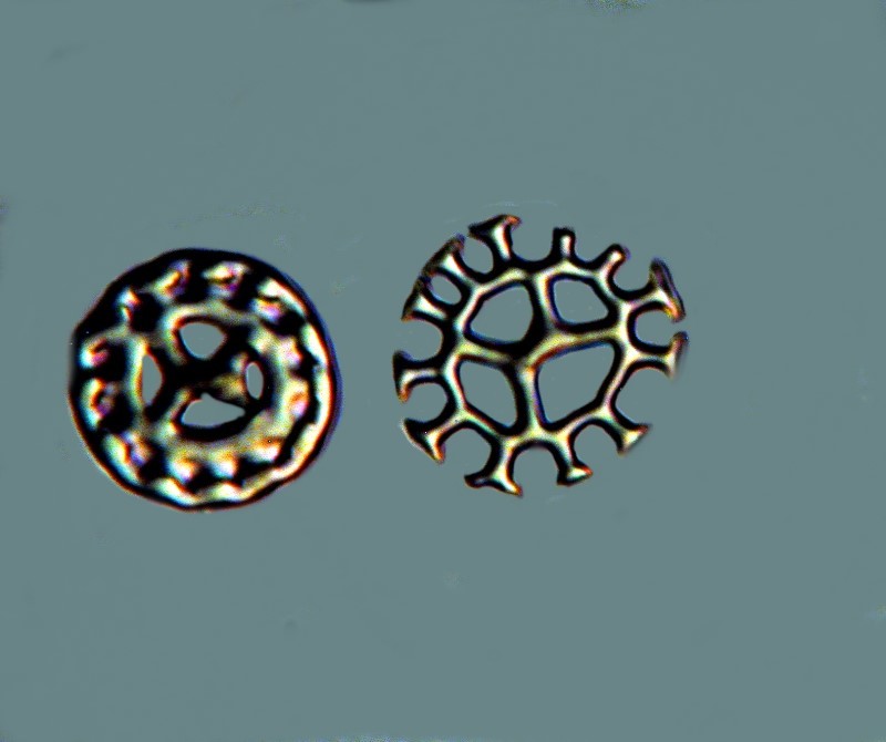
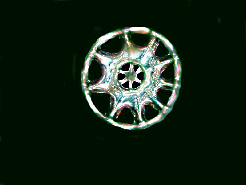
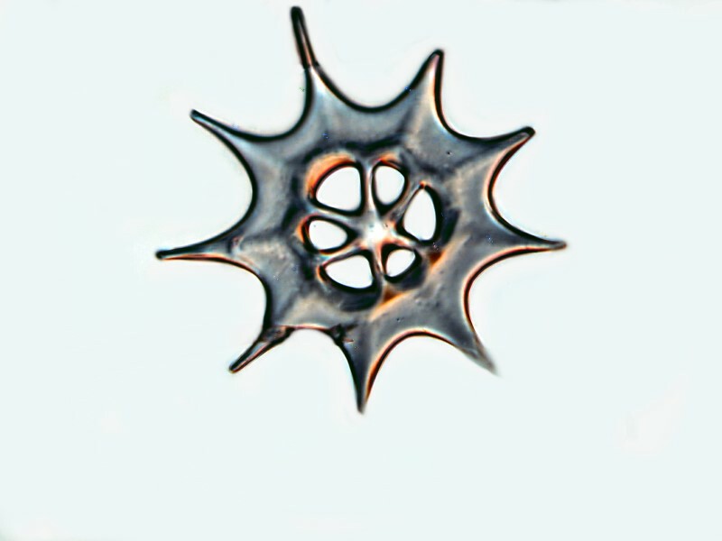
In this next image, we see a larger incomplete “wheel” spicule and 4 small spicules that look complete. These occur in sufficient numbers so that I might wonder whether these are a distinct type or whether further deposition might create large spicules from them, were it not for the fact that the morphology is quite different. The large spicules have 6 central “windows” and 9 outer “windows” whereas the small spicules have 4 central “windows” and 12 to 14 outer “windows”.
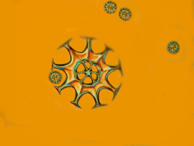
This last image was produced using “optical staining” produced with a Nomarski Differential Interference Contrast system.
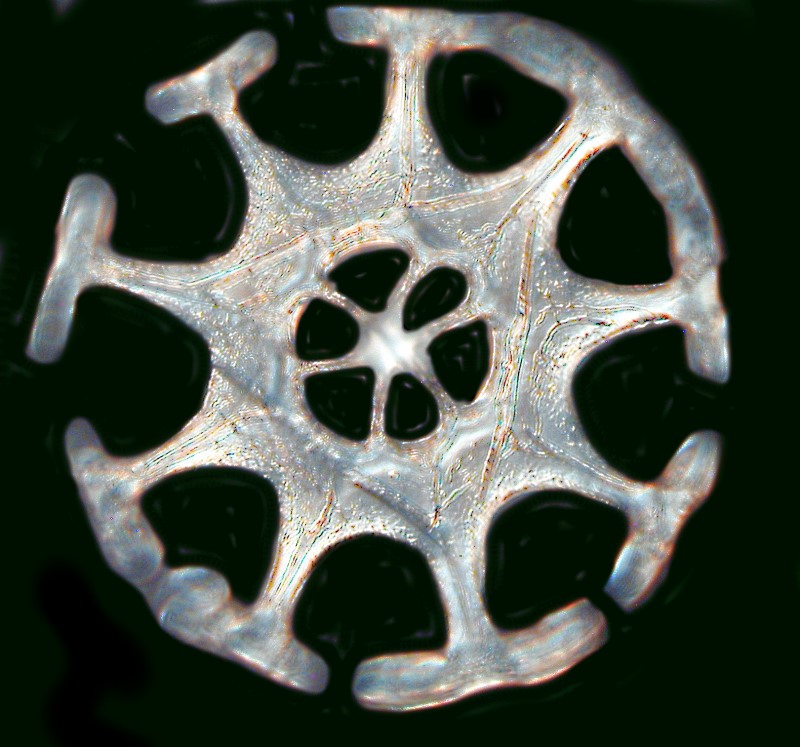
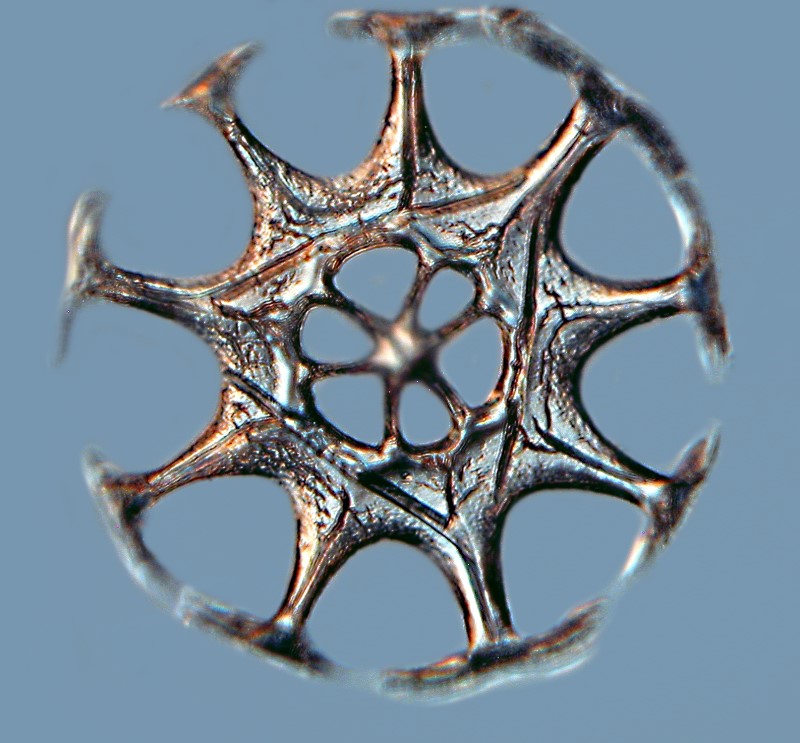
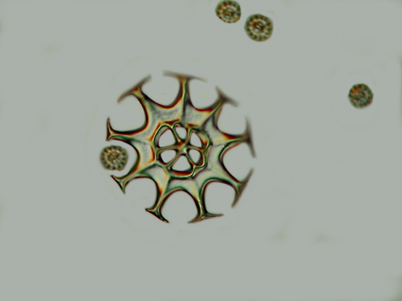
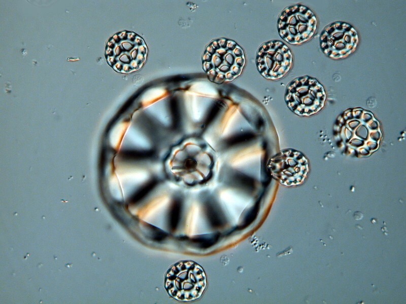
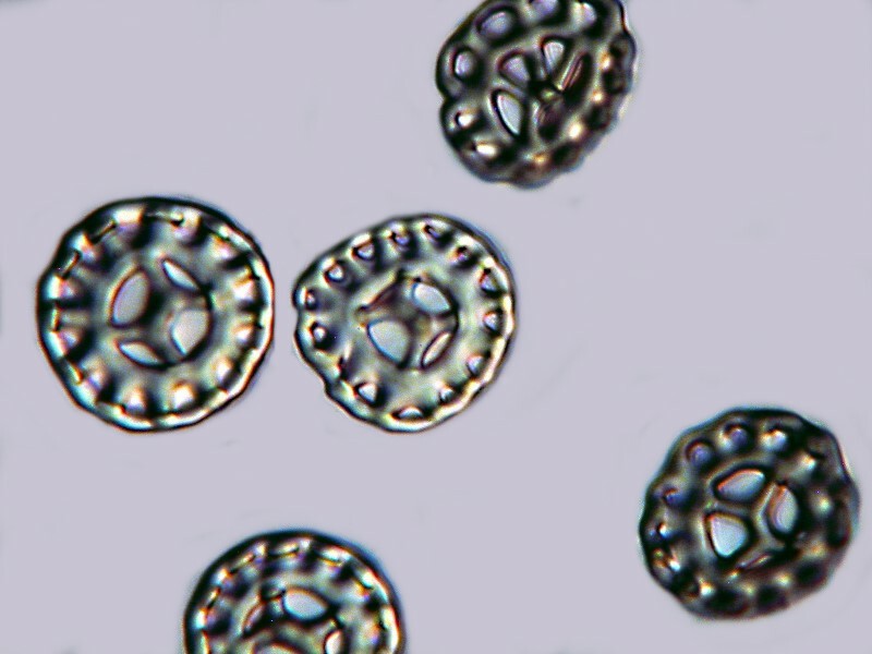
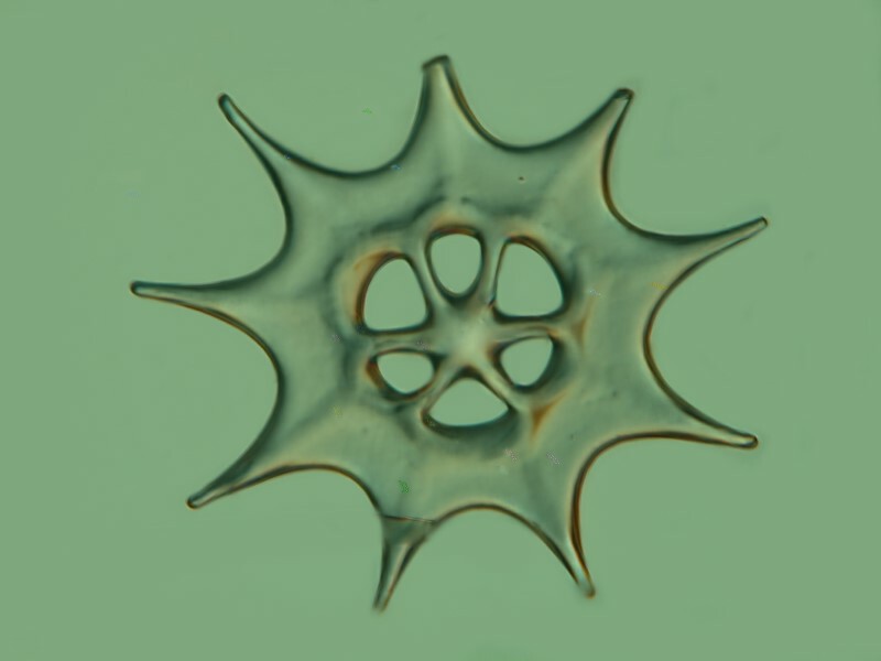
These next images will allow you to compare the large spicules with the small ones and see the differences in morphology for yourself.
Now, having had my curiosity deeply aroused–sounds erotic doesn’t it and why not? As the French playwright, film maker, poet, and artist Jean Cocteau once remarked: “Great literature is that which produces an erection of the soul”, so why not the same with a beautifully intricate puzzle in nature? As I was saying before I interrupted myself–having had a profound stirring of curiosity, I decided that the next task was one which would involve a long, elaborate and, at times, tedious series of steps concerned with taking this organism apart bit by bit until I had thoroughly examined the entire remains. You might retort, “Well, it’s a fairly small organism.” That is, of course, a relative judgment and my intent is to chemically test and extract all the calcareous material for microscopic examination. This will very likely take months, especially since I am easily diverted and am, at this moment, also intrigued by the “sea mouse” Aphrodite aculeata and its hair in particular. Some years ago, a colleague of mine, who was a first-class protozoologist and cell biologist, was studying the nucleatic material in Spirostomum. The problem was finding the right species. There are relatively small species, such as, Spirostomum teres but it has a single ovoid macronucleus and he wanted a species that had a beaded chain macronucleus. An obvious candidate was Spirostomum ambiguum which is a giant in the world of protozoa. I had some cultures of S. minus which is somewhat smaller and considerably less wide and I suggested that he consider them. He did and pronounced them an ideal compromise. He was doing serial sections of these organisms for study with a Transmission Electron Microscope using an ultra-microtome and he told me: “If I had to serial section S. ambiguum, it would be like sectioning an elephant using a razor blade.” So, “small” is highly relative.
That fact was underscored for me again this afternoon when I was looking at a thin sliver of tissue from my Philippine Chiridota. Several days ago, I removed the tissue from the alcohol-preserved specimen and put it aside to dry thoroughly. This was necessary because I want to place it in immersion oil, add a coverglass, and examine it with a compound microscope using brightfield, Nomarski DIC, and polarization. What I found reinforced my resolution to examine as much as is feasible of this specimen in close detail.
For such a small piece of tissue (about 10mm long and 2 or 3 mm wide), there was a fair abundance of the large and small wheel spicules characteristic of this species.
Then, I began to find examples of either damaged or immature forms of spicules–spicules with perfect symmetry but without the outer ring; spicules with the barest beginning of cross bars on the outer projections and all of this reinforces my bipolar, schizophrenic view that what we are seeing are the stages of fairly complex structures which reveal to us intermediate forms and also provide us with information about what happens when pressure is applied to their fracture points. First the images of the incomplete forms.
Then when pressure is applied to a large spicule in immersion oil, it causes it to break apart from the center upward projection outward. This reinforces the idea the these spicules consist of layers or tiers of deposition.
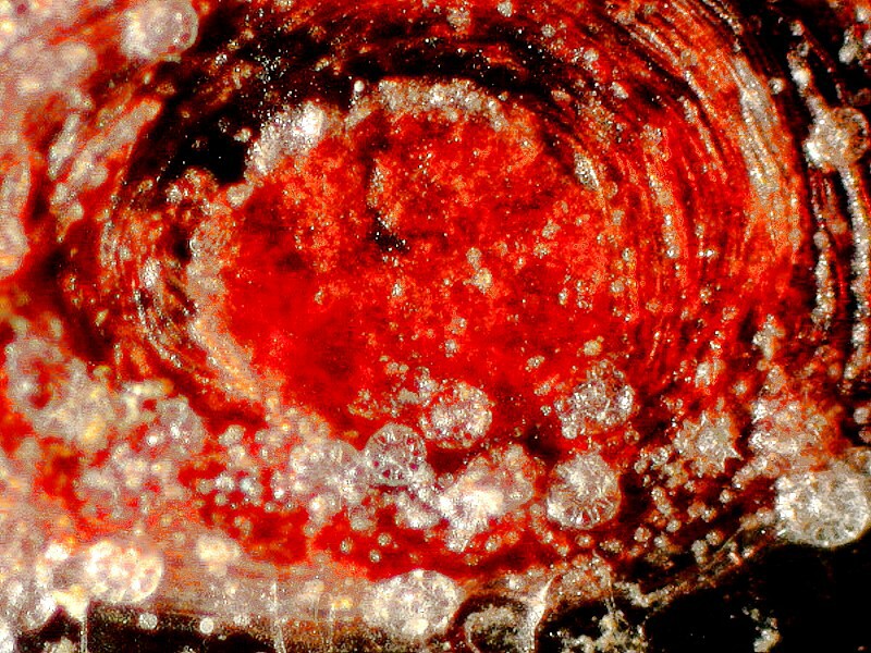
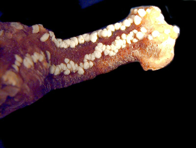
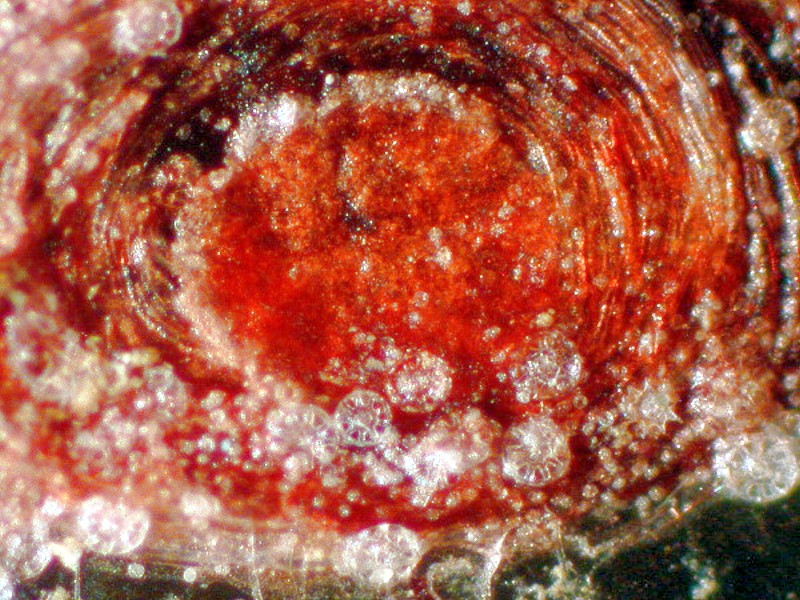
I finally decided to begin examining some larger tissue samples. I began by removing several pieces which were fairly readily accessible as a consequence of the damage to the specimen. Some teasing with micro-dissecting needles and a few snips with micro-scissors provided several pieces which provided very interesting information–and more mysteries. I have been puzzled about the areas of pigment which, as it turns out, are not so much orangish-red as a bright dark red as is evident under the microscope but, much more interesting is the fact that the pigment seems to be concentrated in the tube feet and much of the other tissue is a pale, opalescent white. This is important because now I can tell my friendly Philippine supplier to be on the lookout for some red-footed sea cucumbers that have wheel spicules and maybe I can obtain some intact specimens to help solve some mysteries (and doubtless create some new ones).
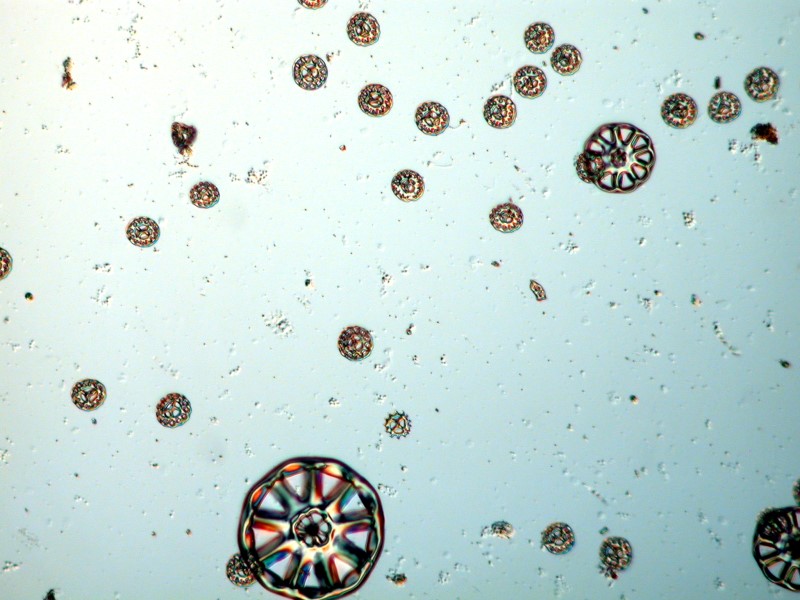
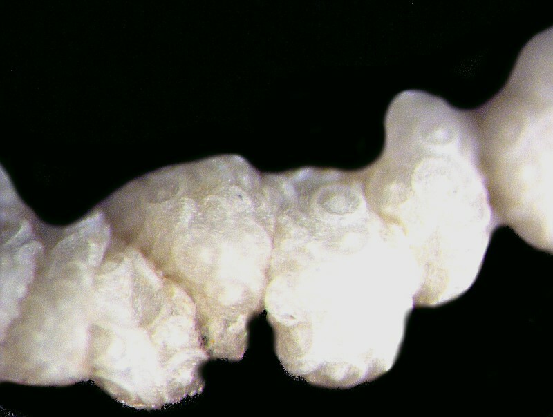
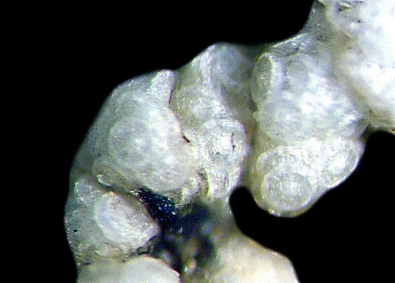
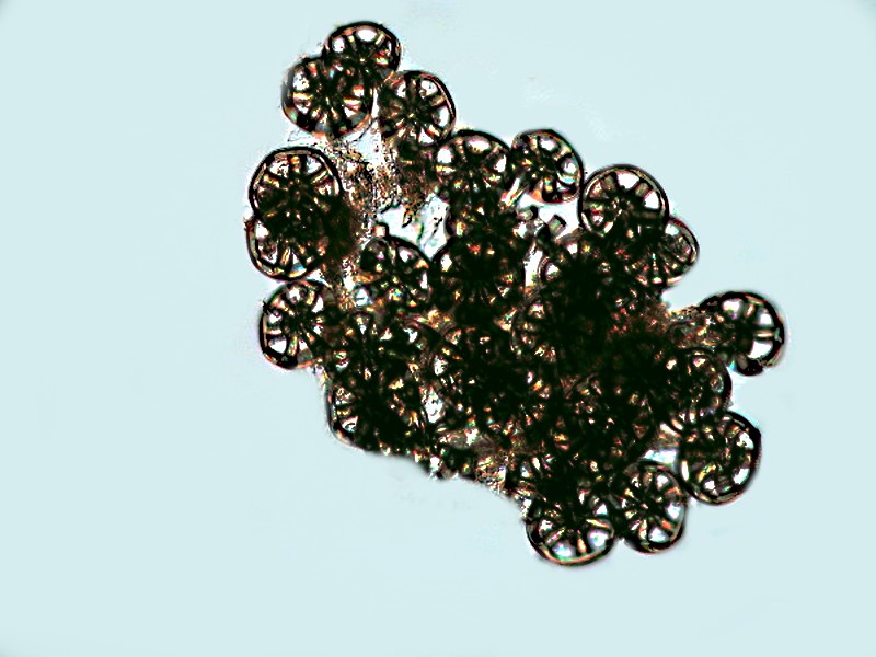
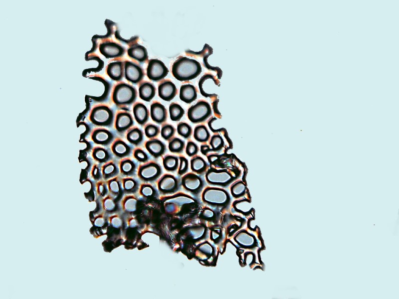
I mentioned previously that the large wheel spicules seem to be widely dispersed over the organism’s surface and the small ones are incredibly abundant especially in the fold along the rows of tube feet. In examining another piece of tissue, I noticed several white clumps covered by thin membranes. As I said earlier, Chiridota typically have clumps of wheel spicules in little pockets subdermally, sometimes randomly, sometimes in rows as can be seem in this specimen of C. laevis from Maine.
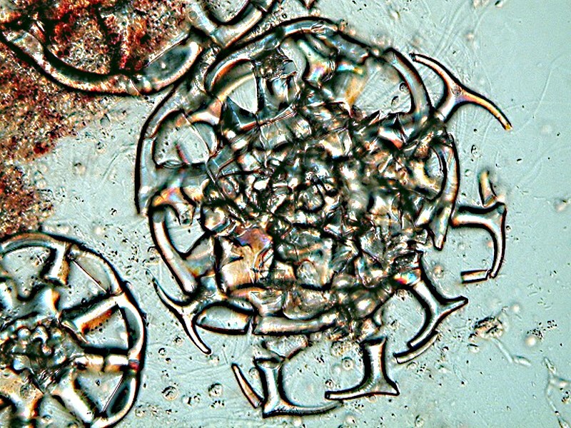
However, in this case, as I zoomed in to get a clearer view of the packets, it became immediately clear that they did not contain the large wheel spicules, but large numbers of small wheel spicules, a significant number of which were not fully formed. They had some fenestrations but, in most cases had not yet developed symmetry and had, in general, a rather grainy micro-crystalline appearance. This was completely unexpected and again tends to reinforce my inclination to regard this genus as demonstrating a series of developmental stages in the formation of spicules.
I cut a piece of the tissue with the packet of spicules still intact and put it on a slide to dry thoroughly. (A convenient way to keep dust off such slides is to place them in a standard size Petri dish.) After 3 days, I added a fairly large drop of immersion oil and a coverglass. It is important to have enough oil to avoid crushing the packet.
However, when you wish to open up the packet to more closely examine the contents, you can exert pressure on the coverglass using a standard dissecting needle until the membrane breaks.
When using the immersion oil on either the large or the small wheel spicules, it is important to let the slide sit a day or two to maximize the possibility that it will penetrate into the fenestrations of the wheel.
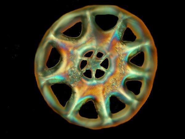
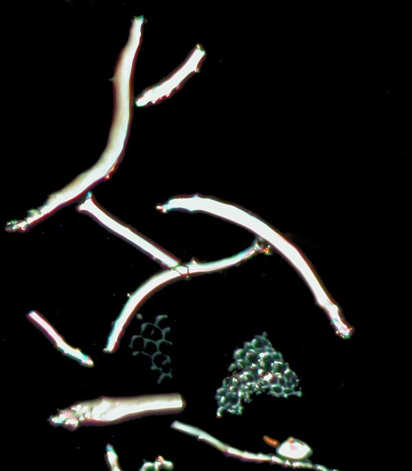
When I put another tissue sample under a compound microscope with Nomarski DIC, I got 2 more surprises: 1) curved rods, some of which had elaborate, but tiny, thorny processes at the terminus and 2) a large wheel spicule which revealed its inner structure in such a clear fashion that I felt it was as though the top or center part was missing. Here is an image of the curved rods.
Immediately, my hyperactive mind leapt to a wild conclusion: perhaps these large wheel spicules are constructed somewhat like diatom frustules. Remember that diatom “shells” are constructed rather like pill boxes with a bottom and a “cover”. However, that analog is too simple to fit the case of these wheel spicules. What I suspect at the moment–based on complete and blind ignorance (following the model of Republican politicians)–is that these are more or less distinct layers of crystalline deposition that constitute the whole, rather like the floors of a low building in which each such floor has its own distinctive character but, all of the floors are interdependent. In general, this fits the sort of model that Hamlin was presenting in Figure 2 as you can see if you go back and look at earlier Part 3 of this essay.
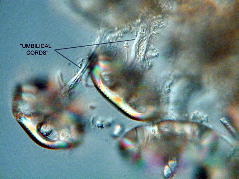
However, even more intriguing are figures 4 and 5 on the same page, for they suggest that there are “cords” that connect the “wheels” in the capsule to a central mass within the packet. After a good bit of careful dissection and searching, I was able to find such “umbilical cords” and here are 2 images of them.
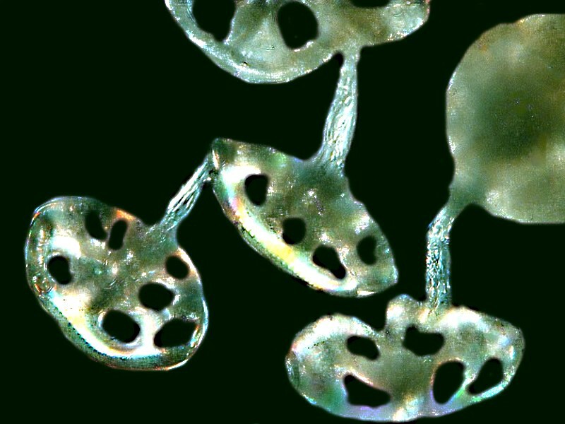
When I look at the dried, large wheel spicules, I am amazed by their vitreous, glistening appearance and when they are facing upward, they rather remind me of a miniature, circular glass tray for deviled eggs. I also find it surprising how many spicules are packed into one cluster.
I removed 7 other small sections of tissue which had been soaking in distilled water, placed them on slides and let them dry. This afternoon, I took a micro-chisel and pried the very top of a tube foot from a slide. I first examined it in its dry state. The top, which has a light reddish pigmentation, looks rather like the top of a circular pencil eraser.
The underside was covered with curved rods.
I decided it was time for some Clorox and as it bubbled away while I was listening to a Mozart Serenade, I watched and waited and using a micro-needle, I tried to push the bubbles away to see what was going on beneath them. The first thing I noticed was some of the rods dropping to the bottom of the slide. The bubbles are persistent and annoying; you push them away, but they immediately return. I watched and waited a bit more and pushed the raft of bubbles again and noticed that some pieces of very delicate fenestrated plate had fallen to the bottom of the slide as well. At this point, I suspected that something quite interesting was emerging and I took a micro-pipet and removed the raft of bubbles. What was emerging was a circular support disk of the lacy, fenestrated material lying just under the top part or “cap” of the tube foot. As I continued to stir and poke, naturally it broke apart, but I can show you some detail of a fragment.
I wanted to examine these under a compound microscope so, of course, that meant rinsing off the bleach–no easy task on a slide when you risk pipetting off the very objects you wish to examine. Usually, I use distilled water rinses but, it occurred to me–why not try hydrogen peroxide. Here a couple of cautionary remarks are in order. A bleach like Clorox contains chlorine compounds and one needs to be very careful not to mix it with the wrong things or you may very well end up generating deadly chlorine gas. Also hydrogen peroxide is a power oxidizer and can produce rapid and violent reactions. As it turns out, this particular combination of Sodium hypochlorite and hydrogen peroxide produces some very active oxygen (hence, the bubbles), water, and table salt. The nuisance of multiple rinsings is still necessary but, sodium chloride crystals are somewhat easier to deal with than sodium hypochlorite crystals. One thing I found out was even with a slide that had not been fully and adequately rinsed, when left to dry, some crystals would form but when immersion oil was added, some of the crystals dissolved enough to allow for some reasonable images with a bit of editing with graphics software.
If the rinsing operation is carried out in a small Petri dish, or better yet, a curved watch glass, then it is easier to pipet off the excess fluid with minimum disturbance of the spicules and if you are sufficiently patient, you will finally get to the point with hydrogen peroxide rinses where there are virtually no salts to leave crystalline deposits. At this point, you can use a micro-pipet to remove and place individual spicules or small groups of them and let them dry on a slide and then either, if indeed there are no crystal deposits 1) mount them in a pH neutral resinous medium or glycerine jelly or 2) if you are not concerned with having a permanent mount and just want to take a series of photomicrographs, then you can mount them in immersion oil.
Since this essay has gotten rather long, I will point out that these techniques will also work for sponge spicules of which there is an extraordinary variety and I hope to deal with some of those in future essays. In the meantime, fear not, I have not abandoned my sea cucumber from the Philippines and Part 5 is already in the works.
All comments to the author Richard Howey are welcomed.
Editor's note: Visit Richard Howey's new website at http://rhowey.googlepages.com/home where he plans to share aspects of his wide interests.
Microscopy UK Front
Page
Micscape
Magazine
Article
Library
Published in the May 2017 edition of Micscape Magazine.
Please report any Web problems or offer general comments to the Micscape Editor .
Micscape is the on-line monthly magazine of the Microscopy UK website at Microscopy-UK .
©
Onview.net Ltd, Microscopy-UK, and all contributors 1995
onwards. All rights reserved.
Main site is at
www.microscopy-uk.org.uk .