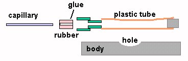
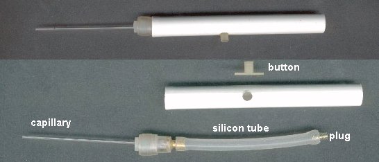
MICRO TOOLS 

FOR MICROSCOPY - II

by Jean-Marie Cavanihac, France
Another tool, the micropipette, is very useful if you want to select only one specimen when you are sorting them with a stereo microscope for example. It's easy to make them, if you can find glass tubes one or two millimeters in diameter. 
I suppose you have a medical assay laboratory, carrying out blood analysis in your town? If you do, explain the purpose of your query, and you can probably obtain "micro hematocrit capillaries". 

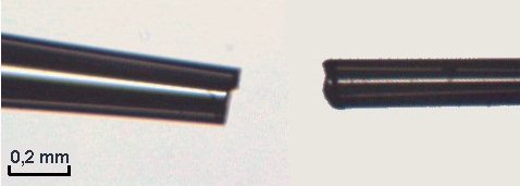
If possible ask for capillaries that are NOT heparinized. (Heparin is used to avoid blood coagulation and some capillaries are coated inside with this chemical). If they are, rinse the tubes well between uses to avoid killing (?) critters. 
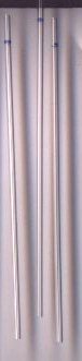
In labs (especially in pediatrics), these 1,2 mm inside diameter capillaries are filled with 100 µl of whole blood  and centrifuged to accelerate cell sedimentation. 

Red and white blood cells separate themselves from the plasma and the length of the red cell portion divided by total capillary length (3") is called hematocrit: usually 40 - 45 % of total length.

(If the result is more than 50 %, you have too many red blood cells and you are probably dopey with E.P.O. ! ).

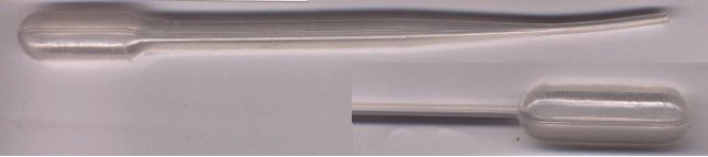
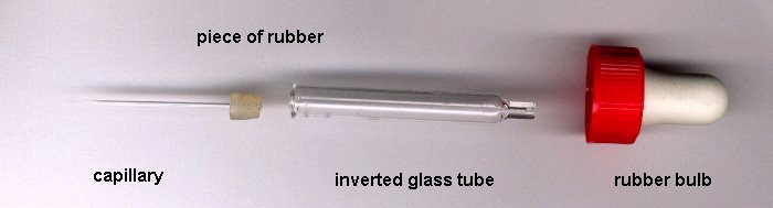
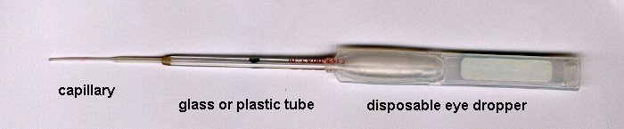
If the biologist is sympathetic, you can ask them for one or two disposable plastic pipettes which are very useful too! (See picture below).
Well, you now have some capillaries: you can use them as they are, but you can also make micro-tipped pipettes. Take a capillary with one hand at each end, use a LITTLE flame (lighter or candle) and heat gently in the middle of the tube, until the glass softens, then stretch it with both hands. 
Tube centre becomes progressively smaller in diameter. 

Stop when the diameter is around 0,3 - 0,5 mm. 

Then break glass at middle - you obtain two micropipettes.

Observe the ends with a low power microscope objective: if necessary polish the tube ends with sandpaper (very fine grade).
Observe the tip from time to time and continue polishing until the end becomes clear. 

You can also RAPIDLY warm the tip in the flame, blowing air through the tip to prevent it closing.

(Un-polished tips in picture.)

Then fit this stretched micropipette onto a disposable pipette or into an eye dropper: the lower one in the picture above is ideal. 
If you use a rubber bulb as shown in the picture, squeeze it slighly just to fill the capillary and NOT the large glass tube, otherwise the specimen may be difficult to retrieve! 
You can also make a sucking device with a modified retractable lead pencil. The plastic tube closed at one end is aquarium air tubing or silicon tubing (see model shops). Drill the rubber eraser from the propelling pencil with a 1,5 mm diameter hole and glue it into its holder - green on drawing, (on which you press normally to make the lead go down). Make a hole in the pencil body to press the plastic tube with your finger or, better, with a button . 
With this device it's more easy to control pressure just to half fill the capillary, and your specimen remains inside.
You can to use, too, a disposable eye dropper fitted with plastic tube (from an old ball pen) as shown in the picture below:


Well, you now have micro-tools.

But how do we recruit micro-workers? You don't need a job advertisement, as you have a means to exploit some microscopic assistance free of charge! If you observe diatoms, you have probably already noted that their shapes can be very different when you look at them in front view or side view. Unfortunately it's difficult to manipulate them, in order to view from both orientations. But, if you add many large protozoans to your slide, they move randomly in all orientations and they knock diatoms, moving them until they are overturned. Thank you gentlemen for your help! 

Add protozoans ONLY to the slide because some of them eat diatoms !

A micro-worker which eats during its working hours!
RETURN TO FIRST PAGE
Comments to the author Jean-Marie Cavanihac are welcomed.

Microscopy UK Front Page
Micscape Magazine
Article Library


All drawings and photographs © Jean-Marie Cavanihac 2001

Published in the November 2001 edition of Micscape Magazine.

Please report any Web problems or offer general comments to the Micscape Editor,
via the contact on current Micscape Index. 

Micscape is the on-line monthly magazine of the Microscopy UK web
site at Microscopy-UK


© Onview.net Ltd, Microscopy-UK, and all contributors 1995 onwards. All rights reserved. Main site is at www.microscopy-uk.org.uk with full mirror at www.microscopy-uk.net.