|
|
A Gallery of Testosterone Photomicrographs ( using polarized light illumination ) by Brian Johnston (Canada) |
|
|
A Gallery of Testosterone Photomicrographs ( using polarized light illumination ) by Brian Johnston (Canada) |
In an earlier Micscape article, I presented several photomicrographs of testosterone taken many years ago using film equipment. Recently I found a very small quantity of the compound, (enough to prepare two slides), and this article presents an overview of the problems and rewards resulting from a study of testosterone using digital camera equipment.
The white crystalline substance has a melting temperature of about 155 degrees Celsius and a melt specimen can be prepared by placing a few crystals on a slide, covering with a cover-glass, and heating very gently over an alcohol lamp. I usually hold the slide about four or five flame lengths above the flame tip and this results in the first melting taking place in about 30 seconds. (This may seem like a long time, but overheating results in tiny bubbles being formed between cover-glass and slide which tend to spoil the final images. The large distance also reduces finger-tip overheating!) Once melting has begun, I raise the slide still higher, and carefully move it in a circular motion in order to ensure that the entire sample has been heated evenly. After melting is complete, the slide is allowed to cool slowly. Fast cooling, for instance by placing in a refrigerator, results in the rather uninteresting amorphous crystal structures that will be seen later in the article. Note that testosterone is considered poisonous and should be handled with care.
Testosterone slide production requires care, and especially patience. Unlike most chemicals that re-crystallize in seconds after removal from the heat source, testosterone usually requires months to completely solidify. Within two or three days however, you should begin to see a thin ring of crystal growth along the edge of the cover-glass. Since this ring often contains excellent viewing or imaging possibilities, it is a good idea to have a look every couple of days to observe the changes taking place.
Keep in mind that being obsessive about cleaning the original microscope slide may actually decrease the number of interesting formations that result during the re-crystallization process. I have found that the tiny particles of dust, and imperfections in the surface glass act as sites for growth, and these often produce interesting images.
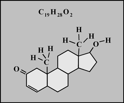
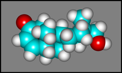
Testosterone is a steroid hormone found in both males and females. The complex molecular structure is shown below. (The second image was produced using "HyperChem" software.)
The images in the article were photographed using a Nikon Coolpix 4500 camera attached to a Leitz SM-Pol polarizing microscope. Crossed polars were used in all images. Compensators, (l and l/4 plates), were utilized to alter the appearance in some cases. A 2.5x, 6.3x or 16x flat-field objective formed the original image and a 10x Periplan eyepiece projected the image to the camera lens.
The photomicrograph on the left below shows a typical field away from the cover-slip edge after about three weeks. On the right is a lower magnification image of the same section of the slide after a month. Notice how another growth front has collided with the first to fill in the black area in the left image.
The thickness of the crystal layer on the second melt slide is different, probably due to my placing a different amount of compound on the slide before melting. This results in a completely different colour spectrum in the image below (which was taken after about two months of growth). The uncrystallized area at this time is very small.
A growing area is shown below. The image on the right utilizes a l/4 compensator, (sometimes called a quarter-wave plate) to produce elliptically polarized light instead of the normal plane polarized variety. Notice that the black radial "fans" are missing in the right image.
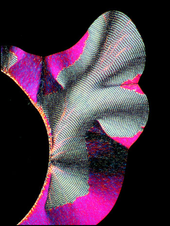
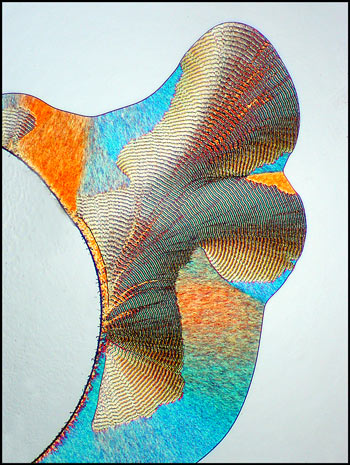
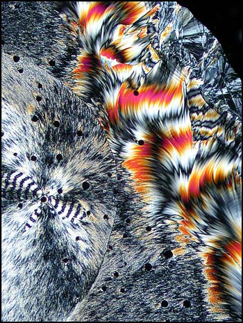
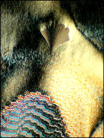
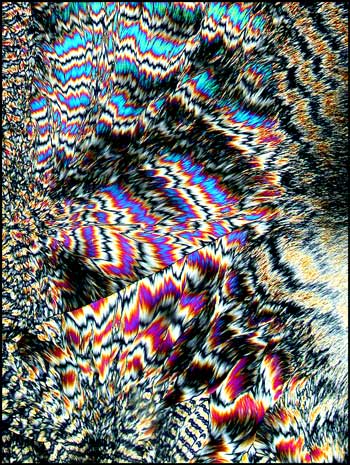
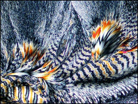
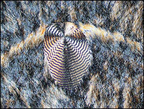
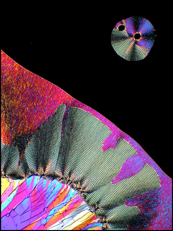
One striking characteristic of most testosterone melt specimens is the presence of colourful repeating bands. These bands grow in concert with the amorphous areas. There seems to be no rhyme or reason to explain their presence in a particular location. Note the distinctive fan-shape at the top of the right hand image below.
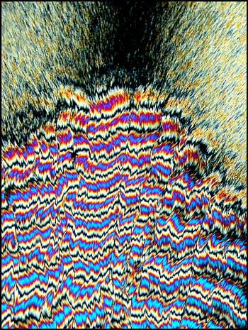
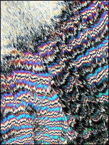
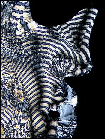
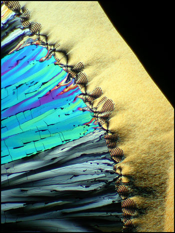
In the following four photomicrographs, taken at a higher magnification, the complex nature of the bands is revealed.
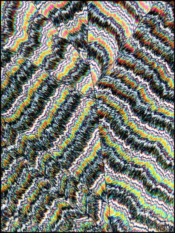
The two images below were taken with still higher magnification.

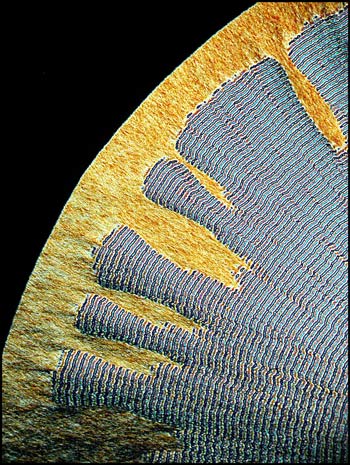
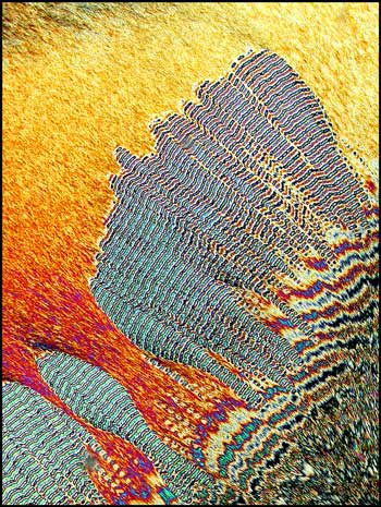
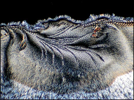
As mentioned earlier, crystals begin to grow at the edge of the cover-glass within a couple of days. This initial growth area is full of photomicrographic opportunities. The following four photomicrographs illustrate this.
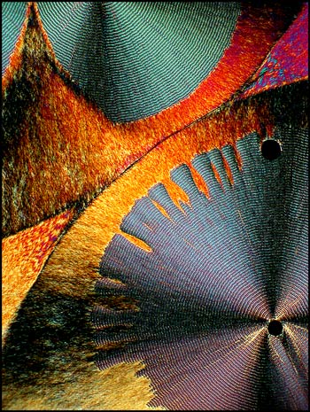
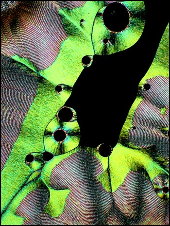
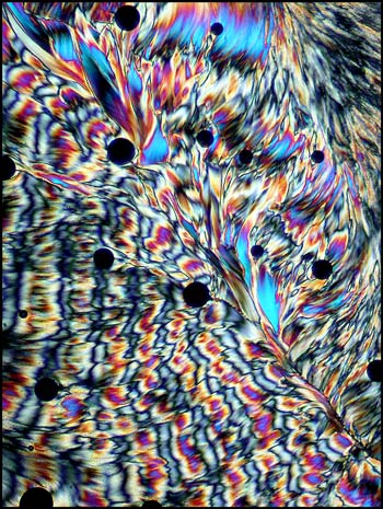
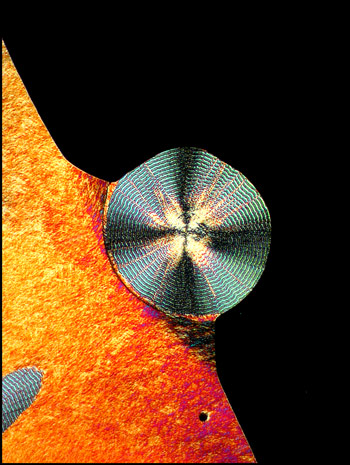
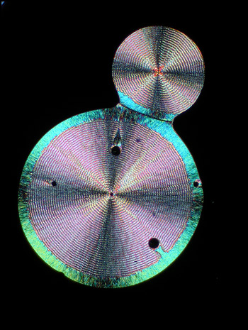
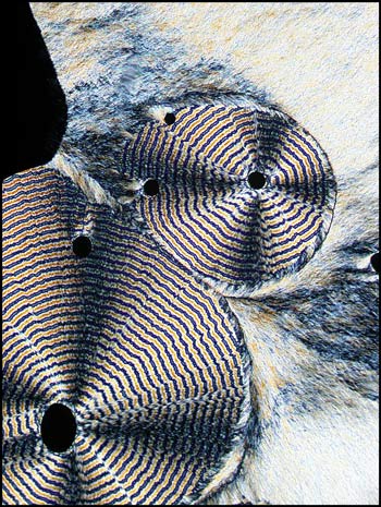
If dust motes are present in the crystal melt, circular patterns may be produced. These tend to be in the central area of the specimen, and therefore don't appear for weeks. The circular patterns may be overtaken by the general matrix, as in the left image, or they may run into one another, as in the right image.
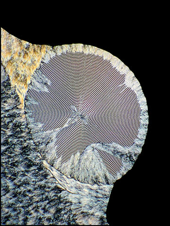
Occasionally the circular form is distorted as in the image below.
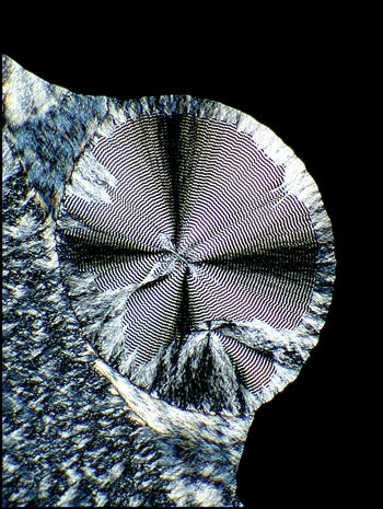
One such circular growth is shown below being surrounded by the feathery matrix. The image on the right uses a quarter-wave plate to remove the interference figure which appears as a black cross in normal polarized light.
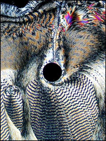
Overheating a spot on the slide during melting will usually produce tiny "bubbles" which contain no crystal material. These spots can act as sites for circular pattern growth as in the image below.
As an experiment, I placed a newly prepared melt specimen in the refrigerator over night to speed up crystal formation. In the morning, the rather uninteresting crystals that can be seen in the bottom left corners of the two images below were present. Upon return to the normal room temperature environment, the very slow crystal growth continued as before.
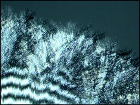
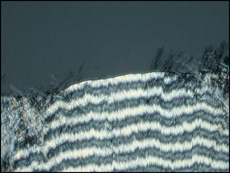
As the crystal front grows, it appears as a sharp line of demarcation between solid and liquid. However, under very high magnification, the fine needle-like crystals that make up the solid can clearly be seen protruding into the liquid.
Strangely, at another point on the growth front, these small needles seem to be missing. I would imagine that they are still present, but orders of magnitude smaller.
Although testosterone melt specimens can be relatively difficult to prepare successfully, and require much patience, they produce an abundance of wonderful image possibilities. How fortunate that I neglected to dispose of an "almost" empty vial two decades ago. The residue remaining in the bottom supplied the material for the two melt specimens photographed in this article!
All comments to the author Brian Johnston are welcomed.
Published in the November 2004
edition of Micscape.
Please report any
Web problems or offer general comments to the
Micscape
Editor.
Micscape is the on-line
monthly magazine of the Microscopy UK web
site at
Microscopy-UK
© Onview.net Ltd, Microscopy-UK, and all contributors 1995 onwards. All rights reserved. Main site is at www.microscopy-uk.org.uk with full mirror at www.microscopy-uk.net .