|
Nineteenth Century British Microscopy and Natural History: Part 2 by Richard L. Howey, Wyoming, USA |
Editor's note: Additional plates added March 2009.
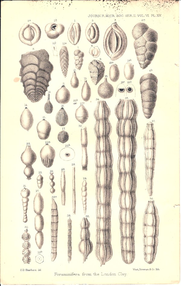
Once again, I want to examine an issue of the Journal of the Royal Microscopical Society, this time the issue from October 1886. The key paper in this issue is by Charles D. Sherborn and Frederick Chapman and is Part XII of a series, this one titled: “On some Microzoa from the London Clay exposed in the Drainage Works, Piccadilly, London, 1885.” I’ll bet this has you all sitting on the edges of your seats. The microzoa in question are foraminifera and ostracods and the article is accompanied by three splendid plates of the forams which I shall reproduce for you here. As I mentioned in the previous article in this series, forams have had significant economic importance because they are indicators of oil-bearing strata. Each plate has over 40 drawings and in the article each one is identified in terms of both genus and species. If you want the list identifying the forams in the three plates, just let me know and for a mere $150 I’ll send it to you.–Just kidding.
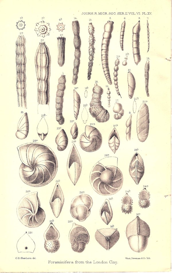
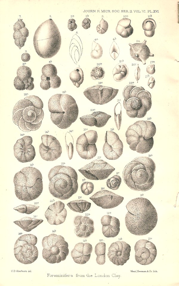
These more than a century old identifications can be helpful indicators in trying to pin down some particular forams you might have, but foram taxonomy is a progressive nightmare with micro-paleontologists juggling species and genera on a monthly basis. Like most scientists, micro-paleontologists (these are wee scientists only about 3mm. tall) invent their own language when they get down to dealing with the finer details. Consider the following:
“Our specimen the first of this ‘genus’ [Cassidulina] recorded from the London Clay, is very small, and we at first hesitated to place it under Brady’s form, but remarking its pear-shaped bulimine-like orifice, and its subrotundate form, we consider it referable to this variety rather than C. crassa d’Orbigny.” The terminological additions over the last century makes definitive identification of species largely the province of the specialist.
The “Summary of Current Researches” follows the same model as the one in the issue we looked at previously. This time under the embryology section for vertebrates, there is a summary of an article titled: “Embryology of Armadillos” which sounds fascinating. The author of the article, which appeared in American Naturalist, Dr. H. v. Ihering reports that South Americans believe that armadillos produce only male offspring.
“He states that all the foetuses taken from two females presented the external characters of males only...The sexual characters are probably like those of hyaenas, in that the female foetus has a clitoris so large as to give her a close resemblance to the male.”
One of nature’s endless experiments which present us with intriguing and bizarre puzzles to solve.
This issue is full of fascinating (to me, at least) summaries and links to the original articles, especially on invertebrates. The late 19th Century was an exciting period for both the serious amateur and the professional biologist, for if one was diligent, thorough, and patient, the chances were quite good that one would come across a new species. There was an enormous amount of taxonomic activity and, in particular, a great deal of interest in phylogenetic hierarchy. Papers appeared frequently describing new organisms as well as other papers that dealt with reorganizing the classification of various groups of organisms. In the 1880s, rotifers and tardigrades were still placed under the category of Vermes or worm-like creatures and they were lumped together with rhabdocoels, nemertines, and even the bizarre Balanoglossus. If you’re not an enthusiast of the aquatic invertebrates, you may be asking yourself: “What in the devil is he talking about? And why should I care?” O.K. It’s your own fault. You asked for a lecture. Please be patient. Let me try to give you a few reasons.
1) Many of these creatures are intrinsically exciting. Some are extraordinarily beautiful; some are bizarrely grotesque; and almost all of them have hidden secrets, many of which can help us develop not only new insights, but new technologies.
Take a look at this elegant creature.
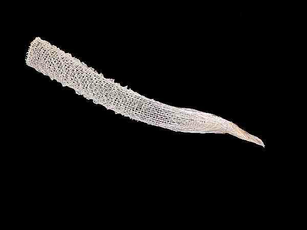
This is the skeleton of Euplectella aspergillum, a deep sea sponge, also know as Venus Flower Basket. It is composed largely of silica (glass) and belongs to a group know as the glass sponges or hexactinellids, so named for their 6-pronged spicules. It anchors itself in the substrate by means of long glass “hairs” which you can see wrapped around the base of the basket.
So what’s so special about this? Well, as it turns out these “hairs” are better light conductors than any optical fibers that humans can make today in the sense that they are stronger and not given to cracking and we all know that fiber optics have enormous numbers of applications. Why would sponges evolve in such a way as to develop an optical fiber superior to what we can make? It’s simple; just to prove that they’re smarter than we are.
Even worse, there’s a large marine worm which many people find grotesque (I find it quite fascinating) which has two kinds of bristles and one of them is –you guessed it–an especially high quality light conductor. In this case, however, it is not made of glass, but complex protein compounds. The way these occur, in the form of cylinders, makes them more efficient that anything we can yet produce. These worms get up to about 8 or 9 inches in length, 2½ or 3 inches in width, and 1½ or 2 inches thick. Underneath all of the bristles and hair on the dorsal surface are a series of scales or plates. They are bottom scavengers and all sorts of detritus accumulates on their backs and micro-algae thrive in this environment as well, giving them the worm-equivalent appearance of a street urchin. However with a good cleaning and brushing, one discovers that the bristles (or setae) and the scales have iridescent properties, although you’d never know it from these images.
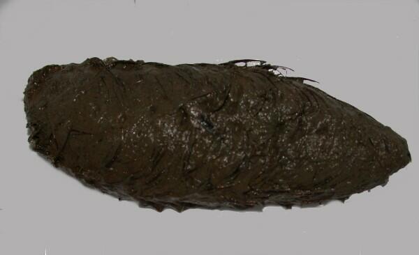
This is a top view and below is the underside.

Here is an image of a different species of bristle worm and you can indeed see the bristles quite distinctly.
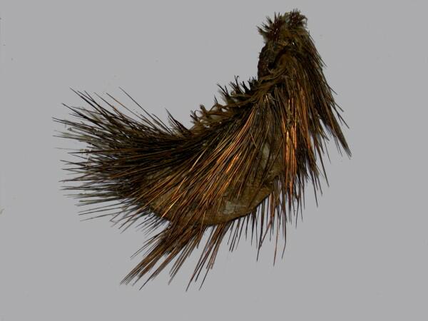
The biologist who gave this remarkable creature its name must have had a wry wit, for it belongs to the genus Aphrodite. For those of you who are Woody Allen fans, we might, from the image above call it Muddy Aphrodite. It has also long had the popular name of sea mouse. The hairs of the sea mouse were a popular object for Victorian slide mounters.
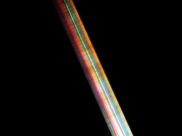
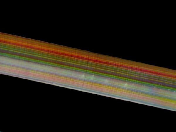
Now, the truly remarkable thing about these images is that they are NOT taken with polarized light; the colors are a result of refraction from the fibers within the hairs!
2) If I believed in reincarnation, the next time around I think I’d become a marine pharmacologist. This is an enormously exciting area of research which is underfunded and under-supported. Let me give you just two examples of this sort of research. There are some jellyfish which possess extraordinarily powerful neurotoxins. If you are severely stung by the tropical jellyfish popularly known as the sea wasp, you may survive for only a few minutes. Am I proposing that this toxin be extracted for use by the military agencies and secret agents? Let me give you a loud and unequivocal NO!!! That is one way to get funding and unfortunately too many researchers, in a wide variety of fields, have made radical ethical compromises in order to get funding from military and “intelligence’ agencies. It is an outrage that politicians, military agencies, religious zealots, and gigantic, impersonal, multinational corporations are determining the course of scientific research in this country. The reason these neurotoxins are important is that some research has shown important possible medical applications for some of these substances. By altering the molecules to eliminate the toxic effects, it is believed to be possible to use these compounds to produce painkillers which are up to 20 times more effective than morphine.
The second example has to do with sea cucumbers which are related to starfish and sea urchins, but bear little external resemblance to them. A substance called holothurin has been extracted from certain species which demonstrates anti-cancer activity. There are thousands and thousands of marine organisms which we know virtually nothing about and it is not only possible, but probable, that a concentrated series of extensive research projects will yield not only new scientific knowledge, but technological and medical innovations as well. Sadly our mismanagement of marine resources is as disastrous as our mismanagement of terrestrial resources and many species may become extinct before we can study them and attempt to preserve and conserve them.
3) Another reason for studying organisms comparatively is in order to determine where they fit in classification models and to learn more about transitional adaptations that took place in the evolutionary process. Clearly mere external appearance is an insufficient means to determine interrelationships. A casual investigation of a tidepool may present you with sponges that look like tunicates and tunicates that look like sponges and yet sponges are truly way down near the bottom of the phylogenetic tree, whereas tunicates are up in that marvelously mysterious borderline area between the invertebrates and the vertebrates.
A fascinating transitional form is the Peripatus which bridges the annelids, the true segmented worms, and arthropods; all those jointed critters like crabs, insects, spiders, beetles, etc.
Such creatures that incorporate features from more than one phylum are especially important in understanding aspects of evolutionary development. This is especially true when we discover living examples. Many of the transitional forms are extinct and we have to rely on the accounts of the fossil records given to us by paleontologists. With fossil material, and in particular, invertebrates, interpretation is a demanding and difficult enterprise. There are some groups of fossils that have been moved over and over, the graptolites and condonts being two significant examples.
However, even with living organisms, preposterous misinterpretations were and are possible. In this issue there is a summary of an article by D.D. Cunningham entitled: “Aerial Habit of Euglenae”–or as I would describe it: Come One, Come All, See the Protozoan High-Wire Acts–O.K., it’s not quite that outrageous, but, nonetheless, the thesis of the article is fairly wild.
I was unclear; the title above is that of the summary; Dr. Cunningham’s article was titled: “The Relation of Cholera to Schizomycete Organisms” published in Science Gossip, 1886, pp. 163-4. We are told: “Dr. D.D. Cunningham states that at almost any season many of the tanks in and around Calcutta are more or less covered by a scum of Euglenae, which is of a bright brick-red colour in the morning, and of a vivid green in the evening, and which is much less conspicuous and defined during the day than it is from sunset to sunrise.”
This is an organism with which I have been on speaking terms, so to speak. This Euglena is an extraordinarily interesting creature and the explanation of the color shifts is truly fascinating. However, before we get to that, let’s hear Dr. Cunningham’s explanation.
“These variations in its characters are dependent on recurrent periodic changes in the condition of the component Euglenae. The definition, and specially the dry dusty aspect of the scum in the evening and early morning, are due to the fact that at these times the vast majority of the Euglenae are aerial, and not aquatic organisms, the cells containing the then encysted and passive protoplasts being raised in various degrees above the surface of the water, and in the majority of cases being entirely removed from contact with it, and projecting freely into the air.”
Another headline of The Micro-National Inquirer:
EUGLENAE LEVITATE
I hope Dr. Cunningham’s diagnostic talents were better than his protozoological ones. What a remarkable inventive and fanciful explanation.
The organism in question is Euglena rubra. I first encountered it about 55 years ago in a small, shallow farmyard pond on my great uncle’s farm. When I examined it, I quickly discovered that not only did it have the expected green chloroplasts for photosynthesis, but red chromoplasts as well.
E. rubra seems to have evolved in such a way that it has a distinct preference for very shallow pools of water which means that thermo-regulation becomes a critical issue. Since there may be very little depth to the water column in such environments, full sunlight may often be able to penetrate through most of the column and these organism may well not have the option of moving to depths where temperature and light intensity are optimal. With this protozoan, nature undertook a remarkable experiment. Not only does it have the traditional chloroplasts needed for food production, but it has the red chromoplasts which function as detectors and a kind of heat shield. When the sunlight becomes too intense, the green chloroplasts migrate through the protoplasm toward the center and the red chromoplasts move out so that they lie just under the surface of the membrane thus providing protection to the chloroplasts. This migration, as a consequence of the variation of light intensity, explains the alternation of the color of the surface of the ponds from green to red to green. How in the world Cunningham ever came up with the idea that the Euglenae had an “aerial” aspect is quite beyond me.
There is also in this section on Protozoa, a 6 line report of the composition of the test (shell) of the foram Amphistegina.
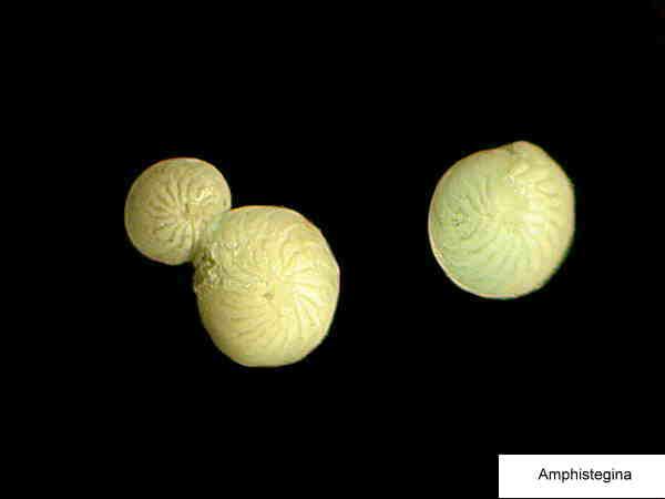
The investigator treated some of the shells with nitric acid which dissolved them, since they are calcareous, and discovered integrated within the foram test, diatom shells ranging from 10 per foram to 150. It is also stated that this is true for other forams such as Orbiculina. It is absolutely astonishing how life exploits space; there is hardly a niche imaginable that some organisms have not managed to make use of, which is a topic for a separate essay.
In the Botany section under Cryptogamia, there is an intriguing little note titled “Underground Algae and Fungi”.
“Herr A. Schneider gives an account of the vegetable organisms found in coal-pits, salt mines, and ore mines in different parts of Germany. They comprise rhizomorphs, Mucorini, Pleosporeae, diatoms, Oscillariacae, palmella-like colonies, micrococci. spirilla, etc.”
Even though these are relatively primitive life-forms, consisting largely of fungi, algae, and bacteria, they provide an excellent example of the point I was making above, for not only are fossil organisms found in such environments, but living ones as well, even though we generally think of such sites as inhospitable to life.
In the subsection under MICROSCOPY on Instruments, Accessories, &c., there are some plates of wonderful old bits of apparatus. Fine 19th Century microscopes and microtomes can be a delight to behold for reasons relating to aesthetics (shiny brass); intricate, ingenious design; and performance. I offer for your delectation Nachet’s Large Microscope.
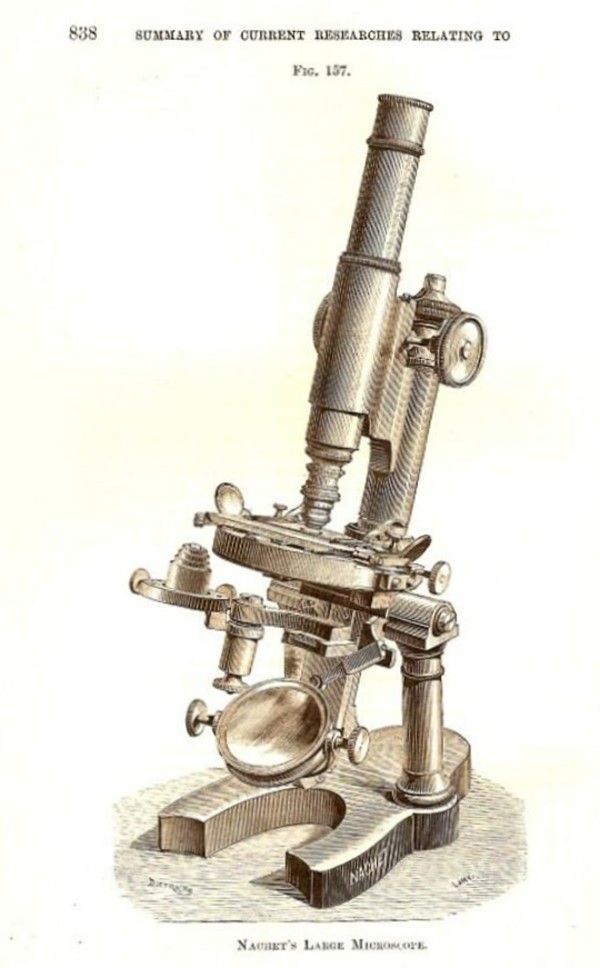
Notice that the fine focus is at the top of the column which connects to the plate with the rack and pinion for the coarse focus; it has a mechanical stage, a condenser, and a mirror that can be adjusted to almost any angle and, in addition, there are 2 small mirrors located on the stage and positioned at such angles that they allow for observing the approach of the objective to the cover glass thus reducing the chance of damaging rare and/or expensive slides.
Nachet also manufactured a microscope “with fixed revolver for Objectives”. This was designed to thwart obstreperous pupils.
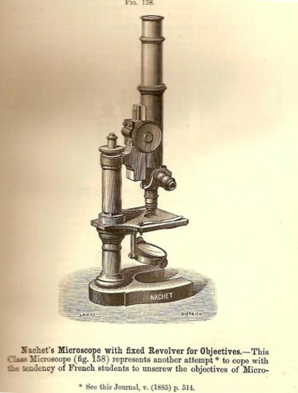
“This Class of Microscope represent another attempt to cope with the tendency of the French students to unscrew the objectives of Microscopes placed in their hands. The objectives are attached to a revolver so that they cannot be unscrewed, neither can the lens-cell be removed.”
Wow, the French are strict; first they invent the guillotine and then a microscope with a revolver to shoot students that try to steal the lenses. And then there was poor Jean Valjean hounded by Inspector Javert just over a loaf of bread. The “revolver” in this case is, of course, what we would call a turret. The problem regarding the mistreatment, vandalism, and theft of microscope components is by no means limited to French students. When I was still teaching, in a period before a demented Dean dismantled the biological stockroom, I was friends with the supervisor and one day we were chatting about the problem of microscope repair. He told me to follow him and we went to an area at the very back of the stockroom where I hadn’t been before and there were shelves and shelves of student microscopes and illuminators which had been damaged or had had parts stolen from them. Most of the damage was, I am sure, unintentional, but this doesn’t reflect well either on the laboratory instructors or the practical intelligence of the student in learning simple procedures.
Microscope manufacturers, like automobile companies, are constantly coming out with new “improved” models, especially in the area of student microscopes where there is a huge market in middle schools, high schools, colleges, and universities. This often means that within a few years, replacement parts for earlier models are no longer being produced. New versions of such “student-proof” microscopes are still being manufactured which shows that, in some respects, students haven’t changed in the last 121 years.
In the late 19th Century, researchers were excited about the application of photography to microscopy and Nachet produced two models, both of them quite elaborate.
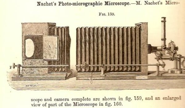
This is Nachet’s Photo-micrographic Microscope. As you can see, it is set up on the principle of an optical bench and the bellows can be extended or retraced to achieve focus on a ground glass plate.

This redoubtable instrument is Nachet’s Photographic Microscope for Instantaneous Photographs. The microscopist keeps his finger on the trigger lever A and when the object is in focus, pulls the trigger, and for an instant the prism is moved aside allowing the light to enter the camera body. A spring lever then returns the prism to its previous position for viewing.
The craftsmanship involved in the production of fine late 19th Century microscopes is astonishing and some of these instruments were not only of extremely high quality, but also highly pleasing aesthetically, since many of them shone with the warm glow of brass. A German maker in Berlin, named Fuess, made intricate petrological microscopes, two of which you can see below.

These are polarizing microscopes which geologists use to examine thin sections of rocks and minerals. The one on the right is especially elaborate and in 1886, anyone who owned one of these possessed a state-of-the-art instrument.
The inventiveness of the 19th Century microscopists was truly impressive. They used such devices as hot stages, Cole’s Self-Adjusting Frog Plate (which didn’t injure the frog) and Compressoria. The compressorium was a wonderfully ingenious and useful device which once again showed the capacity of these early craftsmen to do exceptionally precise machine work. There was a variety of designs, but the basic principle was the same for all of them. Suppose you are working with some active, delicate micro-organisms and you don’t want to risk trying to narcotize them to slow them down. You can put a cover glass over them on a slide and wait for the water to gradually evaporate thus exerting pressure on the organism and holding it in place for close examination. This procedure, however, gives you very little control and can easily distort the organisms. A much more effective and efficient means is being able to place a drop of water on a piece of glass in a compressorium–sometimes it’s a slide; sometimes a cover glass–and then have the capacity to very gradually screw a cover glass down on the drop of water until just the right amount of pressure is exerted to hold the organism in place and this micro-chamber is made in such a way that evaporation is minimized. Rarely an old one comes up for sale and almost no one is making them anymore. I say almost no one, because several years ago, I came across an advertisement for one made by a man in Florida who is a specialist on rotifers. He had only a few left and strongly suggested that he would not be making any more of them. So, I purchased one at a reasonable, but not cheap price. The precision was certainly worth it.
Another accessory that was coming into demand at this time was the microtome. Microscopists are never satisfied with looking at just the surfaces of things; they are like automobile buffs–they want to see what’s “under the hood”. The microtome is a device to make very thin slices of plant or animal tissues. Obviously, if you have something that’s an inch or more thick, it’s virtually impossible to see what it’s like internally. The techniques for making such sections is called microtomy. The thickness of the section is measured in microns and a micron is 1/1000th of a millimeter and there are 25.4 millimeters in an inch so, as you can see, you don’t want to measure elephants in microns. Using a hand microtome, most sections produced are in the range of 30 to 50 microns; using a high quality rotary microtome, beautiful sections of only 10 microns or less can be cut and then there are the modern ultramicrotomes used for electron microcopy and these instruments even have a stereo-microscope built-in for observing the sections as they are cut. These can slice sections that are only a fraction of a micron thick. In fact, top quality ultramicrotomes can cut sections down to 60 to 100 nanometers and 1 micron = 1000 nanometers, so as you can see, we are getting into the realm of the very, very, very thin. For example, a Paramecium can be embedded in a tiny block of epoxy resin (after, of course, proper fixation and hardening) and then the block is placed in the ultramicrotome and dozens and dozens of slices of this small organism can be obtained for observation. I wish we would use our tax dollars to build more microtomes and fewer weapons.
These 19th Century microtomes were marvels of ingenuity with all sorts of wonderful knobs and screws and levers. Below is an image of Becker’s Sliding Microtome.
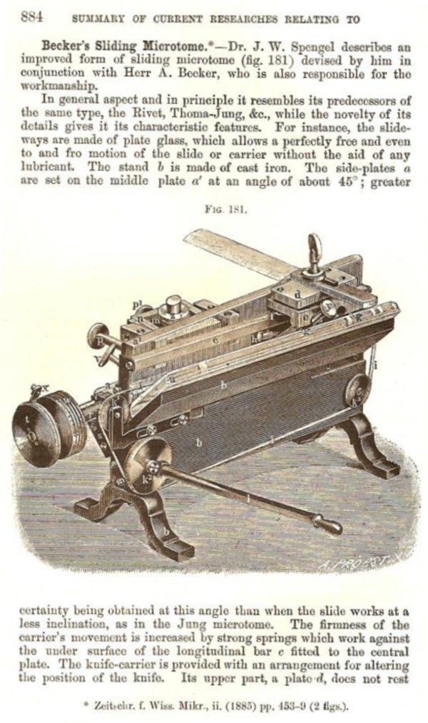
The features of each instrument are described and their particular advantages are noted. For example, regarding this model we are told “the slide-ways are made of plate glass, which allows a perfectly free and even to and fro motion of the slide and carrier without the aid of any lubricant.”
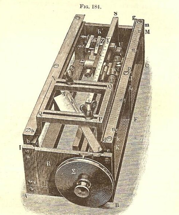
Then there is Vinassa’s Microtome for Pharmacologists. I have no idea why it should be especially for pharmacologists; perhaps they would shave a little bit off of each pill they sold to increase their profit margins. If you notice all the lettered parts, you can see that this was clearly an intricately designed instrument (probably subsidized by the Guild of Pharmacologists).
Finally, there is Weigert’s Immersion Microtome for Large Sections.
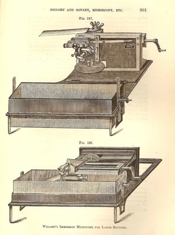
The specimens to be sectioned were placed in alcohol in the trough; however, the instrument could also be used to cut dry sections by turning on its side.
The advertisements at the back are ones for microscopes, camera lenses, mineral and fossil specimens, living specimens microscope condensers and accessories, and one for a book which I want to mention. The volume is The Microtomist’s Vade Mecum by Arthur Bolles Lee and it is a classic for anyone interested in micro-technique and its history. It has gone through many editions and contains hundreds of formulae for fixatives, stains, and their applications along with maceration and decalcification techniques, methods for anaesthetizing animals from protozoa to fish, procedures for imbedding tissue, for examining bones and teeth–in other words, it is a treasure trove of accumulated experiments to find techniques to observe more and more details of a wide range of organisms. In later editions, there are sections on botanical techniques as well.
To me, it continues to be amazing how sophisticated and imaginative 19th Century microscopy was. It is also marked by a kind of dedication and breadth that is sometimes lacking in modern researchers who rely so much on technology and rote procedure that they never really master the marvelous instruments which are available to them.
All comments to the author Richard Howey are welcomed.
Editor's note: Visit Richard Howey's new website at http://rhowey.googlepages.com/home where he plans to share aspects of his wide interests.
Microscopy UK Front
Page
Micscape
Magazine
Article
Library
Please report any Web problems or offer general comments to the Micscape Editor .
Micscape is the on-line monthly magazine of the Microscopy UK website at Microscopy-UK .