I am currently a student enrolled in the Imaging and Photographic Technology major at Rochester Institute of Technology. The images and design for this page were created for the Principles of Photomacrography course offered from the Biomedical Photographic department.
I’ve taken Nature Photography twice in previous quarters during my college career at RIT and fully enjoyed them. During the most recent quarter of the course, I focused my attention on close-up photography and this sparked my interest in getting even higher magnifications; thus, I strongly desired to take this course.
Flowers in particular have always fascinated me for both their beauty and their structure. For this project, the parts of a Carnation will be diagramed, as well as bisected Chrysanthemums will be shown. The bisected images we photographed using a Nikon D70 with bellows attached and a 38mm thimble lens, a vertical copy stand, and a microscope illuminator. For the other images, a Canon 20D with a 60mm macro lens was used. A razor blade was used to bisect the flower.
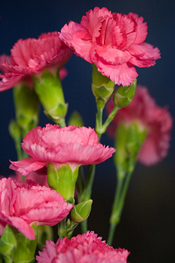
Carnation - Pink & Yellow
- Kingdom: Plantae
- Division: Magnoliophyta
- Class: Magnoliopsida
- Order: Caryophyllales
- Family: Caryophyllaceae
- Genus: Dianthus
- Species: D. caryophyllus
Carnations are flowers that are available all year round, and like the Chrysanthemums following, they come in a wide range of colors and size. Carnations can grow to be 2-3 feet high.
Flower anatomy:
- calyx – the outer whorl of sepals that are typically green, but are petal-like in some species.
- corolla – the whorl of petals which are usually thin, soft, and colored to attract pollinating insects.
- androecium - the stamen that is made up of a filament topped by an anther where pollen containing the male gametes is produced.
- gynoecium - the carpel that contains the female reproductive organs and contain an ovary with ovules, the female gametes.
The Carnation is an example of a flower that has both male and female reproductive organs which can be seen in the following diagram of the different parts of the Carnation:
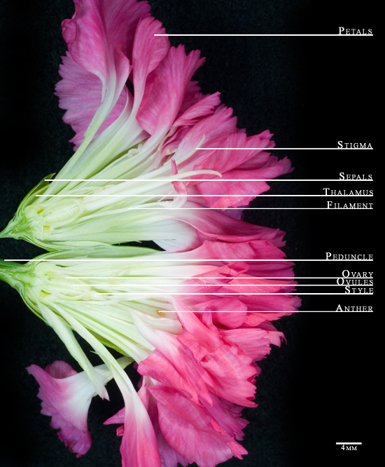
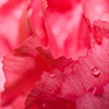
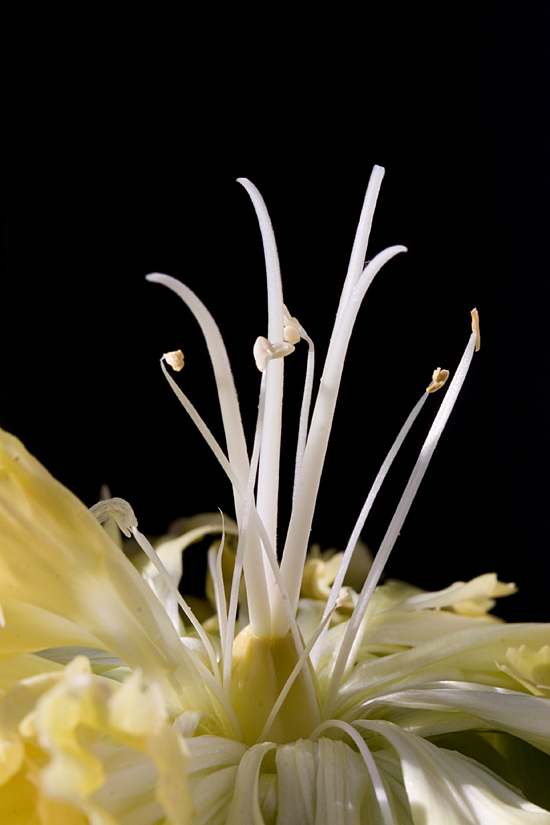
The following photograph was taken to show the male and female reproductive organs of the Carnation as they are, and intact. To achieve this image, given the petals completely conceal the male and female reproductive parts, the petals of the Carnation were folded back. This image, like the full image of the Carnation, was photographed with the Canon 20D with illumination from sun entering the window.
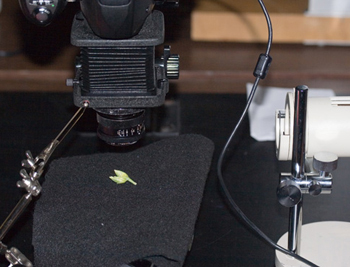
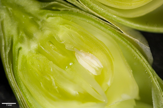
In addition to photographing the full bloom of the Carnation, a flower bud was also disected and photographed using the bellows and Nikon D70. The following is the resulting image taken using axial illumination.
Chrysanthemums
- Kingdom: Plantae
- Division: Magnoliophyta
- Class: Magnoliopsida
- Order: Asterales
- Family: Asteraceae
- Genus: Chrysanthemum
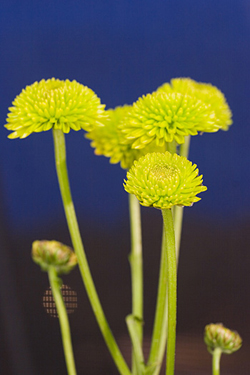
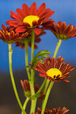
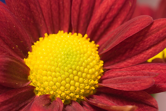
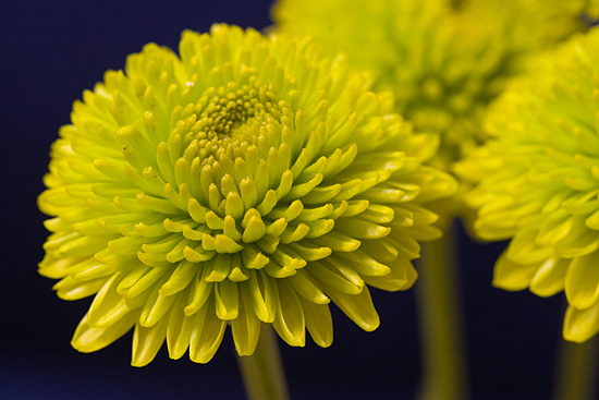
The red flowers, in the image above, are Chrysanthemum Daisies, and the green flowers depicted to the left are Green Santini Chrysanthemums. These photos were taken with the Canon 20D and a 60mm macro lens.
The flowers, like the carnations before, were placed on the windowsill and were photographed using the available light.
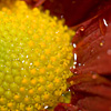
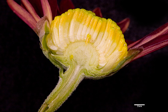
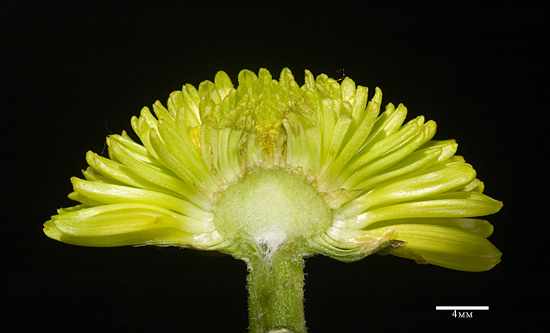
The images below show the flower both before and after it has been bisected with a razor blade.
Lighting Set-up
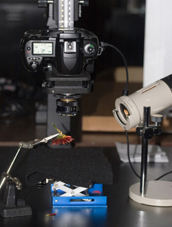 For the images utilizing the Nikon D70, bellows, and copystand, either one of these two lighting set-ups were used: direct or axial illuminated.
The direct lighting as shown on the left resulted in images of higher contrast than the images photographed with axial illumination. To accomplish the axial illumination, a glass slide was angled at 45 degrees, using a 3rd arm, and the light was then shined on this.
If the following thumbnail is clicked, the difference of using these two set-ups can be shown:
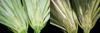
The ovules contained in the ovary of the flower are hard to distinguish in axial illumination, but by using a more direct light, the ovules are separated from the ovary.