|
|
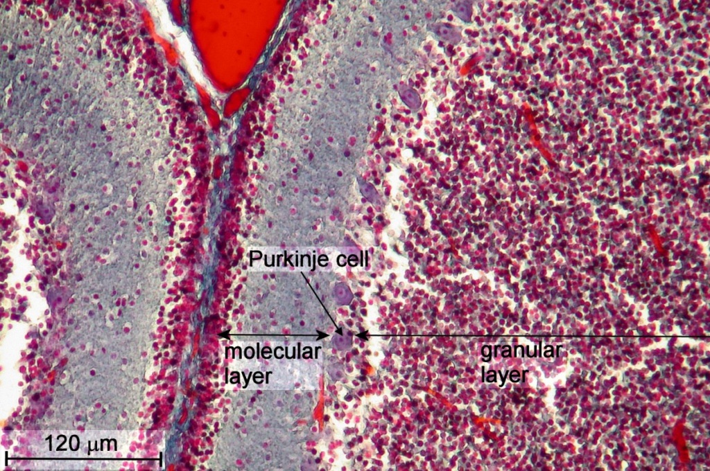
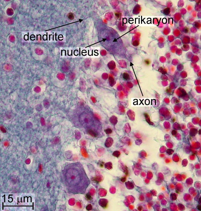
After a short introduction into the anatomy of the human brain, I will look at large neurons, called Purkinje cells, which are located in the cerebellum of the human brain, followed by an exploration of astrocytes, which have been made visible with the famous Cajal/Golgi heavy metal impregnation.
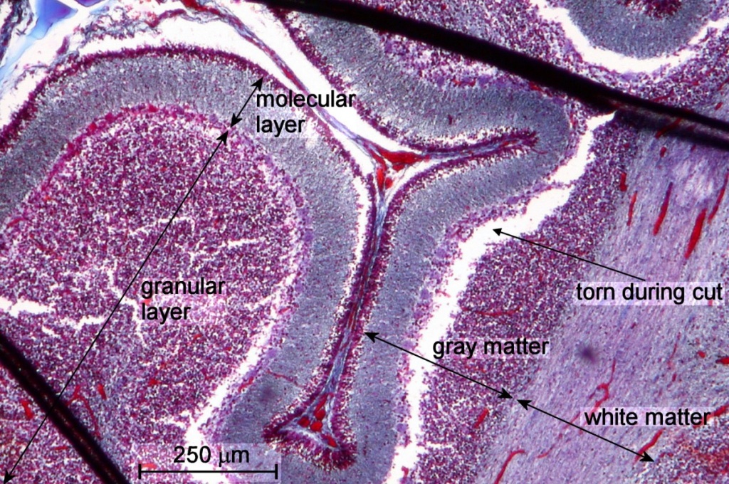
The gray matter of the CNS contains most of the neuron cell bodies, while the white matter of the CNS contains the axons. The white appearance of the fresh tissue is caused by lipid in the myelin sheaths of the axons. Myelin is 80% lipid and 20% protein and formed by oligodendrocytes (type of neuroglia). Myelin sheaths are used to insulate axons. (BTW, a nerve cell with a myelin sheath around its axon is called myelinated.)
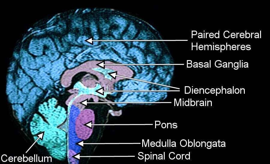
The human brain consists of various major parts, each fulfilling highly complex functions. These parts are paired cerebral hemispheres, which are invested by the cerebral cortex, diencephalon, which is formed by large gray matter structures, midbrain, pons, medulla, and cerebellum. (See Fig. 2 for an overview drawing of a human brain.) The cerebellum is responsible for coordinating muscular activity and maintains posture and equilibrium.
In
the following paragraphs, we look at the microscopic structure of two selected
features of the human brain, Purkinje cells in the cerebellum and one type
of glial cells of the white matter, called fibrous astrocytes.
| Type of CNS Supporting Cells
(Glial Cells) |
Functions |
| Astrocytes |
|
| Oligodendrocytes |
|
| Microglia |
(similar to microphages in blood) |
Let us focus on astrocytes (star shaped cells). Astrocytes provide mechanical support and mediate the exchange of metabolites between neurons and blood vessels. Astrocytes are not the blood-brain barrier. The blood-brain barrier is a protective barrier that works like a filter. It lets certain molecules pass while other substances of the blood are prevented from entering brain tissue. The blood-brain barrier is formed by nothing more than endothelial cells (cells that form capillaries). Astrocytes induce these endothelial cells to form tight junctions with each other.
Astrocytes couple with blood vessels through so-called footplates, which are specialized attachments to blood vessels. There are two different types of astrocytes. In the gray matter, we can find protoplasmic astrocytes. These astrocytes have many short, relatively thick processes. The astrocytes of the white matter, called fibrous astrocytes, are less branched but their processes radiate from the cell body for considerable distance. - To the best of my knowledge, we will be looking at fibrous astrocytes, which are located in the white matter [2].
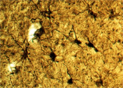
Before exploring these interesting cells, I want to mention a few details about a powerful staining technique used to make astrocytes, as well as neurons, visible. This technique is called heavy metal impregnation. In 1873, Camillo Golgi (1843 - 1926) published a short note in the Gazzetta Medica Italiana about the structure of the gray matter in the brain. He described his success in observing the elements of the nervous tissue after subjecting brain tissue samples to metallic impregnation. The black reaction, Golgi discovered, is based on nervous tissue hardening in potassium bichromate and impregnation with silver nitrate. But Golgi did not correctly describe the microscopic structure of the nervous system. In 1887, Santiago Ramóny Cajal (1852 - 1934) was introduced to the Golgi method. With this method, Cajal was able to correctly describe the nervous system. In 1909, Cajal published his findings in the classic text 'Histologie du système nerveux de l'homme et des vertébrés'. In 1906, Cajal and Golgi shared the Nobel Price for their study of the nervous system.
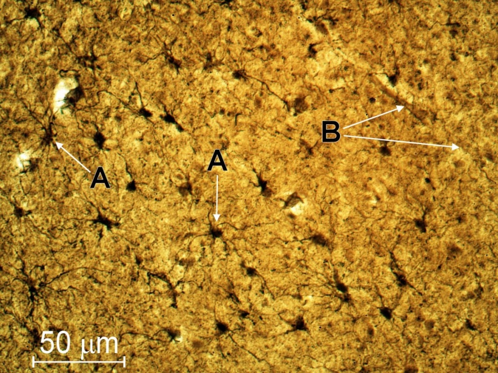
For the heavy metal impregnation technique, thick sections should be used to increase the likelihood of having complete neurons being included in the plane of the section. The technical details of the metal impregnation are not discussed in this article. The reader may read Cajal's standard text [3]. - I was able to purchase slide 93 W 6322 from WARD [4] labeled 'Astrocytes (sect.)' showing astrocytes highlighted by Cajal's gold chloride method. Fig. 4 shows some clearly visible astrocytes (objective 40x). The reader can easily see a network formed by these astrocytes. In the region examined, I was not able to find neuron cell bodies and hence conclude that the region depicted in Fig. 4 belongs to white matter. A blood vessel is also visible in Fig. 4 (indicated by 'B').
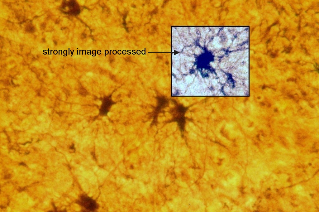
BTW, serious lack of contrast forced me to "upgrade" the digital camera equipment from a Nikon Coolpix 995 to a more specialized CCD camera using a cooled CCD. Although a Nikon Coolpix 995 is a very suitable choice for most bright-field applications of H&E stained histology slides, I could not obtain sufficient contrast for this application. For comparison, Fig. 5 shows an image of astrocytes from slide 93 W 6322, which has been recorded with a Coolpix 995. In Fig. 4, the reader can find tiny circles that are caused by this specialized CCD camera. This is an artifact that cannot be observed with the Coolpix camera.
I want to thank Dr. Fei Liu for many suggestions and stimulating discussions. The technical support of C&N for designing the HTML version of this article is greatly acknowledged. Last but not least, I thank all anonymous supporters who provided assistance and equipment.
Comments to the author, Gregor Overney, are welcomed.
| [1] | G. Overney. Human Histology for Amateur Microscopists, Micscape Magazine 82 (2002), and references therein. |
| [2] | On page 135 in [5], one picture of astrocytes is shown using the Cajal method (Fig. 7.21 b). Unfortunately, the authors leave it up to the reader to identify the type of the astrocytes depicted. |
| [3] | S. R. Cajal. Histology of the Nervous System of Man and Vertebrates (translated N. Swanson and L. W. Swanson), Oxford University Press, New York (1995); S. R. Cajal. Texture of the Nervous System of Man and the Vertebrates (annotated and edited translation of original Spanish text by P. Pasik and T. Pasik), Springer Verlag, New York (2000). |
| [4] | WARDs Natural Science Establishment, POB 92912, Rochester, NY 14642. |
| [5] | B. Young, J. W. Heath. Wheaters Functional Histology, 4th Edition, Churchill Livingstone, London (2001). |
| [6] | Background adjustment (or subtraction) can be performed using either the built-in feature of the Nikon Coolpix camera or with the help of image processing software. For instance with Paint Shop Pro 7.04, using the menu "Colors->Adjust->Levels", the user can easily flatten the color spectrum with respect to any RGB color-value. Of course, post-processed background adjustment can reduce the dynamic range per color channel. For 24-bit RGB, the maximum color range per color channel is 8-bit, or 256 distinct values, per channel. |
Please report any Web problems or offer general comments to the Micscape Editor.
Micscape
is the on-line monthly magazine of the Microscopy UK web
site
at Microscopy-UK.