|
Geometry and Pattern in Nature 2: by Christina Brodie, UK |
Butterfly wings often exhibit a wonderful range of colouring and patterning, which serves both to deter predators and attract a mate. This colouration is created by many tiny scales which grow from the wing's epidermal cells and overlap each other.
The colours and patterns are enough to fascinate most people, but examining the scales in themselves can prove to be doubly interesting. Scale colour and shape may vary considerably in an individual butterfly species; for example, rectangular scales may be found towards the centre, and longer hairlike or forked scales towards the edge of the wing. This is presumably an aerodynamic adaptation to streamline the airflow.
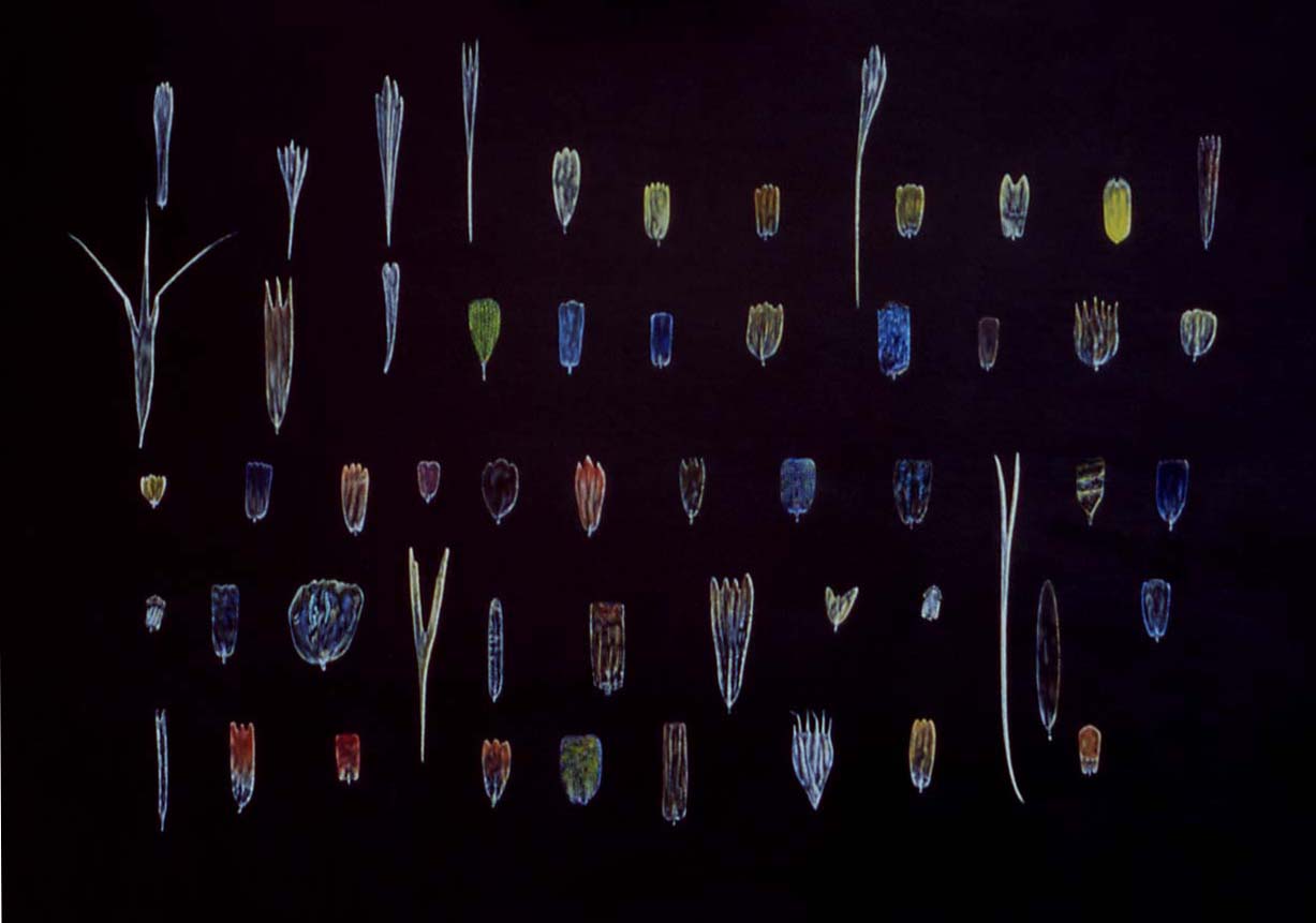
A view of a number of types of butterfly scale is necessary in order to appreciate their differences. I was inspired to create the picture you see below by a plate from the book The Microscope Made Easy by A. Laurence Wells, kindly given to me by a member of the Quekett Club at one of the twice-yearly Langton Matravers meetings. The scales are depicted exactly as they appear, carefully placed in shimmering rows, on a slide specially made by renowned diatomist and slidemaker Klaus Kemp.
From the painting, the considerable range of colours and shapes of butterfly wing scales can be clearly seen. Some of the scales are coloured by pigment molecules within the scale cells, such as uric acid (white), carotenoid pigments (yellow, orange), flavonoids such as quercetin (red, purple), and melanin (brown), the natural colour of the cuticle material from which the scale is made up. The pigment produces colour by selectively absorbing and transmitting, or reflecting, light of certain wavelengths. Each pigmented scale produces only one type of pigment to a greater or lesser degree over its entire area, so that wing patterns are created in a manner similar to those in tilework or mosaic.
However, the iridescent shades of green, blue, yellow, orange and purple, which may show a metallic or silken lustre, are produced by the quite different mechanism of structural colour. Structural colour produces colours that are more brilliant and intense than colours achieved by pigmental colours, through the reflection of light from the scale's surface and internal structure. The colour may change according to the angle the scale is viewed from, producing an effect of banded or glittering, intermingling colours similar to the effect seen when petrol is floated on water. These types of colours may also be called interference colours due to the fact that they are caused by the interference of light in the actual structure of the scale.
The term incident light is that given to any ray of light that strikes a surface, which in this instance we will take to be the thin film of cuticle, the protein-based material that is produced by a single epidermal cell and from which the scale is made. When light strikes the upper surface of the transparent/ translucent film, some of it is reflected back at an angle. The remainder travels to the lower surface, from which more light may be reflected back to the upper surface. The angle of reflection is constant in both instances, but whether or not strong interference colours occur depends on whether the waves are in phase (moving in exactly the same direction) or out of phase. If they are in phase, then constructive interference will occur. The waves will not cancel each other out; instead, there will be an intense reflection of light at that particular wavelength. On the other hand, if the waves are out of phase, what is called destructive interference occurs. The waves cancel each other out, and there will be a considerably weaker intensity of colour emitted from that point on the surface.
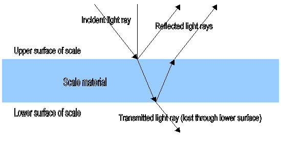
Diagram adapted from one at http://newton.ex.ac.uk/research/emag/butterflies/interference_background.htm
Whether or not a certain colour or type of interference effect is shown in a scale depends very much on the nature of the surface of the scale (low- or high- relief), its thickness, the refractive index (ability to refract or bend, as opposed to reflect light) of the cuticle material, the wavelength of incident light present and the angle from which the light strikes the film surface. For example, only one colour can be viewed when the light strikes the object at a particular angle. The brilliant blue of the Morpho butterfly wing will change from violet through blue to brown (the latter its actual colour, caused by melanin) depending on the viewing angle.
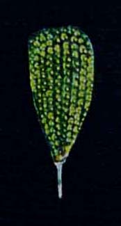
Scales may possess both pigment-derived and structural colour. The colours can blend to produce a colour intermediate between the two. For example, a scale containing yellow pigment might be structurally configured to refract blue light strongly, with the resulting colour appearing green.
If the physical and environmental conditions harmonize well with each other, very pure colour will be created. To enhance the effect even more, the scales are actually made up of several layers of thin films, so that some of the light which might otherwise be lost from the lower surface of the film is conserved and reflected back.
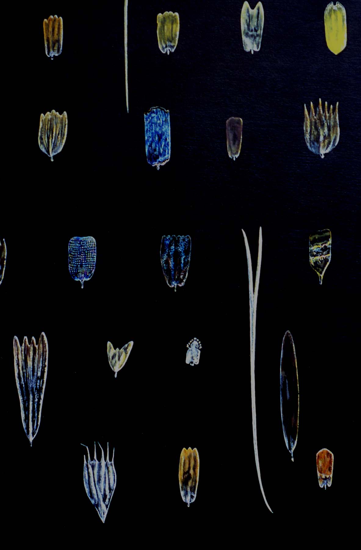
I would hazard a guess that the bright blue scale towards the top centre of the above image probably came from a species of Morpho. The scale below it, at the centre of the image, only shows iridescence in certain patches, being predominantly coloured by melanin. The scale to the left of this, blue with a rainbow-type iridescence, actually shows the same surface features as the metallic green scale depicted below - regular vertical rows of small circular depressions interspersed with ridges - whereas the scale at centre right shows strong diagonal bands of iridescence.
In moths of the Urania genus, scales are comprised of approximately 4 or 5 layers of cuticle interspersed by air pockets and held in position by vertical struts of cuticle. They may show some simple ridging, which aids in the reflection of light at certain wavelengths, but the surface of the scale is comparatively smooth.
In butterflies of the Morpho genus, the layered sheets of cuticle take on a pyramidal three-dimensional structure which has been likened to a Christmas tree. There are literally thousands of these structures on the surface of the scales, and they can only be viewed with the aid of an electron microscope, since the thickness of the cuticle sheets has been measured at approximately a tenth of the wavelength of light. Their presence gives the scale a high-relief surface and a shimmering appearance comparable to that of silk, in contrast to the scales of the Urania moths.
Diagrams and electron micrographs of both types of structure can be found at http://newton.ex.ac.uk/research/emag/butterflies/interference_background.htm
These types of reflective multilayer structures are not shown in all the Lepidoptera. Another type of reflective structure is comprised of a mesh of cuticle with regular, circular holes, which predispose the material to scattering light in a particular direction, and may create a greater consistency of colour over the whole wing. A more detailed description of scale structures can be found at http://newton.ex.ac.uk/research/emag/butterflies/classification_page.htm
As such, I have not been able to assign genera - or species - to most of the types of butterfly scale found in the painting! Maybe some butterfly enthusiasts - or experts - could help?
Source: Iridescence on Butterfly Wings, originally published in Physics Review Sept.1998, by Philip Allan Publishers; see http://newton.ex.ac.uk/research/emag/butterflies/iridesc-text.htm
Klaus Kemp's website: www.diatoms.co.uk
Christina Brodie has recently set up a website to showcase her Micscape and other artwork, which includes botanical and natural history painting.
Visit Christina Brodie's website www.queen-christina.com where contact details can also be found.
Images © Christina Brodie 2004. All rights reserved.
Editor's note: Christina
Brodie has shared a selection of her other beautiful drawings of nature, including
artwork of microscopic organisms, in the Micscape articles below:
Published in the October 2004 edition of Micscape Magazine.
Please report any Web problems or offer general comments to the Micscape Editor.
Micscape is the on-line monthly
magazine of the Microscopy UK web
site at
Microscopy-UK.
© Onview.net Ltd, Microscopy-UK, and all contributors 1995 onwards. All rights reserved. Main site is at www.microscopy-uk.org.uk with full mirror at www.microscopy-uk.net.