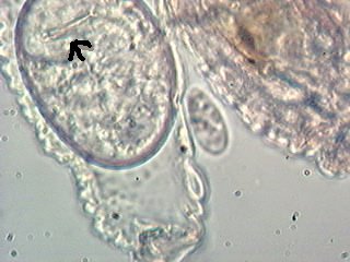
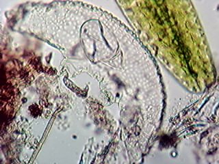
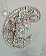
The tardigrade (water bear) Hypsibius (Isosibius) annulatus (Murray) Marcus 1929 found moulting on a temporary mount made on the 8th August was kept under observation. It was examined daily and on the 13th August some structure could be seen in the eggs (fig. 1). On the 14th the eggs were empty (fig. 2). I thought they must have been eaten as the content did not appear to be anything like a fully formed tardigrade 24 hours earlier. However I found what was obviously a juvenile nearby (fig. 3), this measured about 140 µm. It was colourless whereas the adult was brown and it was also much too lively to be described as tardus. I have since found another tardigrade of the same species that is stretched out and lifeless and was therefore able to measure it accurately - 240 µm long. The legs are 27 µm long not including claws. Fig. 2 shows the sculpturing on the surface of the skin very well.
Note the tardigrade on the microscope slide temporary mount was kept under observation for seven days. It has partly dried out, but there are still green and healthy looking desmids and active amoeba to be seen today 26th August.
The photomicrographs were taken using a Busby webcam.
Many thanks to Anne & Alastair Bruce who provided the sample.
Comments by e-mail are welcomed to Bill Ells.
William Ells.
|
|
 |
|
|
 |
|
|
 |