Amoeba proteus in closer detail
by Wim van Egmond, The Netherlands


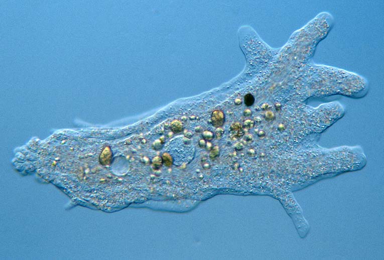
 The enlarged image shows the flowing of the cytoplasm when the amoeba locomotes. At the front end several pseudopods are formed. The typically lobed area at the rear end is called the uroid. In the centre of the cell the single nucleus is visible. Left to it lies a water expelling vesicle. Remains of prey are enclosed in food vesicles. Tiny crystals make the cell look granular.

back to First Page


Page 2: LOCOMOTION AND FEEDING


Page 3: STUDYING TINY AMOEBA
Page 4: SHELLED AMOEBA
 

All comments to the author Wim van Egmond are welcomed.

Find out more about microscopic pond life on The Smallest Page on the web
The Pond Life Identification Kit may help you identifying small freshwater organisms

Visit Wims home page for links to his many webpages on microscopy

Microscopy UK Front Page
Micscape Magazine
Article Library


WIDTH=1

© Microscopy UK or their contributors.

Published in the September 2001 edition of Micscape Magazine.

Please report any Web problems or offer general comments to the Micscape Editor,
via the contact on current Micscape Index.

Micscape is the on-line monthly magazine of the Microscopy UK web
site at Microscopy-UK


© Onview.net Ltd, Microscopy-UK, and all contributors 1995 onwards. All rights reserved. Main site is at www.microscopy-uk.org.uk with full mirror at www.microscopy-uk.net.