|
|
The
development of Physella, |
EMBRYOS OF PHYSELLA sp.
Almost all the freshwater gastropods put their eggs in
jelly-like masses adhered
to different surfaces (stones, leaves and stems of submerged plants).
At the
aquarium they choose generally the glass walls where they are easily
visible
like little discs, in some species, or with a horseshoe shape like in
the
one shown below.
|
|
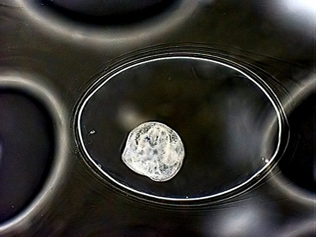
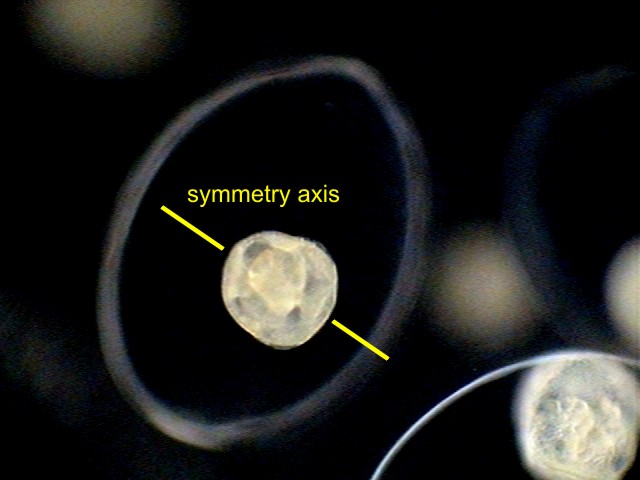
| This picture was taken
using the trick that I name " The Köhler microscope" using
the 10x objective. The longer side of the picture is more or less 1
cm long. |
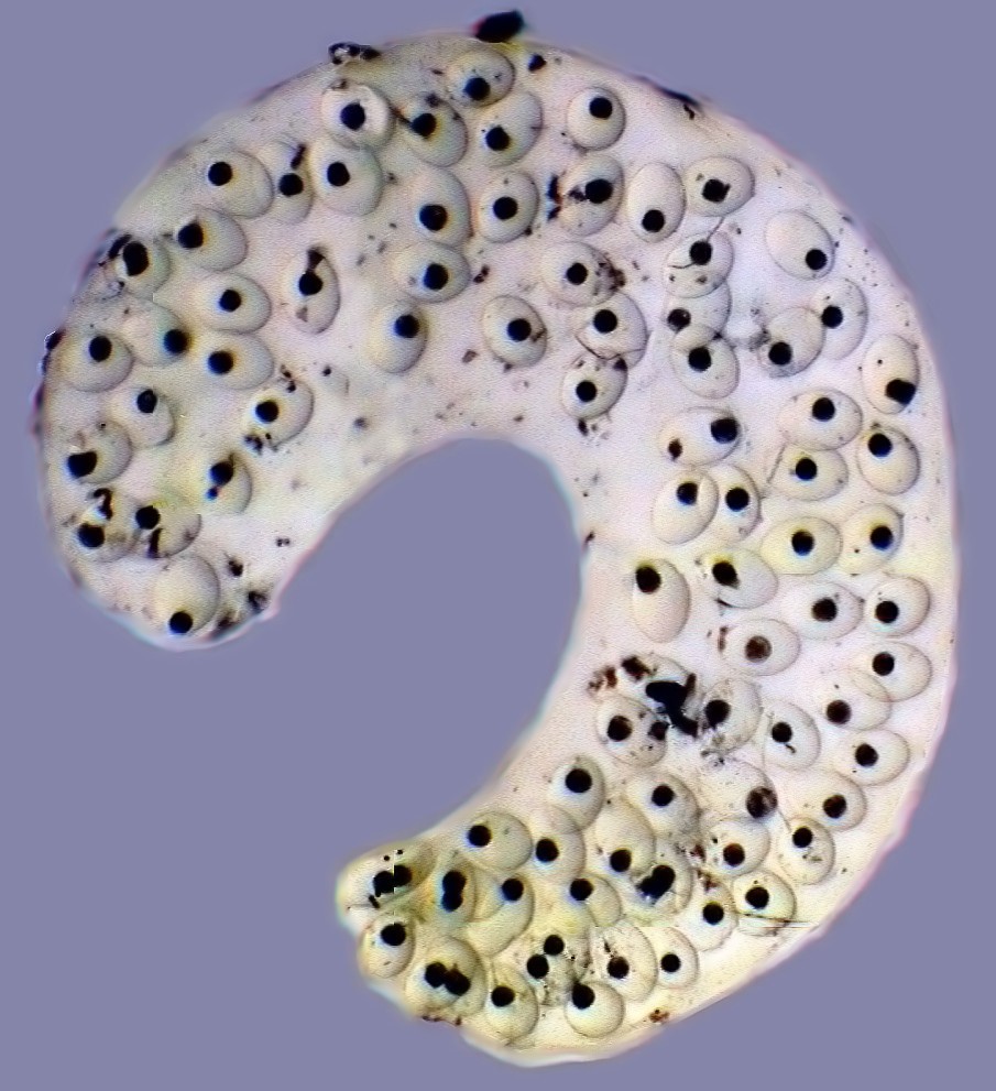
The picture shows a gelatinous ootheca that contains 90
transparent eggs,
which allows us to easily follow the embryo development. The eggs recently
laid have
a cytoplasm full of yolk and with a big nucleus. In just a short time
the
egg divides into two cells, soon into four,
8, 16 cells and form at the end a morula,
that is to say, a spherical
and compact mass of more or less spherical cells (of course the name comes from
the similarity of this embryo with the fruit of the Morus sps. trees, the mulberries).
From now on, it is more
difficult to follow the detailed development, because the most
important
changes happen in the interior of the embryo, and it would need
the aid of
a histologist to identify them.
 |
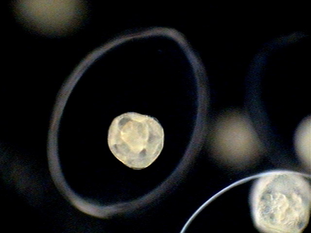
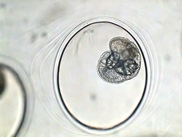
| A very young embryo. |
 |
 |
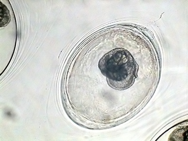
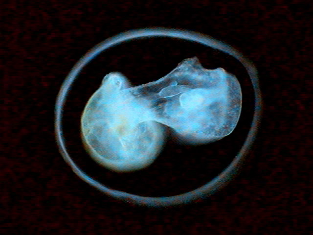
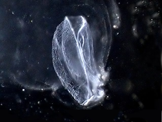
| An
embryo prior to the start of the "torsion" (see text). |
|
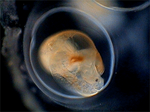
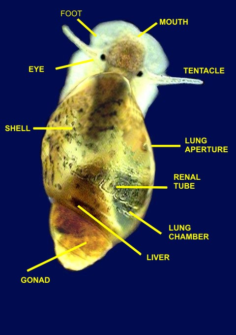
An interesting detail of the development of the mollusks
is that they start
as right and symmetrical, but soon begins a process of
asymmetrization and
torsion of the visceral mass, at the same time that this is being
covered by the
segregated calcareous shell.
 |
 |
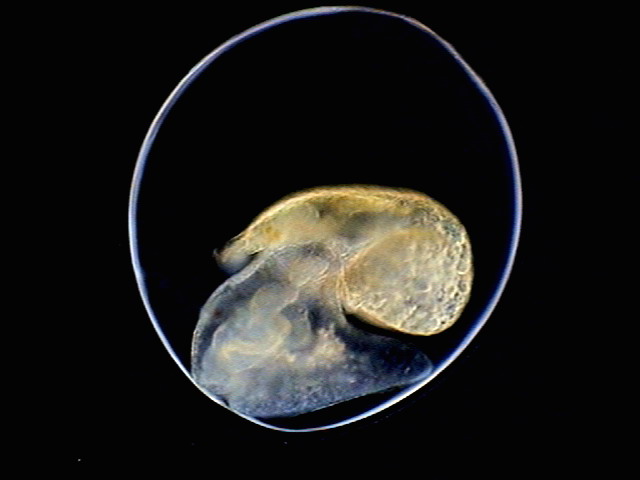
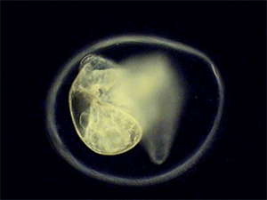
| Two
embryos that have started the torsion of the visceral mass hump over
the flat foot. |
|
The torsion causes the atrophy of the organs in the left
side of the body,
if torsion is to the right, or on the right side if it is to the left.
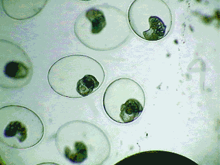 |
| Embryos start to move
inside the shell. |
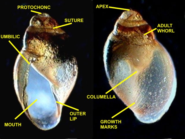
When growing, the shell follows the helical torsion
process generating the gastropods
characteristic spiral. As is logical, with the growth of each
successive turn
the shell constructs a center axis the columella. A beautiful picture
of this
structure is seen in fig. 33.
|
|
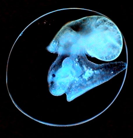
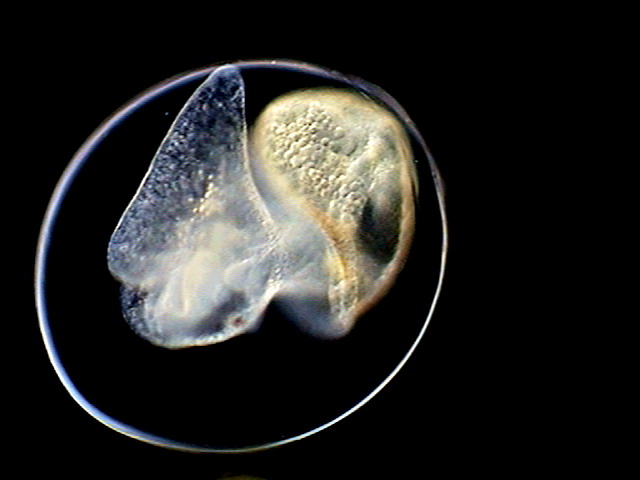
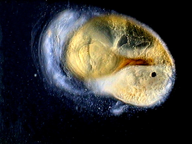
| The
embryo is starting to
secrete its shell over the visceral hump.The head is in frontal view,
with mouth in the center, and the primordium of the left tentacle on
the
uper side.You can see the thickening at the extreme right of the embryo. |
These are two pictures of other more developed embryos.
 |
 |
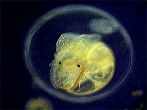
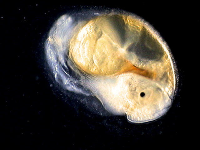
| The
digestive system is well developed in this snail embryo. One can detect the developing radula as a clear band starting at the mouth. |
The
dark spot in the visceral mass is the developing lung. You can see also the primordium of one eye. |
In just a short time the little snails begin to move
within the egg, as the
shown in the following animations.
 |
 |
If you look closely at
some of the more sluggish embryos, you can see the beat of the heart of the
growing
embryo.
 |
Soon they break through the eggs shell and leave it. The
youthful individuals
are beautiful photogenic subjects.
 |
 |
|
Two
pictures of the embryo breaking through the egg shell. |
|
 |
| One empty shell. |
 |
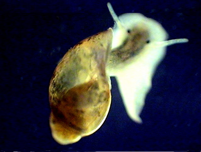 |
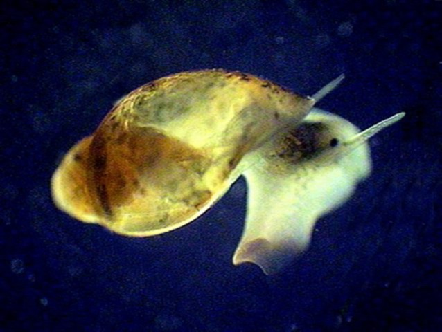
| Exploring
the world. |
|
 |
 |
 |
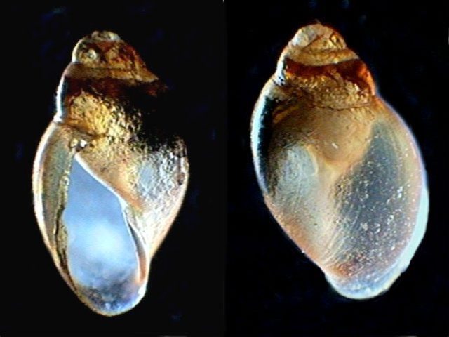 |
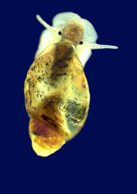
| Morphology
of the conch. The shells are 1 cm high. |
|
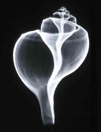 |
Peter Abraham .- Stereo X-rays of sea shells
http://europa.com/~telscope/binotele.htm
Microscopy
UK Front Page
Micscape
Magazine
Article
Library
© Microscopy UK or their contributors.
Published in the September 2007 edition of Micscape.Please report any Web problems or offer general comments to the Micscape Editor.
Micscape is the on-line
monthly magazine of the Microscopy
UK web
site at Microscopy-UK
© Onview.net Ltd, Microscopy-UK, and all contributors 1995 onwards. All rights reserved. Main site is at www.microscopy-uk.org.uk with full mirror at www.microscopy-uk.net.