IDENTIFYING A MYXOMYCETE AND,
A LONG-FORGOTTEN ATTEMPT OF PLACING
THE MYXOMYCETES AMONG THE PROTISTS
Manuel del Cerro and Dietmar R. Krause
Princeton, NJ, USA
FINDING
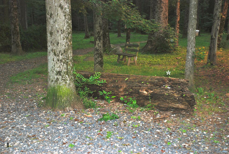
While walking at Rocky Top Dog Park (GPS 40.390673,-74.592737), one of us (DRK) noticed patches of small, brown, fungus-looking formations on the trunk of a fallen tree (figure 1). The finding was reminiscent of a previous one (del Cerro and Krause, 2011) but the structures in question were different and unusual enough to deserve further observation.
Figure 1. The trunk of a fallen tree at Rocky Top. The asterisk indicates the area where unusual-looking brown patches were noticed.
INITIAL EXAMINATION
Even to the naked eye the brown patches growing on the bark resolved into upright growths about one centimeter tall, with very thin stalks supporting a wider upper section (figure 2). Both a hand-held magnifier and a photographic macro lens allowed the visualization of a central vein running the length of the structure (figure 3).
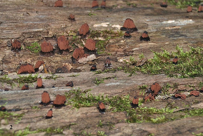
Figures 2. A closer view of the area indicated by the asterisk in figure 1 shows the brown colored elements growing on the bark.
A FEW NEW WORDS
As any specialized human activity, be it sports,, business, or medicine, the study of slime molds or myxomycetes has its own language. This should not be a deterrent to study these fascinating creatures for one needs to know less than a dozen new words to get started. On this subject we do not know more than that.
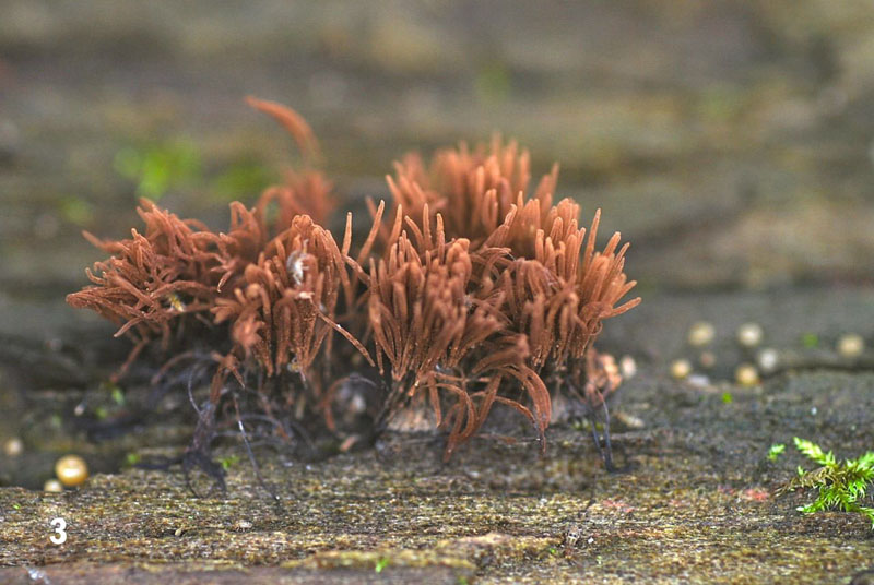
Figure 3. A macro view of the elements forming the brown patches. Compare with the illustration of similar structures in figure 7.
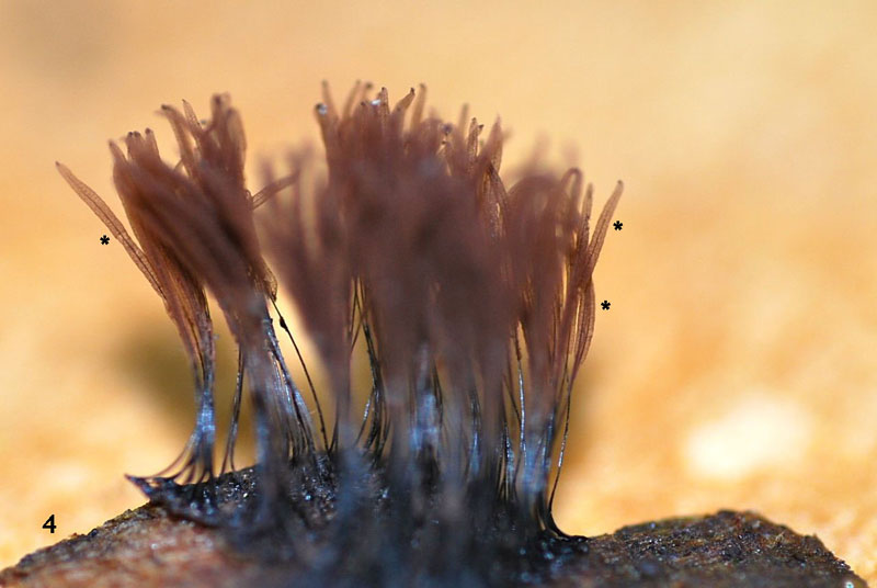
Figure 4. Close-up picture showing both the thin basal stalk (stipe), and the central vein (columella) that runs the length of the upper section of the individual elements (asterisks). Compare with the illustration of similar structures in figure 7.
MICROSCOPY
Examination of the individual brown elements under relative low power (10x or 20x S Olympus plan apochromatic objectives, magnification changer set at 1.0x, and 10x SWHK wide-field oculars), convinced us that we were dealing with the fruiting bodies of a myxomycete (figures 5). This impression was supported by recent experience in dealing with this type of “fungi” (del Cerro and Krause, 2011), and by comparing our images with illustrations depicted in two sources, a modern handbook on the Myxomycetes (Stephenson and Stempen, 1994) and an old Botany textbook (de Buen, 1920; figure 7).
We are then dealing with the fruiting bodies of myxomycetes. The thin, basal portion of these slime molds is called the columella, which extends into the enlarged top section, sporangium. In mature sporangia, like the ones we are observing, most of the structure is occupied by an intricate network of filaments in which spores are embedded, the capillitium (Stephenson and Stempen, 1994; U of Hawaii, 2011). It was interesting that the structures present in our samples corresponded closely to those illustrated in a ninety-year old diagram of a myxomycete (figure 7)
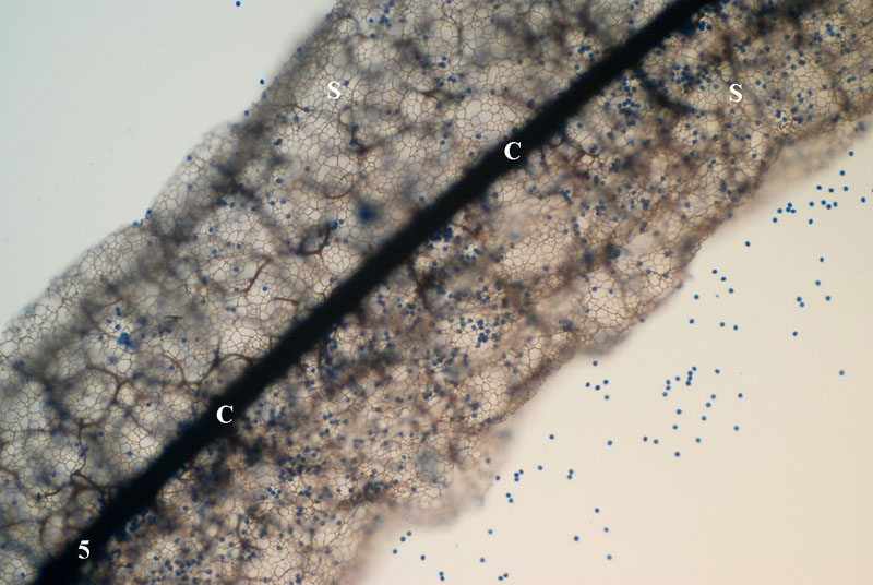
Figure 5. Low magnification view of the sporangium (S) of one of the individual structures seen in figure 4. The dark, central element is the columella (C). The spores still remaining at this time appear as blue dots both inside the sporangium or floating free. The field of view is 725 micrometers wide.
By the time these specimens were collected (early June, 2011), most of the spores had already been released (figures 5 and 6), those remaining showed strong affinity for the cotton blue stain that is part of the lactophenol-cotton blue mixture used to fix, mount, and stain fungal tissues.
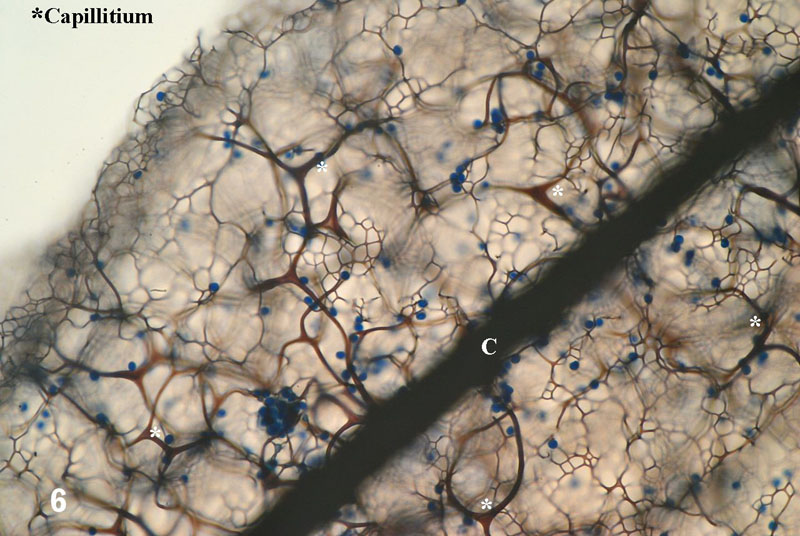
Figure 6. Higher magnification of the fruiting body shown in the previous figure. The dark structure running obliquely across the field is the columella (C). The network of filaments that holds the spores is called the capillitium. Most of the spores have been released, those remaining appear as blue dots. The field of view is 360 micrometers wide.
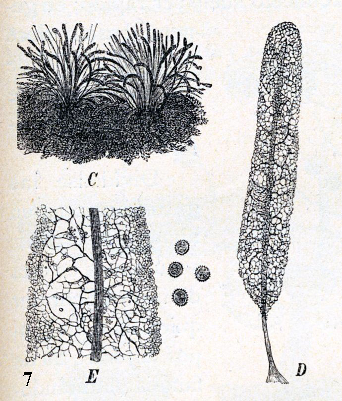
Figure 7. Diagram of the structures found in a myxomycete (from Odon de Buen, 1920).
The spores are round, 7.2 micrometers in diameter on the average, and appear smooth even at the maximum magnification used in our observations (1,500x; 100x objective, 1.5x magnification changer, 10x oculars) (Figure 8).
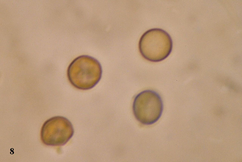
Figure 8. Spores as seen using the 100x oil-immersion objective.
The field of view is 55 micrometers wide.
IDENTIFICATION
Could we with the information at hand try to identify these myxomycetes, at least to the level of genus? Yes. Using Stephenson & Stempen (1994) as our primary guide, and considering the observed features, we propose that we are dealing with the genus Stemonitis. The dark color of the fruiting body suggests that this may be S. fusca. But there is more to this story, see Appendix below.
SOURCES
• de Buen, Odon (1920) Historia Natural Volumen Noveno Botanica. Montaner y Simon, Barcelona, pp 177 and 187.
• del Cerro, Manuel and Krause, Dietmar R., 'Is it an Animal? Is it a plant? Is it a Myxomycetes?, Micscape Magazine, July 2011.
• Stephenson, Steven L. and Henry Stempen (1994) Myxomycetes. A Handbook of Slime Molds. Timber Press, Portland, Oregon, USA, pp 183. [In our opinion this book is an indispensable aid to anyone interested in learning about myxomycetes]
• University of Hawaii (retrieved June 2011). Myxomycota. http://www.botany.hawaii.edu/faculty/wong/bot201/myxomycota/myxomycota.htm
APPENDIX
MYXOMYCETES AS PROTISTS, IN 1920!
We mentioned previously the many changes undergone in the classification of myxomycetes (del Cerro and Krause, 2011). Once included (with some reluctance) in the kingdom “Plantae,” they were removed to kingdom “Fungi” when the fungus were given a kingdom of their own (reviewed by Lynn Margulis, K.V. Schwartz and M. Dolan, 1994). Further studies, showed that: “slime"molds are now known to be quite unrelated to the fungi” (Anonymous, 2011). Modern workers have removed the Slime Molds from the kingdom Fungi and placed them into the kingdom Protista. It is interesting to see how science evolves, but it was equally interesting to find the this “modern” idea was first presented to the scientific community more than ninety years ago. Figure 9 taken from Odon de Buen’s Botanica (1920) illustrates the evolution of a slime mold, while the accompanying text (translation below) advances the proposition that Slime molds should be considered Protists, rather than Fungi.
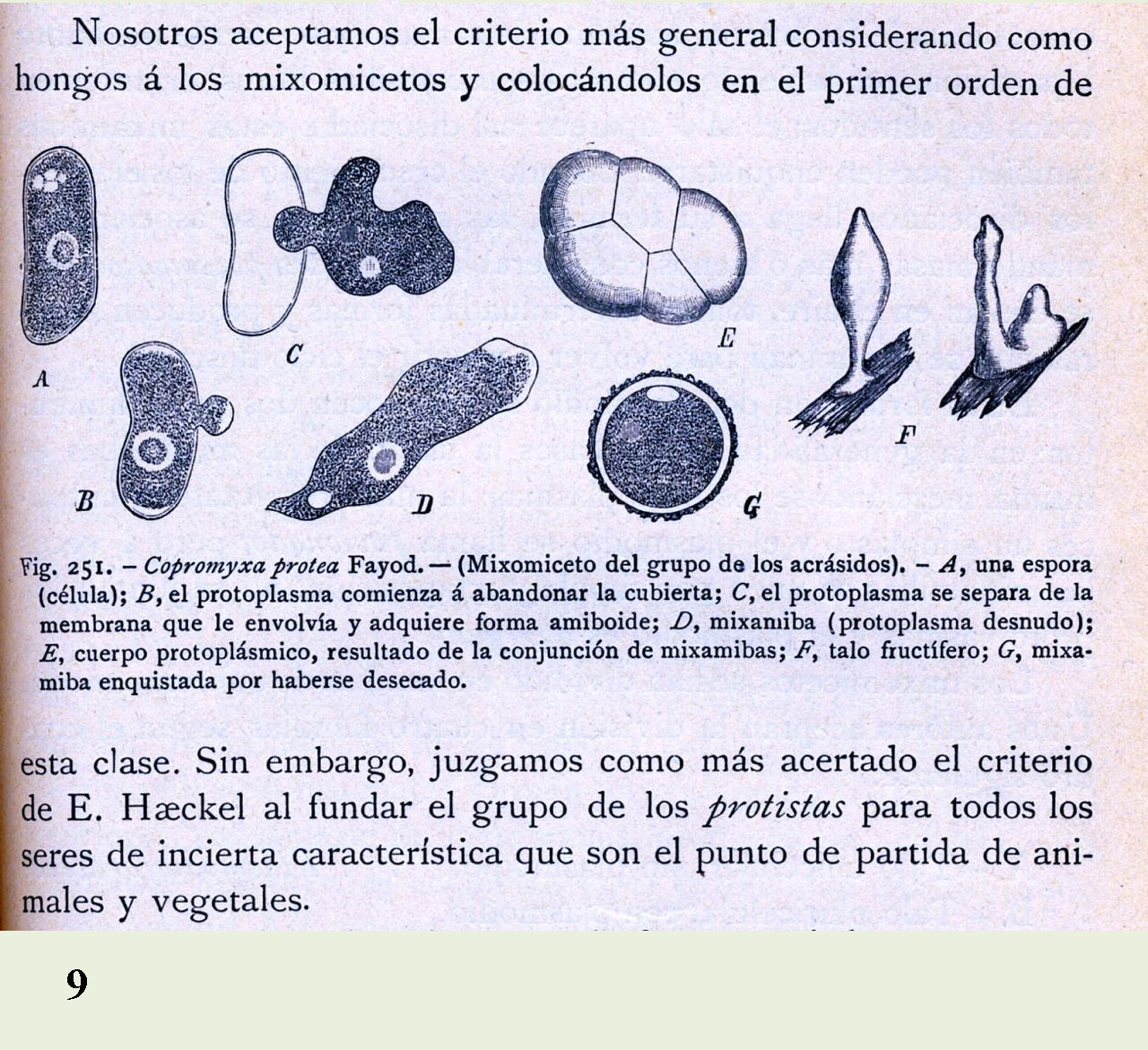
Figure 9. Diagrammatic representation of the life cycle of a myxomycete and text suggesting they could more properly be considered Protists than Fungi (from Odon de Buen, 1920).
A contextual translation follows:
“We accept the general criterium that considers myxomycetes as fungi. However, we judge as more appropriate the criterium of E. Haekel who created the group Protista for all the living things of uncertain character who are the starting point in the evolution of animals and plants.”
The Myxomycetes classified among the Protista in 1920! The great Spanish biologist Odon the Buen (1863-1945) was ninety years ahead of his times.
CONCLUSION
It is fortunate that a leisurely stroll in a dog park allowed us to become more familiar with the myxomycetes and to discover a pioneering and long-forgotten proposition regarding their place in the Tree of Life.
FEEDBACK
Comments to the authors are welcomed (via Manuel del Cerro) will be gratefully received.
APPENDIX SOURCES
• Anonymous (retrieved 2011) Introduction to the “Slime Molds.” http://www.ucmp.berkeley.edu/protista/slimemolds.html
• de Buen, Odon (1920) Historia Natural Volumen Noveno Botanica. Montaner y Simon, Barcelona, pp 177 and 187.
• Lecointre, G. and H.L. Guyader. [Illustrated by D. Visset & Translated by K. McCoy.] 2006. The Tree of Life: A Phylogenetic Classification. Harvard University Press, Cambridge, Massachusetts.
• Margulis, L., K.V. Schwartz, and M. Dolan. 1994. The Illustrated Five Kingdoms: A Guide To The Diversity Of Life On Earth. HarperCollins College Publishers, New York.
Microscopy UK Front
Page
Micscape
Magazine
Article
Library
Published in the September 2011 edition of Micscape Magazine.
Please report any Web problems or offer general comments to the Micscape Editor .
Micscape is the on-line monthly magazine of the Microscopy UK website at Microscopy-UK .
© Onview.net Ltd, Microscopy-UK, and all contributors 1995 onwards. All rights reserved. Main site is at www.microscopy-uk.org.uk .