|
Pickled Plankton, Polychaetes, Platyhelminths, Phoronids, Pisaster, Porifera, Placozoa, Protozoa, and Pyrosoma by Richard L. Howey, Wyoming, USA |
After long critical and careful consideration, you may have concluded that I love literate alliteration. What I really want to discuss in this essay are the marvels and mysteries one can discover in preserved samples, especially those of marine life. Although more and more of the human population already inhabits or is migrating to coastal areas, there are still many of us for whom a trip to the ocean is a major undertaking. For me the closest seacoast is 1,100 miles away. Over the years, I have collected along shores in Maine, California, and Oregon, have contacted marine biologists who kindly sent samples from Hawaii, Alaska, Mississippi, Alabama, California, Texas, South Carolina and even the Antarctic; and I have purchased preserved specimens from Florida, Maine, California, the Philippines, and Thailand–mostly by means of research grants.
This afternoon, I opened up a pint plastic container from Maine with all kinds of bits and pieces in it. One thing I have learned over the years is to always examine carefully the broken odds and ends, the detritus, and the micro-muck. The broken pieces often save one from having to sacrifice intact specimens and in the detritus and bits associated with other organisms, there are all sorts of wonders to be unearthed. (Why can’t we say “unwatered”?)
I took a shallow, white plastic dish about 4 inches square of the sort that airlines used to use to serve “food” in before the Bush administration crippled the economy and made people choose between fuel and “food—at ever increasing cost. I took a pair of forceps and extracted 10 or 12 small pieces from the larger dish and transferred them out of the alcohol into water in the smaller dish. It was getting late in the afternoon, so I only had about ½ hour to look at these bits and pieces. I found several pieces of arms from brittle stars and a central disk from one; 3 small polychaete worms with lovely, iridescent setae (bristles) extending out of each of the dozens of “feet”; nematodes, a nemeritine, a ferny bryozoan with numerous specimens of Spirorbus shells on it; a small worm that looks rather like Glycera, the “beak thrower” which was preserved with its proboscis extended; a colonial tunicate spreading over a shell fragment; 3 nudibranchs; a wee Mytilus (purple mussel) shell; and then there was a nearly spherical lump about 1 ½ inches in diameter and it had all kinds of intriguing small things attached to it–at least 3 species of tiny crustaceans, some small worms, bits of bryozoa, a few centric diatoms, and some odd thing with tapering projections of what appeared to be tissue. The only way in which I will have even the remotest chance of determining what this strange spheroid is, is to slice it open, but I don’t want to do that until I have carefully removed all the creatures I can that are attached to its surface and try to photograph as many of them as I can for future reference when trying to identify them.
As it turned out, as I began removing the organisms, I decided that I could use a small stiff brush to get rid of detritus from the surface. As I did so, this small spherical creature began to seem familiar and the projections turned out to be instrumental in triggering my memory. I checked the wonderful Ralph Waldo Miner book Field Book of Seashore Life, and it was indeed what I thought– a small tunicate called Boltenia echinata. Solving one puzzle like this is quite gratifying, especially when one is confronting 47 other organisms that one may very well not succeed in identifying beyond some general rubric like “polychaete worm”.
I also will save as much of the detritus and adhering muck as possible for later disassociation and examination under higher magnifications to look for diatoms, spicules, forams, etc. It is astounding how much information can still be gleaned from organisms that have been maimed and distorted by the action of preservatives. The great 19th Century expeditions might well have had a naturalist aboard, but the great quantities of material being collected demanded that most of it be quickly preserved, often in bulk, to prevent decay.
Let’s use the occasion of this essay to just ramble and explore a bit beginning with some protists. Diatoms and other micro-algae are very likely to show up because of their extraordinary adaptability and exponential reproductive potential. Diatom “shells” are primarily composed of silica and so are very hard and can survive even the ministrations of mad diatomists who boil them in concentrated acids. Many types of micro-algae have quite resistant sheaths of organic compounds including cellulose.
First, let’s look at a few diatoms:
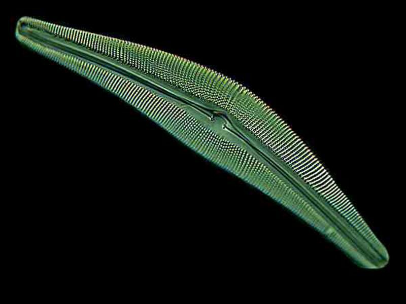
This first one is a pennate type of the genus Cymbella:
As you can see there are all kinds of tiny “pores” through which protoplasm moves creating a slow gliding motion of the organism through the water.
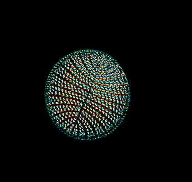
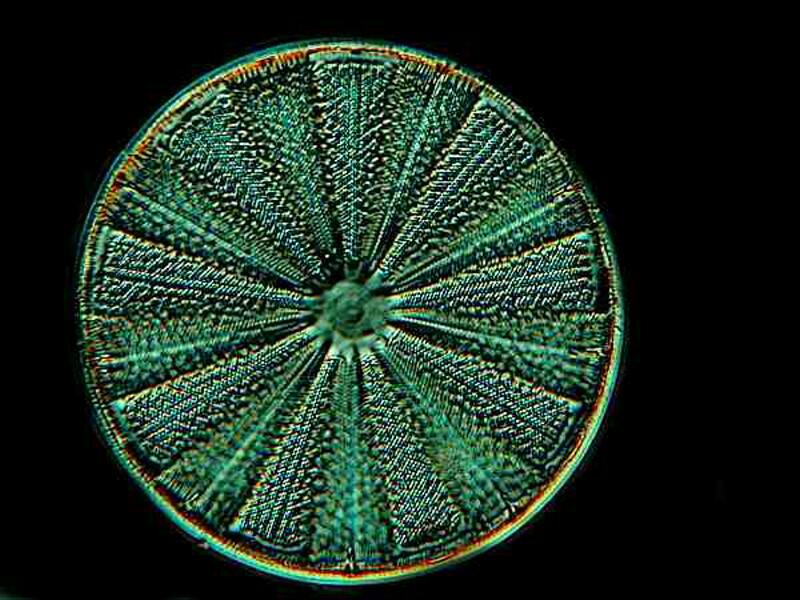
Another common type is the centric diatom which can have a considerable variation in pattern. Here are 2 examples and the second one which is wheel-like is especially attractive to my mind.
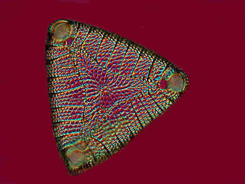
A somewhat less common type is the triangular form and below we have an example of Etogonia.
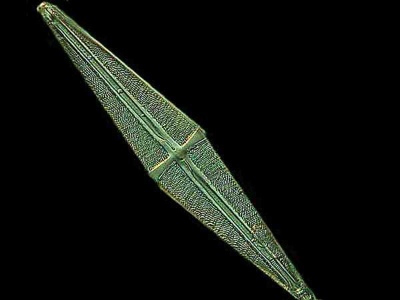
Another common, but striking genus with its elongated central cross is Stauroneis:
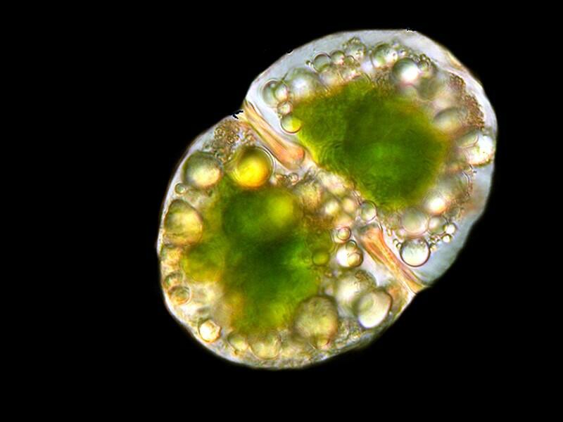
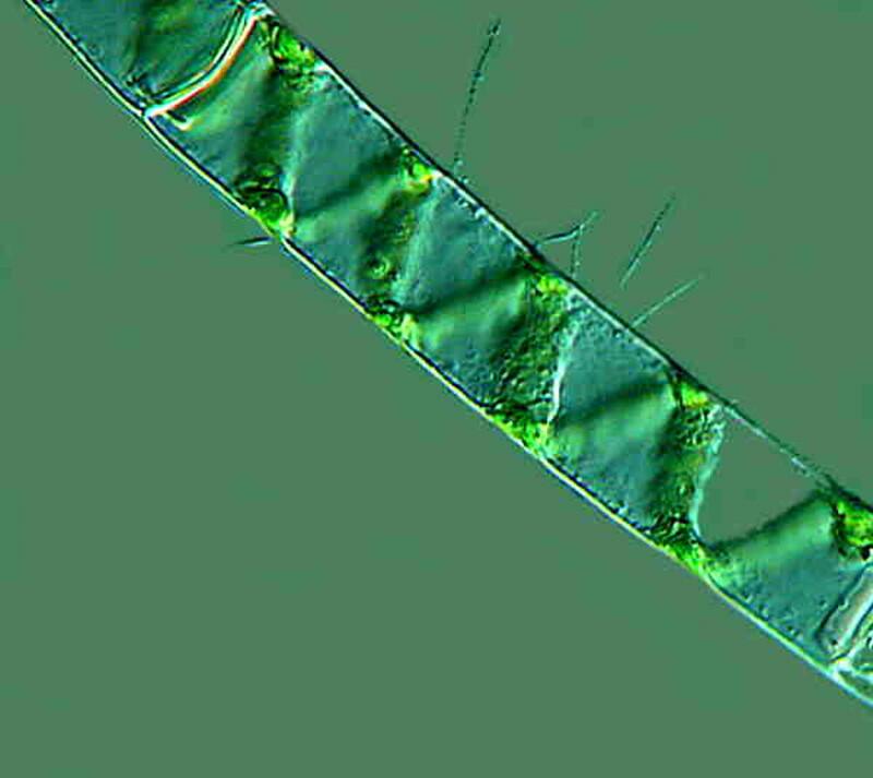
Next we will look at 2 examples of algae. The first is a desmid and the second is a lovely filamentous algae with a coil of chloroplasts running through the transparent, encasing tube. It is of the genus Spirogyra.
A very interesting type of micro-algae often survives the preservation process well, provided the liquid is not acidic, and these are the calcareous algae in which the tissue is overlaid with layers of calcium carbonate which is often reddish, pink, or white.
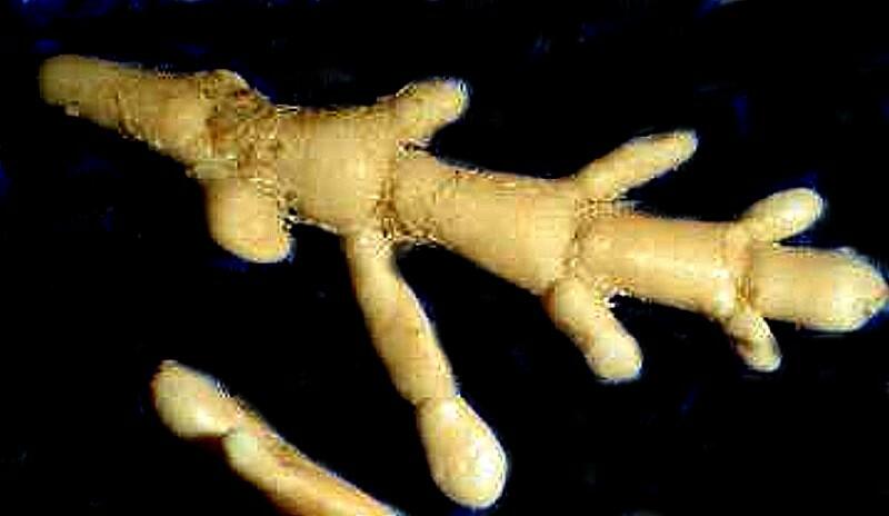
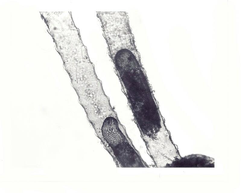
Protozoa are much harder to find in preserved samples with 2½ exceptions. Radiolaria (siliceous), Foraminifera (calcareous), and certain flagellates and ciliates that have organic “tubes’ or “vases” in which they reside. Folliculina is a fairly common example. When alive the organisms have a slight bluish-green cast and an elongate “U” shaped structure covered with cilia extend from the end for food gathering. These organisms are highly contractile and here, in an old Polaroid image which I took years ago, we can see the upper part of 2 separate tubes and a portion of the contracted organisms.
In fairness, one should also mention the Acantharians (closely related to Radiolaria) which have skeletons composed of Strontium sulfate.
Porifera (sponges) usually preserve well, are highly opportunistic, and will colonize virtually any niche that provides currents of water containing nanoplankton. In studying the micro-structure, one again has to be concerned about the preservative having a neutral pH. There are many forms of sponges that have calcareous spicules which in an acidic medium will etch and gradually dissolve. So, my recommendations are 1) when feasible, transfer specimens out of formaldehyde (or any other acidic medium) and into alcohol, 2) if that is not feasible, buffer the formaldehyde to keep it a neutral or slight alkaline pH, 3) if you have several pieces of the same material, I suggest preserving at least one in strong alcohol (but please don’t use single malt), place another piece or two in a tube of distilled water, and another one or two in small tubes or plastic boxes and let them dry.
As for Placozoa, don’t even bother looking for them in preserved samples; finding them in live samples is tricky enough. In this entire phylum, there is only one species, Trichoplax adhaerens which is an intriguingly strange organism and may represent a transitional form from the protists to the Metazoa. If you want to know a bit more about it, you can look at my short article on Micscape or if you want to know quite a lot more about it, you can read Karl Grell’s article in Microscopic Anatomy of Invertebrates, Vol. 2.
Platyhelminths are, of course, flat worms. If you’re interested in the verbosity of insects–etymological entomology–then you know that “plateau” means “flat place” and “platypus” means “flat cat”. (Just kidding.) In general, flatworms don’t preserve well; in fact very few worms or worm-like creatures are easy to preserve well. For almost 2 centuries, naturalists have tried to devise effective techniques for narcotizing and preserving such creatures, as well as other contractile organisms, with only moderate success.
One of the reasons that samples that have a rich assortment of organisms which have been rather haphazardly preserved are of such a source of fascination for me is that, over and over again, I encounter those happy accidents, those serendipitous occasions when, in spite of all the odds, I find superb examples of organisms in splendid condition when they should be massively distorted. Mind you, this is not something that you should rely on in general and if you are interested in preserving specific groups of organisms for study, you should by all means learn the basic protocols for preserving them.
Platyhelminths, phoronids, sipunculids and other worm-like creatures can be narcotized and preserved reasonably well and studied to good effect but, one must always remember that there is no substitute for investigating these creatures live if at all possible.
In the title of this essay, I mentioned Pleurobrachia. These creatures belong to the phylum Ctenophora which consists of indescribably beautiful, transparent, luminescent multi-colored jellies that make Times Square, Las Vegas, and all the other Neo-Neon cities look like the monstrous vulgarities that they are. Virtually every preserved organism is but a pale reflection of what it was in its living state, but ctenophores are dazzling. Go to this site and look at some photographs of living specimens or better yet some video.
These fascinating beings are ones which very, very few of us will ever see in their natural habitats. So much of the life on our planet lives in hidden, secret places and, as a consequence, we often miss wonderfully mysterious marvels.
Phoronids are small worm-like creatures often grouped with entoprocts and ectroprocts (Bryozoa). Like many of the rather unusual organisms in small phyla, their placement taxonomically has been an issue of disagreement. They are exclusively marine, build chitinous tubes, and have tentacles for feeding. There are only about a dozen species. You can see some images of them here:
Polychaetes, on the other hand, occur in vast number and in a large variety of species. Larval forms of polychaetes are abundant in plankton, but are often very difficult to classify. There are, however, a few species that spend their adulthood as members of the planktonic community and one of the most interesting is Tomopteris and, to my eye, visually pleasing.
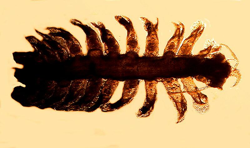
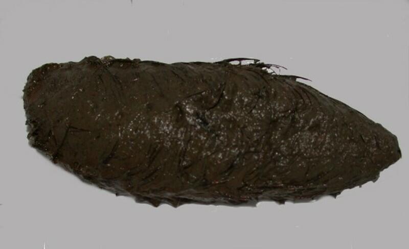
If you’re squeamish about worms, then you might not want to have a “sea mouse”, (Aphrodite) but they are fascinating and I have written about them elsewhere, so I won’t repeat myself, except to say that they are not candidates for any beauty contests. I’ll show you the dorsal view first and then the underside where you can see the segmentation which demonstrates that it is a true worm.
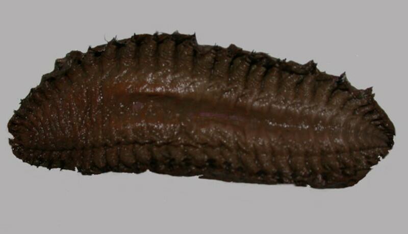
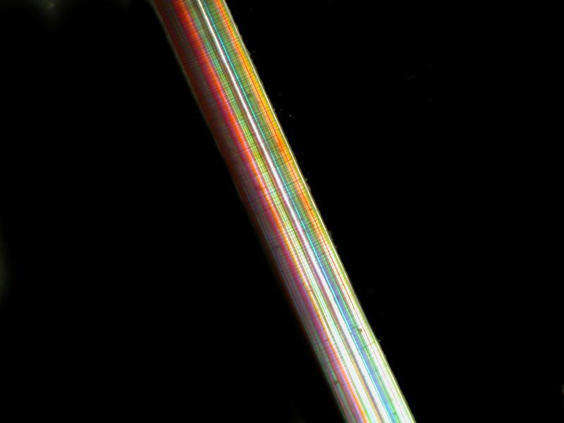
However, here under all this muck and slime there is still some beauty hidden. Sea mice have setae (bristles) which are remarkably efficient transmitters of light, even more efficient that our best fiber optics and here is one that has been cleaned and photographed under polarized light.
Some of the polychaete worms are quite beautiful, especially the Sabellids which include the “feather duster” worms.
A wonderful group of creatures which appear somewhat worm-like are the nudibranchs which, in reality are mollusks and are among the most stunningly colorful and splendiferous organisms on the planet. Explore these links, because they reveal not only the richness, but the remarkable variety of these remarkable creatures.
Another splendidly colorful group is the starfish and they have remarkable powers of regeneration. Many people are familiar only with common dried specimens sold in craft and shell shops which are typically a rather dull brown and give virtually no sense of the extraordinary variety and subtle ranges of color. However, be warned, there are suppliers and merchants who sell dried specimens which have been dyed and sometimes in grotesque, phoney, fluorescent hues which have none of the delicacy and subtlety which nature presents us with.
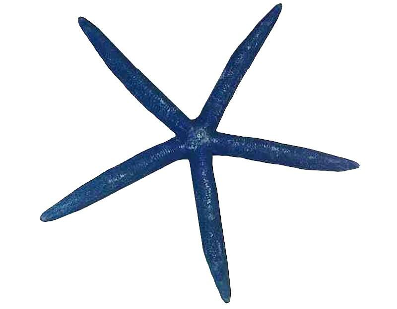
There are blue starfish, ones that are bright red, orange ones, purples one, lemon-yellow ones, black ones, white ones, some that are a lovely milky beige color with brilliant scarlet spines, and among the Dermasterias the so-called “leather stars’ named for the texture of the skin membrane not because they are into sado-masochism and leather bars, there is often a display of intricate, variegated color patternings.
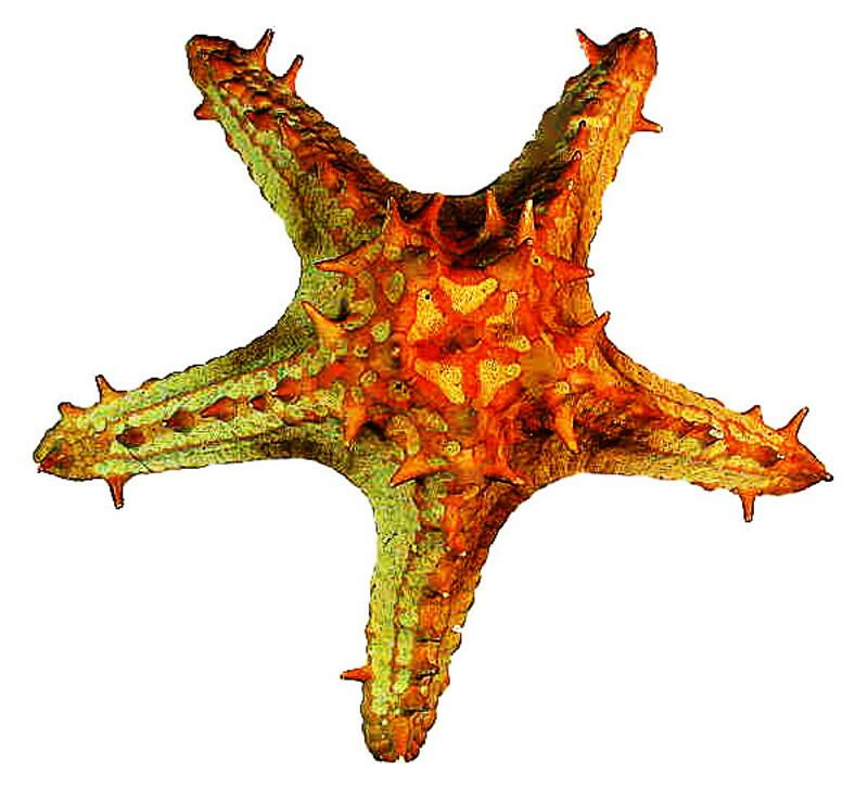
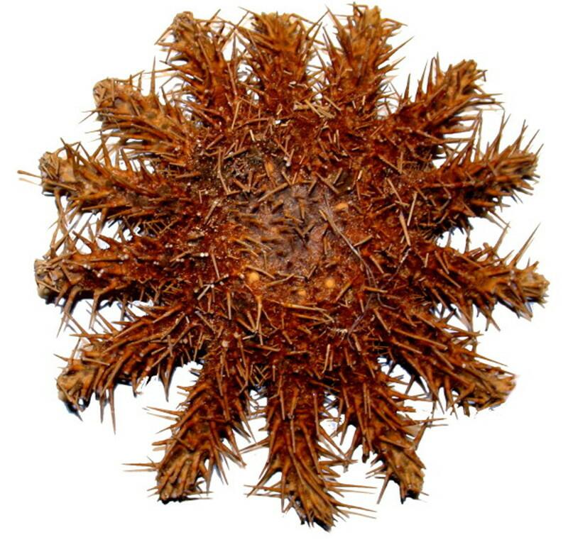
Here is an example of a tropical thorny starfish with brightly colored spines.
And here we can see a specimen of Linkia laevigata which even though it is dried has retained its natural color; in other words, this is NOT a dyed specimens
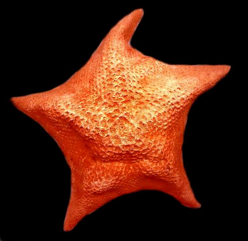
Next is a bright orange “Bat star” which as you can see on close observation has some unusual “plates” which form the outer surface.
Finally, here is a formidable creature, Acanthaster plancii, the “Crown of Thorns” starfish which has devastated large sections of tropical coral reefs. It feeds on coral and its population has greatly increased in the last couple of decades in significant part due to the over collecting of one of its major predators, the large gastropod Triton whose shell collectors prize. Efforts to control this starfish have been difficult in part because the spines contain a toxin which not only discourages predation, but makes it a challenge for divers to collect and destroy them from heavily infested reef areas.
Regarding regeneration, commercial oyster fisherman in the 19th Century learned about this extraordinary phenomenon in an economically painful way. Many starfish are predators on shellfish and satisfy their appetites in an astounding fashion. They attach to an oyster or clam with their tube feet and use the powerful muscles in their arms to pry open a small slit between the two valves (shell halves, thus the term, bivalve) and then evert their stomach through this narrow opening and digest all the delicious internal parts leaving behind a useless pile of shells from the point of view of the oyster fishermen.
In the title, I mentioned Pyrosoma, (a pelagic tunicate), which means “fire body”. This incredible creature 1) appears like a long, slender glassy nearly transparent gelatinous tube, 2) is composed in significant part of cellulose compounds even though it is an animal, 3) is colonial and can be several feet in length, and 4) possesses bioluminescent bacteria so that if you were to run your finger along its surface on a ship’s deck at night, you would observe its reaction as a luminescent stripe.
These are truly very strange and fascinating creatures as you can see by exploring some of the images here:
The ocean is full of countless mysteries which can be explored, living or preserved, in many ways using simple tools, a good stereo-microscope, a good compound microscope, and a few basic reagents. In return, you will reap a lifetime of pleasure, learning, and wonder.
All comments to the author Richard Howey are welcomed.
Editor's note: Visit Richard Howey's new website at http://rhowey.googlepages.com/home where he plans to share aspects of his wide interests.
Microscopy UK Front
Page
Micscape
Magazine
Article
Library
Published in the September 2012 edition of Micscape Magazine.
Please report any Web problems or offer general comments to the Micscape Editor .
Micscape is the on-line monthly magazine of the Microscopy UK website at Microscopy-UK .
© Onview.net Ltd, Microscopy-UK, and all contributors 1995 onwards. All rights reserved. Main site is at www.microscopy-uk.org.uk .