|
Note on 'diatom dotting' at low magnifications. Resolving Stauroneis phoenicenteron. by David Walker, UK |
In my September 2021 article I shared studies of resolving S. phoenicenteron with lower mag / NA objectives by imaging at 400 nm to increase the resolution. This is easily done on a stand with a 100W halogen lamp (if bulb not UV blocked) by placing an interference filter on the field lens. Resolving less demanding diatoms at lower objective mags can be fun and instructive. They are useful subjects to check the quality of the entire optical train as well as demonstrating some basic principles of microscopy such as condenser quality / iris setting, light wavelength effect on resolution and the quality of a photomicrography setup.
The pores were 0.63 µm between pore centres. Using a typical equation (see Footnote 1) this suggests an NA of 0.53 is required to resolve the pores. The diatom pores were successfully resolved with a Zeiss Neofluar 16/0.4 objective on a Photomicroscope III at 400 nm (NA increased to 0.55 theoretically) but was unsuccessful using a 10/0.45 Nikon CFI planapo objective on an Eclipse 800 stand. The 400 nm filter should allow resolution of pores with the 10/0.45 but struggled to capture an image because the 1.3 Mpixel camera owned had insufficient resolution, see image below. The Eclipse also had no Zeiss Optovar type mag changer and a 2X C-mount relay lens was not enough to fill the camera field beyond third of sensor width. The photo port of the Eclipse also does not respond to increased mechanical tube length to increase the area covered on the sensor. The screen capture below when image zoomed to 200% shows the problem.
Opticstar PL-130M 1.3 Mpixel camera. Stauroneis phoenicenteron. Nikon Eclipse 800, ach-apl condenser, slight oblique, Nikon CFI 10/0.45 planapo, 2X relay lens on photo port.
Screen capture of 200% screen mag.
The Opticstar PL-130M was showing its age (bought 2007) so have recently upgraded to a 6.4 Mpixel monochrome camera, a ZWO ASI178MM, 1/1.8 inch CMOS. This, like the Opticstar is primarily sold for astronomy but the manual notes it is equally suitable for microscopy. Astronomy software is provided with the camera but prefer the simplicity of Marien van Westen's Micam v1.6 software* which works well in Windows 10. The first image below shows how small a field the diatom occupies with a 10X but also shows the field of the ZWO camera with a C-mount 2X optical relay. The second image shows that the ZWO was able to resolve the diatom to pores. (*For reasons unknown the later software versions do not recognise my two cameras.)
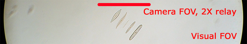
Nikon 10/0.45 full visual field of view to the black borders, and ZWO camera field with 2X C-mount optical relay. The coarser striae are well beyond visual observation.
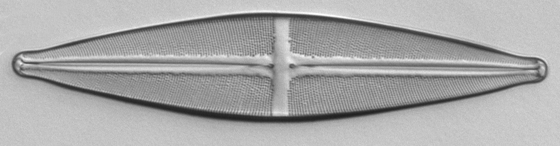
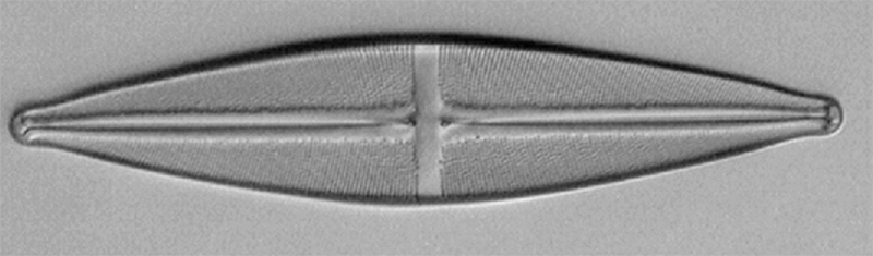
ZWO ASI 178MM 6.4 Mpixel camera. Same 10/0.45 objective, brightfield, oblique, 400 nm filter. Crop at 1:1 of the full image.
The diatom still occupies a small field on the camera sensor but the increase in Mpixels from 1.3 to 6.4 is now sufficient to resolve the pores.
Footnotes:
1)
The textbooks discuss various forms of the equation linking resolution R (nm
or µm)
to wavelength l (nm
or µm)
and NA of objective.
The equation R = 0.61 x l /
NA was used.
Acknowledgement: The author used the invaluable 'Test Slide version 2.0' (Diatom Cubed mountant) supplied by Stefano Barone of Diatom Lab.
Comments to the author David Walker are welcomed.
Published in the September 2022 edition of Micscape.
Please report any Web problems or offer general comments to the Micscape Editor .
Micscape is the on-line monthly magazine of the Microscopy UK web site at Microscopy-UK
© Onview.net Ltd, Microscopy-UK, and all contributors 1995
onwards. All rights reserved.
Main site is
at www.microscopy-uk.org.uk
with full mirror
at www.microscopy-uk.net
.