|
Amoeba proteus in different spot-lights |
Illumination and contrast techniques for the light microscope |
|
Bright field |
|
|
|
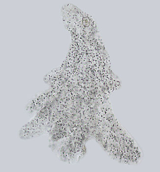
This photograph was taken with a X25 brightfield water immersion objective. Note the
lack of contrast compared to the phase and DIC methods. Bright field microscopy is used when there is enough contrast in the subject matter or artificial staining techniques are employed. However, when an object of low contrast to the background is being viewed, such as protozoa, very little of the subject matter can be made out. When photographing protozoa such as the amoeba very little in the way of information can be gained from this type of illumination, certain structures can be made out but phase contrast and DIC give a much better impression of the animal. |
| In order to get round this problem, biologists and histologists have used counter staining for over one hundred years; and this helps to differentiate the various tissues and organelles that can be found in a variety of subjects that would otherwise be rendered invisible. This method does have its drawbacks. The cells are usually killed and therefore cannot be studied whilst moving around their natural habitat. Bright field microscopy gives incredible views of algae, desmids and prepared slides such as those used by histologists. | |


Back to An Introduction to Microscopy
Microscopy
UK Front Page
Micscape
Magazine
Article
Library
text & Image © Steve Durr 1999
Web design: Wim van Egmond
© Onview.net Ltd, Microscopy-UK, and all contributors 1995 onwards. All rights reserved. Main site is at www.microscopy-uk.org.uk