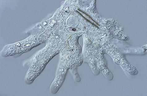
| This photograph was taken with a X40 Nomarski objective or differential interference contrast objective. It can be seen from the photograph the amount of information that this technique reveals when used correctly. | |
|
The DIC set-up consists of: -a Polarizer. -a Wollaston prism. -the Object. -Wave train. -Objective. -Wollaston prism. -Analyzer. -Eyepiece. |
|
| The advantage of using a DIC set-up is the 3D-pseudo effect that it gives and also,
unlike phase contrast there are no halos around the subject. The need to kill the cell does not apply with this
system and is therefore useful to biologist's and medical researches. The Wollaston prisms have been built from
two birefringent prisms with their crystallographic axis forming a right angle. The light is split into two rays
by the Wollaston prism. Some of the rays pass through the specimen and are retarded, from there the rays are projected
into the objective where the second Wollaston prisms are found. The rays are then recombined and are brought to
a similar direction where they can interfere. By using a rotating stage the effect can be varied. This system is no better than any of the others (phase, brightfield and dark field) but helps to give another dimension to the organism that is being studied. This equipment is very expensive but some amateurs are lucky enough to get to use this type of contrast equipment. There is method to get images that resemble D.I.C. By shifting the condenser from it's axis so called Oblique illumination enhances the contrast and also the resolving power of the microscope. |
|