 |
Illumination
Variants |
A report on an
interesting variant of oblique illumination
( by Paul James, UK)
The following 'notes' are based on simple experiments using an ordinary bright-field/phase binocular microscope, and oblique illumination with coloured light. The success of reproducing the same effects with your own microscope(s) may well depend on small but significant variations of optical design in the substage condenser and NA of the objectives used, and most importantly the suitability of your slides.
Serendipity?
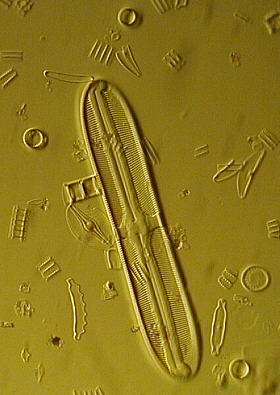
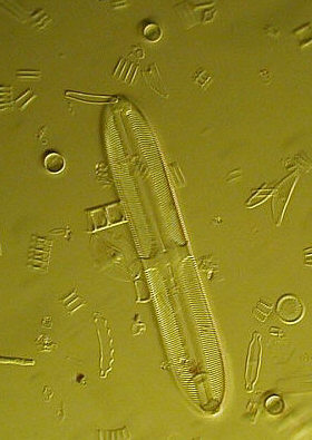
Whilst fiddling with my substage condenser during a session of darkground illumination, I stumbled across an interesting and rather unusual situation which fully caught my attention. I was looking at a slide of diatoms using a simple patch stop and a yellow filter, when I noticed at a critical point in the focussing of the substage condenser, a remarkable change in the appearance of the image. Owing to the limitations of my camera, the photo below barely conveys the beautiful smoothness and subtlety of detail, but does reveal some of the solidarity, and waxy/plasticky like texture I saw.
|
The diatoms in the field appeared utterly solid in form, as if I was peering at the sea bed strewn with diatomic bits and pieces, so smooth was the creamy waxy like 'floor'. Areas of the field showed subtle variations in depth, the diatoms and debris were revealed in bitingly sharp form, and oddly some small pieces appeared depressed in the 'floor', contrasting with the beautifully side lit larger pieces and whole diatoms. In short it was like looking at a fossil made from injection moulded plastic!
A theory on the cause of this beautiful phenomenon?
At first I was unable to identify the mechanism causing this striking effect. Might it be purely psychological? The effect without the filter was not as convincing, and I imagine that the coloured light helped to establish in the observers mind that the 'space' between the subject matter on the slide was solid and quite real. Some colours conveyed this solid fossil effect better than others, but it was just a matter of time before the mind adjusted to the 'new' colour. Being able to photograph the effect, albeit as an inferior image proved to me at least that this was not entirely a quirk of the mind, and that the psychological 'input' was marginal.
Naturally, having stumbled across this, and satisfied myself that I could repeat the settings, I tried other slides and woefully found that few could be induced to show the same type of effect so dramatically, and if they did, they were inferior to this J D Möller slide (circa 1890).
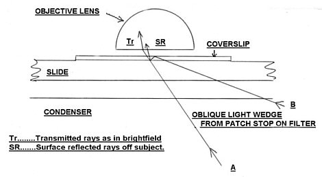
What could bring about, what appears to be a mixture of transmitted and reflected light, in such proportions as to bring about such a convincing illusion of solidarity ?
A clue was that this effect was only apparent with the x 40 and x 20 objectives, as with the substage setting undisturbed, lower powers produced fairly legitimate darkground images. The larger NA of the higher power object glasses accepted the more acutely angled light not entering the lower power objectives, which may very well hint at to what is actually happening.
Could total internal reflection be occurring ? So that the light which descends from the top surface of the cover slip, re-illuminates the subject from above ? Hence the combination of 'surface' and transmitted illumination brings about the overall solid surface and shadow effect. I could be wrong of course?
|
The diagram above is only a representation of what might be responsible for the illusion of surface illumination? Those rays passing through the sector near B which are the most oblique, could probably be reflected back off the upper surface of the coverslip because of total internal reflection TIR. |
One very convincing indication that the likely cause of this 'surface' illumination is TIR, was the fact that the highlights could be 'extinguished' by reducing the substage iris a fraction, which left commonly observed oblique views with diffraction images. Once this surface illumination disappeared, the illusion also vanished. Thus, only when the most oblique rays were cut off, did the sensation of solidarity in the image disappear.
 |
 |
Iris fully open |
Iris closed slightly |
Setting up
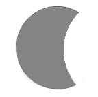 |
A simple patch
stop similar to this, will produce oblique rays across
the subject when placed in the substage filter tray. Ideally the 'cusp' should be thin and sharp too, simply achieved using thin metal or opaque plastic sheet. Cardboard will produce respectable results for initial trials. |
Decentre your condenser by 1-2 mm or so in any direction, and with the condenser below it's normal position for bright-field, focus the objective ( 20x-40x ) on a suitable area of the chosen slide (preferably strewn diatoms). Then set up your 'scope with the patch stop shown on above left with a colour filter, a yellow photo type is ideal. Patch stops work best if placed in the filter tray directly beneath the condenser. Open the substage iris diaphragm fully, since the condenser needs to be operating at maximum aperture. The filter, if of awkward size can be introduced into the optical path anywhere between the substage and light source.
 |
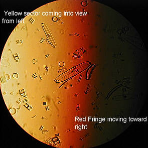 |
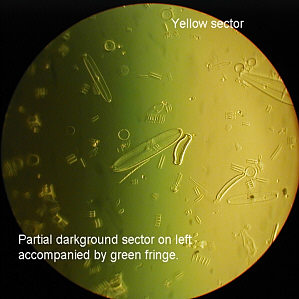
Raise the condenser slowly. The field will probably be dark initially, but a reddish fringe will appear, sliding across the field, followed by yellow light and finally to a possible green fringe bordering on what appears to be the edge of a dark-field sector.In the photo's above this is shown as movement from the left to right, but it can be in any direction depending upon the orientation of the patch stop. Hopefully, you should see the subject matter change from a dark reddish bright-field, with diffraction borders around the subject, to the solid waxy/plasticky form and then become slightly 'backlit' as in darkground. The ideal position of the condenser is such that when in position, the whole field should be the chosen colour, yellow here, when using the 40x objective, and mostly, if not all with a 20x objective.
If the substage iris is closed very slowly, the highlights or what appears to be surface illumination of the subject disappears leaving the familiar bright-field, albeit oblique.
The area we are trying to use is of course the yellow zone (or whatever colour you have chosen).
Here the diffraction fringes of the subject should appear absent, but the whole field and subjects should appear to be highlighted from what appears to be a low setting sun!
Problems
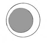
If the condenser can't be decentred:-
Some mirror supports can be moved out of the optical path, and automatically offset the incoming light to produce a similar effect. Or use an off-axis stop as shown below, experiment by altering it's position.
 |
If no impressions of surface solidarity are observed, think about the following :
Experiment by altering the dimensions of the patch stop, and it's position, rotate if necessary.
Change the objective, or substitute one if possible for one of a different NA.
Check that the iris diaphragm in the substage is fully open.
If you don't use a colour filter, the effect is still there, but psychologically, a colour helps to 'identify' a 'solid floor' base, and reveals subtle shadow variations on the slide's surface.
Try decentering the substage condenser a little more or less.
Observe another slide, as some are unsuitable. Uncluttered subjects work best.
The cover slip/mountant properties/ factor
It could well be that your slide of diatoms is not suitable? Try others. Some subjects are not so suitable, and can be difficult producing the nice 'fossil' images. What does appear to be important is the refractive index of the mountant and the subject, and also how intimate the bond is between them. I think Calcareous subjects, especially the optically transparent types work best. Perhaps the most critical aspect is the cover glass/mountant thickness?
Photographs
My Olympus 830L digicam was used to take the photo's using a Wild fluorite 40x (NA 0.85) and 20x (0.65 NA). A 40x achromatic (0.65 NA) objective produced similar results, although with slides of water immersed subjects the latter proved to be superior in this situation.
Gallery
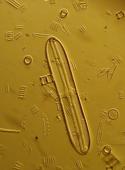
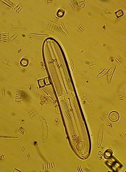
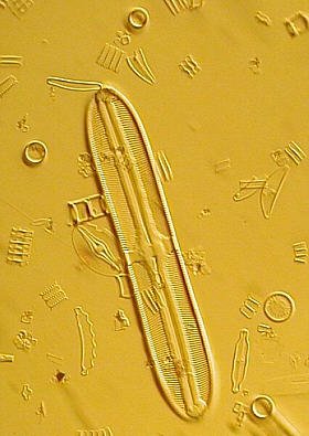
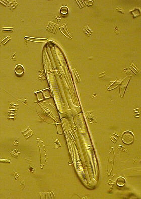
The four photo's below show how varying light source intensity, and rotating the patch stop can bring about different 'views' of the same image. I found that observing in a darkened room with lower light source intensities gave very convincing and satisfying images. x40 fluorite objective.
 |
 |
 |
|
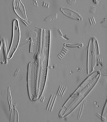
Here's a greyscale version (a yellow filtered image converted to greyscale in image software) of a similar subject using precisely the same technique, and there's a different but still similarly convincing portrayal of the diatoms and debris (40x fluorite objective).
This greyscale version may lead some to think that coloured light is not necessary to show the effect, but photographing the image without a filter, and indeed observing such, reveals a much brighter background with less convincing images. However if the light source is reduced considerably when working with no filter, the effect re-establishes itself more convincingly, due I believe to colour introduced from the 'cool' light filament, and subtle surface depressions in the slide not seen with brighter illumination?
 |
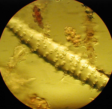
Below, the photo shows a sponge spicule at 400x, and clearly demonstrates the capability of surface examination, albeit in small layers owing to the restricted depth of field of the 40x objective operating at full aperture. Notice here that the lighting effects are different, which is caused by the different optical properties of the subject and mountant. The interface between mountant and subject can be glimpsed in parts too.
 |
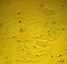
This shot of epithelial cheek cells below was taken using a slightly reduced iris aperture in the substage condenser, which reduced the 'surface illumination' and brought about a better balance, and importantly, allowed an achromatic objective of lower NA (0.65, 40x) to yield a more convincing result.
 |
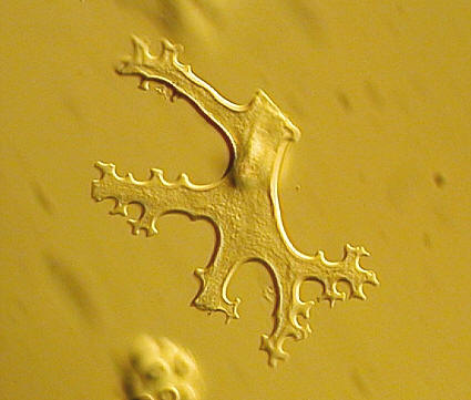
Below, 'bracket' form spicule revealing surface texture. 40x achromatic objective ( 0.65 NA )
 |
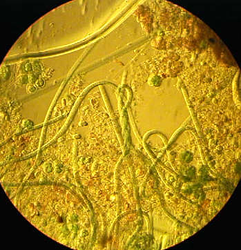
Tangled algae below reveals partial success of this method. (x 20 fluorite 0.65 NA )
 |
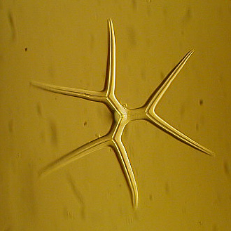 |
Well defined spicule above (x 40 achromat 0.65 NA)
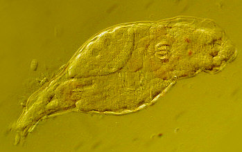 |
'Resting' rotifer above displayed wonderful detail of 'digestive processes'. x20 fluorite.
Pro's
Offers an alternative method of assessing the external features of subjects.
Can reveal detail and 'form' hitherto not easily observed with other basic methods.
Is a great joy to observe some subjects when set up properly.
Con's
Can be difficult to find the right settings of condenser, patch stop, objective and subject etc...
Restricted subject matter. Most diatoms, spicules and other calcareous forms fair well, and also some protozoan subjects, but still never the less dependent on mountant, overall clutter and thickness of preparation. Insect parts, stained tissue sections and other non transparent subjects do not seem to support the effect as easily.
Viewing with a monocular is not as convincing as with a binocular stand.
Summary
The success of this method, whilst being fundamentally 'normal' oblique illumination, depends on all the factors mentioned above being set up correctly, though this will vary from instrument to instrument. Probably the most important aspect in achieving these memorable views, is that the subjects lay on the slide evenly, in the same plane. Thick, cluttered preparations are disappointing.
Good viewing !
| Comments welcomed to Paul James. |
Editor's note: other Micscape articles covering
aspects of oblique illumination are by John Wojtowicz
and by Dave
Walker.
Published in the February 2000 edition of Micscape Magazine.
Please report any Web problems
or offer general comments to the Micscape Editor,
via the contact on current Micscape Index.
Micscape is the on-line monthly
magazine of the Microscopy UK web
site at Microscopy-UK