In January 1998 Micscape asked
'Can you identify these tiny structured objects
on the hairs of a fly leg?'
We now believe they are 'brochosomes',
which in themselves seem a mystery ...... and a possible challenge for
the amateur to push his 'scope to the limit
 Do you remember our 'What are these mystery objects' query in last months
magazine (January 1998)? Click here if you didn't
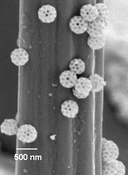
see this article. Essentially they were tiny structured objects only 0.5
micron across (500 nanometers) found on the hairs of fly's leg (shown right).
The originator of the query, Frank Placido (UK), hadn't found a satisfactory
answer and sought the help of Micscape and our readers.
Do you remember our 'What are these mystery objects' query in last months
magazine (January 1998)? Click here if you didn't
see this article. Essentially they were tiny structured objects only 0.5
micron across (500 nanometers) found on the hairs of fly's leg (shown right).
The originator of the query, Frank Placido (UK), hadn't found a satisfactory
answer and sought the help of Micscape and our readers.
The image has been on our site all month and has attracted
some interesting emails (thanks to those who responded). However, if anything,
the responders were as mystified as we were, and we were beginning to think
we couldn't supply an answer for this month's issue, February 1998.
But an email from Mike Dingley in Australia, a regular
Micscape contributor, has set us on what we believe is the right track
to solving the mystery. He said he thought they were brochosomes which
apparently are insoluble solid objects found in the excreta of leafhoppers
(a common group of insects in the family Homoptera). Frank also remarked
in his original query that a colleague had mentioned the objects were something
that sounded like this term.
I did a quick web search for 'brochosome' and found a
web site with an image of brochosomes that look almost identical in shape
and similar in size (estimated from the web image displayed) to Frank's
mystery objects.
We have become intrigued with these brochosomes, as they
raise even more questions. e.g.
-
what is their composition
-
why do leafhoppers excrete them
-
do other insects excrete them
-
are they created by the digestion process, or passed through
the system from their diet (they are mainly sap suckers)
-
apparently leafhoppers are well known vectors of bacterial
and viral diseases of plants, are the brochosomes related in anyway to
the transfer of disease
-
are there people actively researching their form and function
We have a few leads already on some of these queries, and
we are hoping to put together an article on the information we find. We
will acknowledge all material submitted and the copyright does of course
remain yours.
One final thought, some amateur microscopists with
the finest optics on their microscope like pushing their light microscopes
to the limit. This is often achieved by resolving the fine features on
the silica frustules of diatoms. The
brochosomes are typically 0.5 micron across (possibly larger) but not far
from the limit of resolution with a light microscope with green light (ca.
0.22 micron).
Is it possible to find, recognise and photograph brochosomes
on leafhoppers using a light microscope? If the size range of brochosomes
extends to say 1 micron, the surface features would be of the order of
0.25 microns apart, right at the limit of resolution. But if the contrast
is there, is their just a chance of resolving some detail e.g. with
a deep blue filter? Let us know your thoughts on this, even better have
a try and see if you succeed!
Compiled by Dave
Walker
See the following month's Micscape article
for a fuller explanation of brochosomes.
Acknowledgements
Many thanks to Frank Placido, of the University of Paisley,
Scotland for first submitting the query and image. This was a fascinating
query and has sparked an equally fascinating follow-up on what are very
intriguing objects. Also thanks to Mike Dingley and his colleagues in Australia
for finally pinning down what the mystery objects are most likely to be.
© Microscopy UK or their contributors.
Please report any Web problems or offer general comments
to the Micscape
Editor,
via the contact on current Micscape Index.
Micscape is the on-line monthly magazine of the Microscopy
UK web
site at Microscopy-UK
WIDTH=1
© Onview.net Ltd, Microscopy-UK, and all contributors 1995 onwards. All rights
reserved. Main site is at www.microscopy-uk.org.uk with full mirror at www.microscopy-uk.net.