 Micscape's
interest in brochosomes arises from our January Image
of the Month 1998 (shown right) generously submitted by Frank Placido,
UK and the follow-up in the February 1998
issue.
Micscape's
interest in brochosomes arises from our January Image
of the Month 1998 (shown right) generously submitted by Frank Placido,
UK and the follow-up in the February 1998
issue.
and a challenge for the amateur microscopist?
A summary of the information provided to Micscape prompted by our January 'Image of the Month' , with thanks to the contributors listed in the Acknowledgements.
(This is a long text document, after viewing the image and links
you may wish to save for off-line reading if of interest).
Micscape's
interest in brochosomes arises from our January Image
of the Month 1998 (shown right) generously submitted by Frank Placido,
UK and the follow-up in the February 1998
issue.
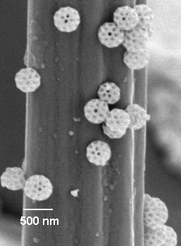
Brochosomes are tiny structured objects excreted by leafhoppers. The brochosomes shown right on the hairs of a fly's leg are only 0.5 micron in diameter (500 nanometers).
Since learning what these objects are, Micscape became very intrigued with them, and asked some open questions in last months follow up (and in relevant newsgroups), to see if our readers could tell us more. We were delighted to receive some very interesting responses from readers world-wide, and we are pleased to summarise this information below, as we are sure it is of interest to others.
Roman Rakitov, Russian Academy of Sciences is one of the few people currently researching into brochosomes. He has taken the trouble to provide a very useful summary which we reproduce in full (with his permission).
Dear Mr. Walker:
I study brochosomes since 1990 - their morphology, morphogenesis, diversity, adaptive significance, role in evolution of leafhoppers as well as related behaviours and morphological specializations of these insects.
The theme of my PhD-thesis was "Brochosomes and related peculiarities of morphology and behaviour of leafhoppers". Besides some published results (see references in the end of my letter), I have a couple of papers in preparation and a lot of unpublished SEM and TEM images. As far as I know, in recent years, only Dr. Max Day from Australia and I worked on brochosomes (BS), so it is hardly surprising that you failed to find anything about them on the Web.
The list of references given below covers the majority of published evidence about BS. Unfortunately, I have no access to the Web and have not seen your on-line magazine, but one colleague of mine has re-sent me your call for information on BS. I hope the following brief information will be helpful.
Sincerely, R.A. Rakitov, PhD, Russian Academy of Sciences.
Brochosomes are minute secretory particles produced in huge quantities by the Malpighian tubules of leafhoppers (Hemiptera, Cicadellidae). Each of the four Malpighian tubules has a dilated middle segment consisting of specialized BS-producing cells. These do not seem to be involved in any kind of fluid / ion transport like typical excretory Malpighian tubule cells but resemble typical protein-secreting cells.
They are filled with a number of vacuoles containing mature and developing BS, eventually released into the tubule lumen. Both genesis and chemical nature of BS are rather obscure. Scarce ultrastructural and histochemical evidence (Smith, Littau, 1960; Gouranton, Maillet, 1967) seem to be out of date. They suggest BS to be originated from the Golgi complex and composed from lipids and proteins. Freshly moulted leafhoppers (adults, in some taxa also nymphs) excrete the BS-containing fluid from the anus and transfer it with their legs onto the fresh cuticle.
Using their specialised spiny legs they distribute BS across the surface of body and appendages (Navone, 1987; Rakitov, 1996) making a sort of fine coating - similar to coatings of waxy particles in many other insects (Rakitov, 1995). This coating is extremely hydrophobic, what allows to suggest the water-repellence is at least one of the functions of BS. Other hypothetical functions of BS, e.g. dissemination of pheromones (Day, 1993), are not supported by any available facts.
BS are produced, at least by the adult stage, in all the major subfamilies of leafhoppers, including the apparently most primitive. Very many species produce similar spherical BS of about 0.5 micron in diameter with characteristic latticework surface, looking like giant clathrin-coated transport vesicles. However, generally, the size, shape and structure of BS vary among leafhopper species (Rakitov, 1995). I was able to find the ones producing BS with average diameter of 4-5 microns or even still larger elongated stick-like BS (in prep.).
In some leafhopper genera BS provide valuable characters for identification and classification of species. BS can easily be rubbed off the leafhopper cuticle and contaminate any insects collected together with leafhoppers (directly or through the net, vial walls, pincers etc.). This has led to erroneous accounts on BS found in mosquitoes and other insects, including leafhoppers' relatives Cercopoids (Tulloch et al., 1952; Frost et al., 1994). Treehoppers (Membracidae), the nearest relatives of leafhoppers, exhibit similar post-moulting behavior, coating themselves with some Malpighian tubule secretions (Rakitov, 1996). However, I believe that the evidence for production of BS in this family (Gouranton, Maillet, 1967; Day, 1993) are also incorrect.
BIBLIOGRAPHY
Arzone A. 1986. Brocosomi: origine, forma, funzione, Atti Accad. Naz. ital. Ent., Rendic. Anno 34. P. 59-71.
Cheung W. W. K. & Purcell A. H. 1991: Brochosome production in the malpighian tubules of the leafhopper Euscelidius variegatus (Homoptera: Cicadellidae): an electron microscopic study. Proceedings of the 49th Annual Meeting of the Electron Microscopy Society of America, pp. 264-265.
Day M. F. 1993: Brochosomes of Australian Cicadelloidea. Proceedings of the 8th Auchenorrhyncha Congress (Delphi, 1993), pp. 10-11.
Day M.F., Briggs M. 1958. The origin and structure of brochosomes. J. Ultrastruct. Res. Vol. 2. P. 239-244.
Frost A.S., Gardner J.S., Nielson M. 1994. Scanning electron microscope study of brochosomes of Cercopidae. Proc. Elctr. Micr. Soc. Amer. Vol. 52. P. 358-359.
Gouranton J., Maillet P.-L. 1967. Origine et structure des brochosomes. J. Microscopie. Vol. 6. N. 1. P 53-64.
Navone P. 1987. Origine, struttura e funzioni di escreti e secreti entomatici di aspetto ceroso distribuiti sul corpo mediante zampe. Ann. Fac. Sci. Agr. Univ. Torino. Vol.14. P.237-294.
Rakitov R.A. 1992. [The leafhopper Vilbasteana oculata (Lindb.) coats its cuticle with a secretion of the Malpighian tubules] Zool. zhurn. Vol.71. No.5. P.49-57 [in Russian]. [English translation: Entomological Review. 1993. Vol. 71. No. 7 P.148-157].
Rakitov R.A. 1993: Brochosomes as an outstanding specialization of Membracoidea. Proceedings of the 8th Auchenorrhyncha Congress (Delphi, 1993), pp. 12-13.
Rakitov R.A. 1995. [The covering formed by brochosomes on the cuticle of leafhoppers (Homoptera, Cicadellidae)] Zool. zhurn. Vol.74. No.1. P.19-32 [in Russian]. [English translation: Entomological Review. 1996. Vol.74. No.9 P. 90-103].
Rakitov R.A. 1996. Post-moulting behaviour associated with Malpighian tubule secretions in leafhoppers and treehoppers (Homoptera, Membracoidea). Eur. J. Entomology. Vol.93. P.167-184.
Smith D.S.,Littau V.G. 1960. Cellular specialization in the excretory epithelia of an insect. Macrosteles fascifrons St l (Homoptera) J. Cell. Biol. Vol.8. P.103-133.
Tulloch G.S., Shapiro J.E., Cochran G.W. 1952. The occurrence of ultramicroscopic bodies with leafhoppers and mosquitoes. Bull. Brooklyn Entomol. Soc. Vol. 47. P. 41-42.
Vidano C., Arzone A. 1984. "Wax-area" in cicadellids and its connection with brochosomes from Malpighian tubules. -Mitt. schweiz. entomol. Ges. Vol.57. No.4. P.444-445.
**********************
TSI Particle Instruments Division, Minnesota (US) have also very kindly provided some information and an image of brochosomes which we have been allowed to reproduce (courtesy of Ron Grogg, TSI).
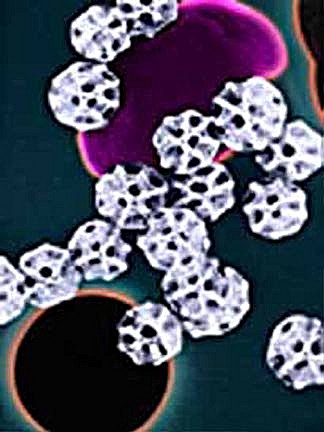
Image supplied by G. Yamate.
(Not to be reproduced without the permission of TSI).
Text courtesy of TSI Incorporated, Particle Instrument Division web site.
The "micro-whiffle balls" displayed in the header at the top of this page were originally described in our paper catalog as "unidentified, carbonized hollow spheres collected in the air around St, Louis, Missouri, USA." The image was prepared from a 27,000x secondary electron image produced with a scanning electron microscope by G. Yamate of the IIT (Illinois Institute of Technology) Research Institute.
A more complete description of these mystery particles
was
subsequently sent to us by Dr. E. Keith Bigg from
Australia. He
writes:
They are "brochosomes". These are solid inclusions that occur in the excreta of the very common insects known as "leaf hoppers". The messy little beasts wipe their excreta on their wings and the brochosomes are dispersed when it dries and they fly.
You can find a picture in Nature 224:715, 1969 (by R.D. Wiffen and M.J. Heard) and its identification in Nature 225:199, 1970 (by A.C. Neville and D.S. Smith). I found large numbers of similar particles in air samples about 10 years ago and was eventually directed to the above references by Bill Birch of the Australian National University at Canberra and by Max Day, a retired scientist who was still working on their nature and function.
Thanks to Dr. Bigg for this interesting update.
(Micscape Editor's note: TSI acknowledge Dr. Bigg for identifying the TSI sourced particles.)
************************
Here are some other valuable comments we have received:
Dave Britton, Australia remarks:
Fascinating question Dave. I have encountered similar
objects inside wasp-parasitised nymphs of Psyllidae (also in the insect
order Homoptera, as are the leaf hoppers). I presume they are also present
in unparasitised individuals of these insects as well, but the "mummys"
left after the wasp emerges from the dead psyllid are transparent, making
the crystalline objects inside visible. I thought at first that they were
eggs, but the variation in size of the objects had me puzzled. I have no
further explanation other than all of these sap sucking insects have a
problem with too much sugar and water in their diet, as sap contains very
low concentrations of protein. This means that most Homoptera produce vast
quantities of waste product. Crystallization of sugar waste may be a more
efficient means of dealing with this waste product.
Dave also notes: Please bear in mind that I
am still not 100% sure if the objects I was looking at are the same as
your brochosomes, but the crystalline appearance, presence in a homopteran,
and size range is not too far out for it to be the case.
Thomas Parker, Marine Biology lab, CSDLAC remarks:
Greetings:
I have enjoyed your site for some time and was curious also about these "brochosomes". You can find some good intro material in the classic text "Insect Physiology" by Wigglesworth - see page 559 and 569.
Apparently these structures are protein waste products formed as dodecahedrans. This text also sites a paper in J. Ultrastructure Research (2) 239-244, 1958 (Day M.F., Briggs M.) on the formation and structures of brochosomes. Most university science libraries would probably have this periodical in the holdings.
***********************
Final thoughts: As we remarked last month, some amateur microscopists with the finest optics on their microscope like pushing their light microscopes to the limit. This is often achieved by resolving the fine features on the silica frustules of diatoms. Roman Rakitov remarked that the brochosomes can be found from 0.5 - 5 microns across. In principle, it's feasible that the smaller ones could be just seen with the finest optics and set-up, but if the larger brochosomes at 4-5 microns are widespread, they are well within the capabilities of more modest optics owned by many hobbyists.
So do brochosomes offer a challenge for the hobbyist: Is it possible to find, recognise (resolve the detail?) and photograph brochosomes on leafhoppers using a light microscope? Why not this summer have a hunt for leafhoppers. (Consult a local insect guide for what your local species look like, the plants they occur on and regional areas they are found). You may have to experiment with methods for removing any brochosomes present. As a suggestion try a fine artists brush to brush the cuticle and legs of the leafhopper, then dab the brush on a dry clean slide and cover with a cover slip and observe at progressively higher magnifications.
We've described this story and how it unfolded in some detail over the last three Micscape issues, because we believe it is a classic illustration of the value of the Internet and the Web for both amateurs and professionals to contact each other world-wide and share knowledge.
Compiled by Dave Walker, general comments welcomed.
Acknowledgements
Many thanks to Frank Placido, of the University of Paisley, Scotland for first submitting the query and image. This was a fascinating query and has sparked an equally fascinating follow-up on what are very intriguing objects. Also thanks to Mike Dingley and his colleagues in Australia for finally pinning down what the mystery objects are most likely to be. Thanks also to the contributors who responded to our February query and/or newsgroup posting. i.e. Roman Rakitov, Dave Britton, Thomas Parker and TSI Particle Instruments Division for permission to quote from and link to their web site and image (courtesy of Ron Grogg of TSI). Also thanks to Manuel Morales who independently told us of Roman Rakitov's research interest.
Please note: To avoid the contributors receiving unsolicited email or post by spammers scouring web pages, we have omitted contact details. Please contact the Micscape Editor (see below) for specific contacts if you would like to follow up comments up with the contributors, who I am sure would be pleased to discuss their interest in brochosomes.
Micscape is the on-line monthly magazine of the Microscopy
UK web
site at Microscopy-UK