The QX3 in Macro Mode by |
The author uses the recently introduced and popular IntelPlay QX3 computer
microscope in macro mode to illustrate the local flora around his home.
It has not rained here for many months, and the ground is dry and dusty. I went out to look for some wild flowers but with very scant results. It is amazing how the trees can keep growing and produce their flowers during a dry spell, but the grass is brown and the smaller plants are suffering.
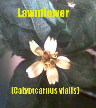
In my yard I usually have a lot of lawnflowers (Calyptocarpus vialis), but now I have to look in shady places to find any. Many times I have found the flowers inhabited by very small insects (see previous article), but they are not to be found now.
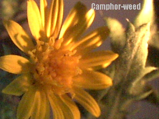
On my local walk I found another plant in flower on the slope of a canal bank, the camphor weed (Machaeranthera phyllocephala). I took home a specimen of this flower and those from three very typical trees for this region to examine them with the QX3 microscope.
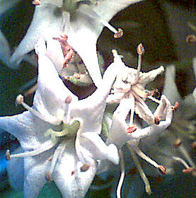
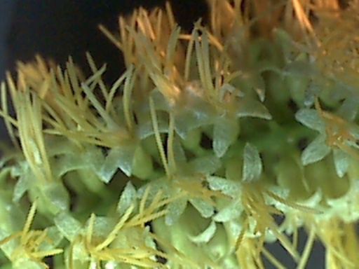
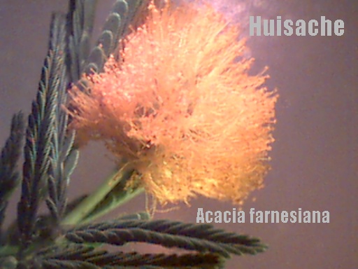
The famous mesquite tree (Prosopis juliflora glandulosa) produces flower spikes about 2 to 3 inches long with many flowers on it, whereas the huisache (Arcacia farnesiana) has flowers in the form of a ball and is a tree with very many thorns. Another thorny tree is the horse bean (Parkinsonia aculeata).
Flowers of the mesquite tree
Flowers of the horse bean tree
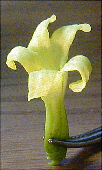
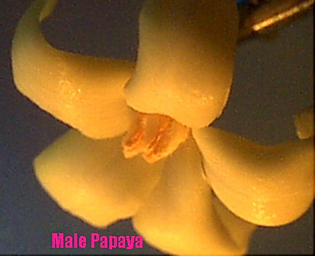
Back in my yard I picked one flower from a male papaya tree, and one from a female. The male flower is smaller and I could just take a picture with the QX3 using two fiber-optics lights with yellow filters, to correct the color. The QX3 built-in light makes it too pink. The female flower is too big for the QX3 so I had to use my Olympus digital camera.
Image left. Female flower of the papaya tree taken with the Olympus D450 zoom digital camera.
Image below male flower taken with the QX3.
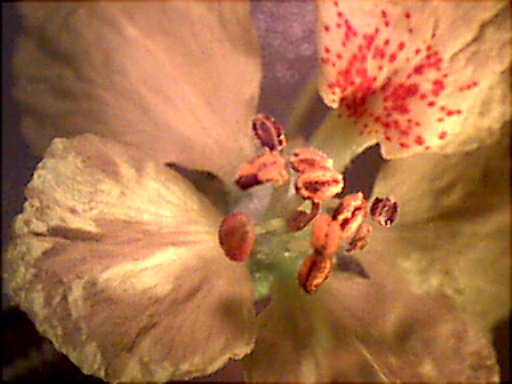
The flowers of Ehretica anacua, commonly called the anacua or sandpaper tree are shown below. It grows to about 30 feet tall and the leaves are rough and feel just like sandpaper. The flowers are in bunches and I have several trees in my yard.
After taking the above pictures, it rained for two days, relieving the dry conditions. All nature is rejoicing and we are commenting on the nice weather we are having now.
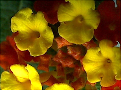
A very pretty weed is the lantana (below) which has small yellow and red flowers. This grows wild but sometimes it is sold at the nurseries.
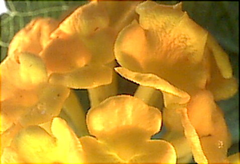
The all-yellow variety of lantana (below) is very similar, even the leaves, but it is a denser bush.
The Mexican heather is a low growing bush, and used for ground cover in yards. The little flowers shown below are about 10 mm across.
All the pictures above were taken with the QX3 at 10X magnification, except the female papaya flower, which was too big for the microscope. Most pictures I imported into another graphics program to crop and enhance them by applying filters. Care has to be exercised when taking a picture with the QX3 in not using too much light, because that increases glare. Also not to enhance the contrast and sharpness too much, or else the texture will look rough. The top light of the QX3 turns any yellow color into pink, and so I just turn it off and use a fluorescent desk lamp, helped sometimes by one or even two flashlights. The flashlights help light the background, where the desk lamp leaves shadows. The desk lamp often gives the effect as if a ray of sunshine lights up that area.
Comments to the author Rudolf Baumueller welcomed.
(Note the identification of the flowers to species are provisional).
An article by the author describing a project for a home-made fiber-optic light source is here.
Microscopy UK Front Page
Micscape Magazine
Article Library
© Microscopy UK or their contributors.
Published in the April 2000 edition of Micscape Magazine.
Please report any Web problems or offer general comments to the Micscape Editor,
via the contact on current Micscape Index.Micscape is the on-line monthly magazine of the Microscopy UK web
site at Microscopy-UK