An Overview of Human Cells for Light
Microscopists
A 3D modelling article by Mol Smith 2010 Please Donate to our Appeal
to Fund the Creation of 3D Models for Microscopic Entities! Please give the pages
in this article time to load: medium size video files involved.
The Human Cell: the miracle of life Since there are quite a few 3D models
in existence to help illustrate the internal structure of individual human cells, I thought an article about cells
would be of interest to light microscopists - especially as we only get to see them as stained, flat, one-dimensional
objects. I think that an in-depth discussion of each cell, is far too specialised an area for me to cover here, but hopefully I can point out a few things on interest.
Possibly, seeing the cells as 3D objects close-up, will provide greater insight into what can only be described
as the miracle of life.
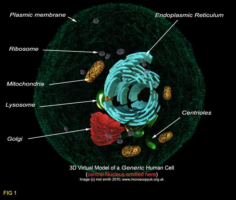
A Generic Human Cell Each cell in the human
body (and animals) is specialised but most will contain structures and processes common to all. Fig. 1 below, which
has been derived from an inexpensive 3D model, clearly shows some of the more important processes. A brief explanation
of the function of each of these follow Fig 1.
Plasmic Membrane This is the cell surface
membrane, completely encasing all internal processes. Made of two layers of lipid (bilayer) sandwiched between
two protein layers, it forms a partially permeable barrier - controlling exchanges (gas) between the cell and the
exterior environment. Numerous proteins are also present in the membrane acting as sensors for taste and hormones,
as are tiny pores to control the essential entry and exit of irons, e.g. chloride, sodium, potassium, and calcium.
Endoplasmic Reticulum There are two types of
Endoplasmic Reticulum: rough and smooth - the former, so called because of the presence of ribosomes on its surface,
displaying a 'pebbled' surface in a scanning electron microscope image. Both types are flattened membrane-bounded
sacs called cisternae. The 'smooth' type is the site of lipid and steroid synthesis. The 'rough' ER (ribosomes
on surface) transports ribosomes through the cisternae.
Ribosomes These small organelles,
consisting of a large and a small submit, are made of RNA and proteins in approximate equal parts. They are the
site of protein synthesis, holding in place various interacting molecules.Long
protein chains are formed at the the intersection of the large and small submit.
Mitochondria The fuel-cell of living
cells, mitochondria combine sugar and Oxygen to provide ATP (Adenosine triphosphate*wiki) - the power source for living entities.They
are about the size of an average-sized bacterium.
Lysosomes These are simple spherical
sacs containing digestive enzymes, and they are concerned with breaking down structures of molecules.
Golgi Commonly called the Golgi
Apparatus, this stack of flattened membrane sacs (cisternae), continuously forms at one end of the stack and buds
off as vesicles at the other. Their function is the processing and transport of many cell materials including enzymes.
Centrioles These are small hollow
cylinders (0.3µm - 0.5µm long) which occur in pairs within the cell cytoplasm (originating in an area
called the centrosome). Each tube is constructed of 9 triplets of microtubules, and is thought that adjacent triplets
may be attached to each other by fibrils. The centrioles replicate themselves at the beginning of nuclear division,
and the two new pairs migrate to opposite ends of the spindle: the structure on which the chromosomes become aligned.
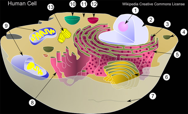
The diagram below shows
a generic human cell cut-away to reveal the internal processes described above. this diagram should help you relate
the cell components to my 3d model image above.
Please note: the diagram
was authored by MesserWoland and Szczepan1990 15 October 2006(2006-10-15), created with Inkscape, based on the
graphics from en wiki. http://en.wikipedia.org/wiki/File:Biological_cell.svg
Permission: (Reusing this file) copyright Multi-license with GFDL and Creative Commons CC-BY-SA-2.5 and older versions
(2.0 and 1.0)
Diagram of a typical animal cell. Organelles are labelled as follows:
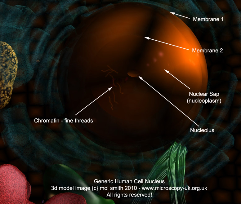
The Cell Nucleus I have omitted the cell
nucleus from my model image above for reasons of clarity. The image below shows a cut-away model representation
of the Cell Nucleus. Note:
I have made the nucleus membranes semi-transparrent so you can peer inside the nucleus itself!
The Nucleus is the largest organelle
within the human cell. it is enclosed by an envelope of two membranes which are perforated by nuclear pores (not
visible in the model above). It contains chromatin - an extended form of chromosomes during interphase, and a nucleolus,
which manufactures RNA.
Life Span of Human Cells It is interesting to
note that with the exception of most (not all) cells of the brain, your entire body, and what constitutes it, is
completely replaced a number of times throughout your life-time. Cells throughout the body continue to renew themselves
by a process of replication. This is done at a different rate depending on the life-span of the cell type. The
brain is the only organ where this process of replication does not occur (except for the cells - neurons - in the
hippocampus). Fortunately, although the number of neurons within the brain begin to decline due to cell death from
around the age of 28 years, it is hardly noticed until our very late years due to the presence of around 100 billion
Neurons at birth.
Here is a list of some of the cell life-spans of the human body
Sperm cells 2-3 days
Here is a tiny animated movie I made showing the generic
human cell spinning.
We have taken a fair look at a generic huuman cell hand can gain a good appreciation of it through the diagrams
and 3D models used. Now we can take a similar look at a few of the specialised cells in the human body using similar
techniques. Let's take
a closer look here at a Neuron!
Published in April 2010 Micscape Magazine.
Please report any Web problems or offer general comments to the Micscape Editor.
Micscape is the on-line monthly magazine of the Microscopy UK web site at Microscopy-UK