An Overview of Human Cells for Light
Microscopists
A 3D modelling article by Mol Smith 2010 Please Donate to our Appeal
to Fund the Creation of 3D Models for Microscopic Entities! Please give the pages
in this article time to load: medium size video files involved.
The Human Neuron Cell Without doubt, the human
brain is considered the most complex structure in the explored Universe. Over 100 billion cells compacted into
a space no larger than a small melon, this organ is the seat of sophisticated awareness and a reality-modelling
system second to none. It cannot be compared to a computer, even though many analogies are incorrectly made, because
almost every single cell component is a unique processor plus memory device. Unlike computers, the brain continually
readjusts its hardware: new physical connections are constantly being made as redundant ones are broken. The magic
component in this truly outstanding achievement of evolution and nature is a cell called a Neuron.
Unique Cell Neurons in the brain
(with one exception - in the hippocampus) cannot replicate themselves, and are therefore incapable of renewal,
which makes the neuron unique when compared to all other cells in the human body. Neurons form a complex web (a
network) with multi-flexible-connections achieved via tiny tentacle-like structures (dendrites), almost touching
at their tips. The microscopic gap between any two connections is called a synaptic gap, and it is across this
space, that tiny messenger chemicals travel to appropriate chemical receivers on dendrites at the far side.Processes which conduct impulses towards the main cell body are called Dendrons,
and those which conduct impulses away from the cell body are called Axons. Different types of neurons (nerve cells)
exist in the human body: uniploar, bipolar, psuedounipolar, and multipolar. These cells are future supported in
the brain by Neuroglia,
cells which are ten times more numerously packed around the neurons throughout the central nervous system. These
are thought to be involved in memory processes and their function is to encode information in the form of RNA.
I have a used a single 3D model of a Neuron to create an artist's view of a section of a Neural Network composed
of brain cells. This movie below represents a static section of the human brain, minus supporting tissues, chemicals,
and blood vessels.
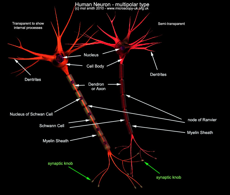
Now, let us take a closer look at a single Neuron.
I have used a virtual 3D model to produce two cells below. They are the same cell but I have made one of them more
transparent so you can look at the hidden processes inside.
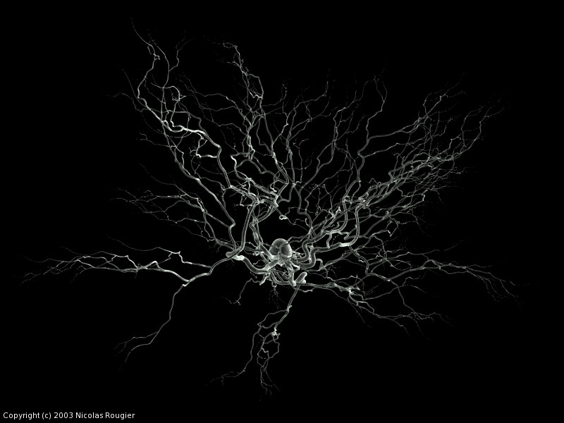
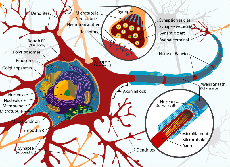
You might wish to compare my model with the SEM
image of a similar Neuron below, and then see the detailed diagram beneath it. Both these images are from Wiki
and are used here under the collective commons licence. Please refer to wiki for reuse permissions.
I think one of the true miracles
of the human brain is the fact that the Neuron uses both electrical and chemical processes to facilitate communication
between cells. This ultimately produces human thought. Let's take a look at this process...
Published in April 2010 Micscape Magazine.
Please report any Web problems or offer general comments to the Micscape Editor.
Micscape is the on-line monthly magazine of the Microscopy UK web site at Microscopy-UK