|
or thereabouts. by Roland
Mortimer,
|
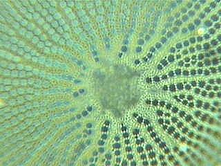
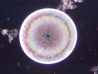
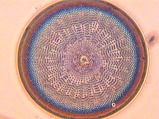
Surface of Coscinodiscus showing the fine detail. Phase illumination. |
Part I, Part II, Part III, Part IV and Part V.
As I mentioned in previous articles, there is not one standard type of illumination which best suits the study of diatom frustules. There are many parameters to be considered, e.g. thickness of the slide and coverslip, thickness/refractive index of resin in the preparation, thickness of specimen, even the optical properties of the slide and cover material etc.
Bright-field probably being the most used is not really the best system, on the other hand, it was used for more than a century prior to dark-field, phase etc. with very good results. Dark-field can show detail which most other illumination systems do not, phase can show detail which dark-field obliterates, but what about the unknown and untried? Most of the images I've included in this article were taken with a CCD colour digital camera and used in combination with a Heine condenser and phase objectives, but in reality, some are not exactly in phase mode but somewhere between phase and dark-field. Others were taken out of phase in direction of bright-field to show the variations which can be obtained with a little bit of 'messing around'. I also used green, yellow and blue filters to alter the contrast a little.
The images which are in fact in phase contrast were taken using an apochromat x90 pV oil immersion objective n.a. 1.15, these objectives denoted pV were made by Leitz for use specifically with the Heine system although standard phaco (phase contrast) objectives can be used if centred correctly. If you have even one phase objective but no condenser for it, try using it in conjunction with a dark-field condenser raising and lowering the condenser slowly and observing the effects, try different coloured filters too, of course a fairly bright light is required.
I use a 12v/50w halogen lamp in my system which fairly heats up things but gives the required illumination for dark-field and phase. This system is not limited to diatoms of course, I've video taped blood in movement in dark-field, phase and intermediate levels of each; watching leucocytes move around like amoebae under this form of illumination is really beautiful as the light used seems to 'shadow' the cells and give them solidity. Many bacteria too can be increased to a very reasonable size and observed under this shadow-giving technique on the computer monitor or on T.V. using the CCD camera. The finely detailed structure on the surface of Cosconodiscus is particularly beautiful under these conditions (shown in title).
The importance of observation under various forms of illumination cannot be overstressed, as even the most famous microscopists in the past have reported and published details of the diatom frustule surface which the electron microscope has now proven incorrect. This can be caused by aberrations introduced by dirty lenses, incorrect adjustment of iris etc. One must observe the diatom frustule in bright-field making sure all the correct adjustments have been made, then re-check in dark-field or phase, only adjusting the fine focus. Reference to good drawings to confirm what is seen is in fact 'real' is a very good practice. Most publications will give sizes in microns but is not a very good guide as all diatoms diminish in size as a natural result of their style of reproduction, therefore many sizes may be found within the same species. This is sometimes misleading in identification as one may think it is an entirely different species or even genus.
Comments to the author Roland
Mortimer welcomed.
Image gallery by the author
(Diatoms shown are to illustrate
effect of lighting variants; identification to genus/species
in progress. Objective
used and some typical sizes in microns given.)
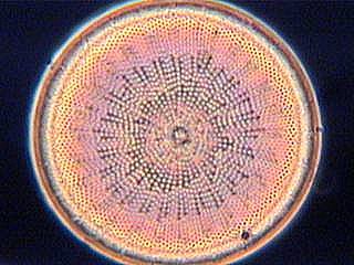
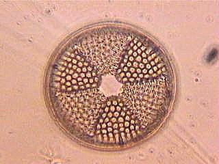
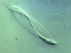
Phase illumination
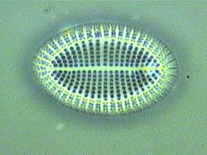
60µm x90 |
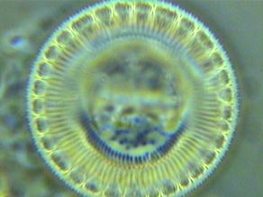
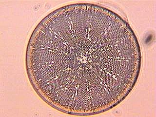
40µm x90 |
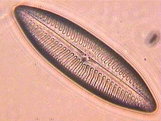
Dark-field illumination
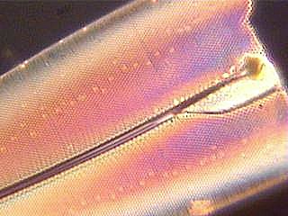
Fragment of Pleurosigma |
80 µm x20 |
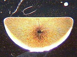
750 µm x10 |
80 µm x45 |
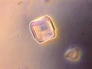
Variations on phase illumination
x45 |
x45 |
80µm x45 |
80µm x45 |
x45 |
x45 |