| Circular
Oblique Lighting (Part 3)
Some final odds and ends By Paul James (uk)
|
| Circular
Oblique Lighting (Part 3)
Some final odds and ends By Paul James (uk)
|
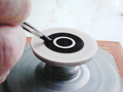
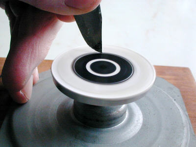
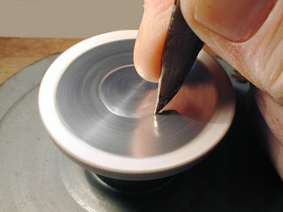
A useful tiny lathe head/potter's wheelWhilst it is quite possible to print an annulus using a printer and appropriate software, or use a mounter's turntable, I chose a method which also has other uses, like this mini 'potter's wheel' which I made from an electric motor rescued from a defunct photo-copier. It's a DC motor unit, and when powered from my variable 0-6 volt AC microscope illumination transformer, through a series rectifier diode, will make this revolve at a useful 60 rpm upwards. A small platform/faceplate mounted on the spindle acts as a support for whatever needs to be rotated for painting or cutting etc..
In these images can be seen the process of painting the annulus, and also 'cutting' it out with a sharp knife. The clear sheet material was temporarily attached with 'Copydex' then revolved at about 750 rpm, which makes for sharper paint edges. The diameter of the ring and its thickness was lightly marked before black acrylic paint was applied whilst spinning, which results in accurate concentricity :-
 |
 |
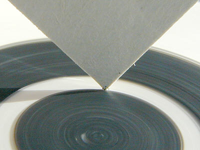
This annulus was painted and cut in about 2 minutes. The paint boundaries can be precisely 'sized' and made perfectly circular using the corner of a piece of thin cardboard which is allowed to rub gently on the rotating sheet against the unhardened paint edge to be trued up or widened. The cutting left slightly rough edges, but was perfectly circular and is therefore of no importance. The 'Copydex' rubs/peels off the sheet with ease.
 |
Pie casing aluminium foil can be temporarily stuck down with 'Copydex' before cutting in a similar manner, though some might prefer to just score the circle then cut with scissors after :-
 |
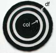
Below we have an example of a painted annulus which separates the DF/COL zones so keeping any internal reflections from lens edges to a minimum. The DF component can, if required, be cut off cleanly by the field stop iris, or substage iris depending upon the annulus's position.
 |
Final notes on position of stops/annuli and condensersI have found that the improvements of detail contrast etc. using COL can be brought into effect with a stop/annulus anywhere between the field lens and the condenser top lens! The variation of effects however is almost infinite, being compounded by the condenser type and its focal length and NA. Yet very colourful and striking effects can be created by just the field lens stop, and subtle condenser focussing :-
 |
My favourite setupAfter much experimentation and deliberation, I find the most interesting and versatile configurations for the Wild M20 are stops in the filter tray......ie 20 mm for the x 40 Fluotar (NA.0.75) and wider for the apochromat (NA.0.95). The variations of image lighting are impressive, since both substage iris and field iris can be altered to suit the subject observed, from pure COL to COL/DF and finally very delicate DF.
In one simple setup therefore, exists an almost endless series of lighting permutations subject to the settings of the substage iris, field iris, and condenser focus.
Memorable observations
In concluding this final article on COL I'd like to describe a few moments during four observing periods which were memorable to say the least. I include for each the relevant technical details.
1) Rotifer ( Red Dot ) Zeiss photomic 1.....using x 10 achromat objective with the internal x 100 phase annulus, which provides a unique balance of COL and DF..............."The rotifer stretched out from the mucilage over a clear caramel background and was highlighted by the DF component, and of course the COL contrast improving effects. Its main body positively stood out from the background with immaculate detail, whilst the fanned myriads of highlighted particles issuing beside the 'wheels' sparkled in the DF component. The workings of its interior were abundantly clear and were displayed in crisp detail".
2) Ciliate....... Spec. as above......."A small unidentified solitary ciliate came across the field and tumbled very slowly through the COL/DF plane like a space satellite. Its cilia sparkled like the rotifer, but its features were transparently crystal clear......all the vacuoles and nucleus clearly mapped out in this magic image, as I slowly followed its descent with the fine focus and stage controls. Using the x2 Optovar position enabled me to see more detail even with this x 10 optic. I think this was certainly the most spectacular rendering of such a protozoan I have ever seen, and one of course I shall always remember".
3) Resting soil Amoeba + bacteria....Wild M20 with Zeiss x 40 apochromat and x 16 Zeiss compensating eyepieces. 22 mm stop in filter tray........ "The bed of the slide was covered with an even layer of bacilli each clearly showing its nucleus and body end tapering. This clarity was not apparent in phase contrast, nor brightfield, or standard oblique. The detail of fine debris between the cells was amazing. Scanning further I found a resting amoeba of about 70-80 microns across. It reminded me of a textbook diagram, so clear were its internal features".
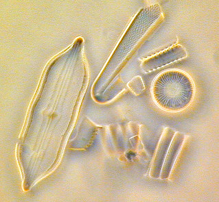
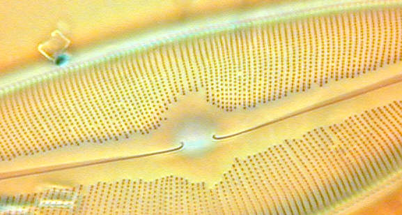
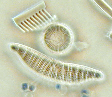
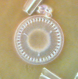
4) Strewn Diatoms on Moller slide................Specs as above ........"This slide of diatoms, which I have seen through various microscopes over the last 42 years, appeared as never before. All the diatoms seemed to be made from porcelain, with the highest contrast dotting and other minute detail I'd ever seen. I noticed too that some improvements were brought about by off setting the stop very slightly, which gave more solidarity and conviction to these beautiful images. So captivated was I that half an hour passed quickly, and it felt that at last I was seeing these beautiful skeletons in their full glory".
Final miscellaneous thoughts
COL does not like thick mountant or water gaps between coverslip and slide in my experience so far with the higher power objectives, but seems to be tolerated with the lower NA optics. The contrast inducing effects with these low power lenses are also less intense, but they still elicit fine imagery of appropriate subject matter when mixed with some DF.
Collimation of all optical components is vitally important in the first instant , though offset stops can bring some benefits in certain situations.
Condenser focus can be extremely critical in some setups, and a subtle change of position can not only alter colour tinges but more importantly reduce resolution quite dramatically.
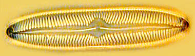
Here are cropped examples all using x 40 objectives, and employing various settings of condenser focus, both iris controls, and stop positions.
 |
 |
 |
ConclusionIf you are a keen 'pond water' observer, and or enjoy the beautiful structures of diatoms, then COL must be taken seriously as a definite leap forward in image enhancement and subsequent observing pleasure. It can disappoint with unsuitable mountant or excessive water gap thickness in the higher powers, and poor alignment of optics, and objects which do not suit it.
However, it is the cheapest and simplest method of improving contrast of the finest detail that your objective can muster, and does this at maximum aperture every time. Such is the improvement of perceived detail with the best objectives, that an eyepiece of at least x15 is required to see this detail comfortably.
The original concept of COL, i.e.. a ring of oblique light flooding the specimen, has been enhanced with the option of DF mixing, and remains for me a powerful and versatile illumination tool for a limited number of subjects. Within this observing range I feel that there is nothing to compare with COL/DF. For general routine observations brightfield and phase contrast are the general workhorses, but for the very best imagery, with moderate eye friendly brightness, and with speedily induced variations too......... COL/DF reigns supreme.
| All comments welcomed to Paul James. |
Please report any Web problems or offer general comments to the Micscape Editor.
Micscape is the on-line monthly magazine
of the Microscopy UK web site at Microscopy-UK
WIDTH=1