 |
| Principal of
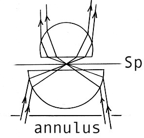
COL illumination.
The cone of light converges on the specimen at Sp. |
| Circular
Oblique Lighting (Part 1)
An introduction to what may be considered to be a superior form of illumination to brightfield By Paul James ( uk ) |
A while back I wrote briefly about an interesting illumination setup involving the use of normal, non phase objectives illuminated with the cones of light from annuli in phase condensers etc. Having experimented further, I have come to the inevitable conclusion that for many subjects, and in particular protoplasmic cellular specimens it is superior in many ways to brightfield, and even standard phase contrast in some circumstances.
Technically speaking it is 360 degree Oblique Illumination OR Circular Oblique Lighting For brevity's sake I shall refer to it as COL illumination.
|
| Principal of
COL illumination.
The cone of light converges on the specimen at Sp. |
COL illumination.The essential ingredients are similar to standard phase contrast, in that the phase ring of the objective, and the na. of the ascending light cone from the annulus coincide precisely to elicit the phase contrast effects. The essential difference with COL illumination is that the objective is not a phase optic, although phase objectives can be used, providing its internal annulus is smaller than that issued from the condenser. Under these conditions a rather unusual effect is noticed, where the field's illumination intensity can be somewhat diminished, and increased contrast is noticed with many specimens. It particularly emphasises contrast in fine detail, which is caused by partial phase shifts of light. The success of this phenomenon seems to depend on the subtle relationship between the na of the objective and ascending cone. It appears to work well with the higher na objectives, though contrast improvements still occur with the low powers.
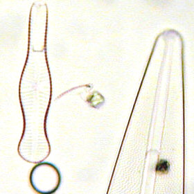
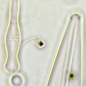
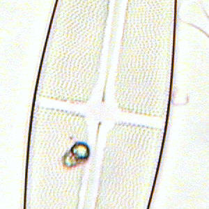
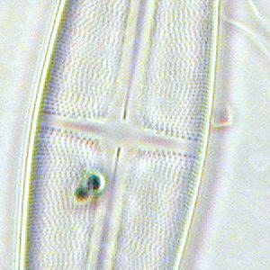
The four images below give some idea of the differences between BF and COL respectively. Note that the images are from the small central zone of the ccd, and are unsharpened. Noise spoils the otherwise smooth COL background.
Brightfield
.....................................................COL
Diatoms (mounted) x40 Fluorite with x100 phase annulus
 |
 |
 |
 |
COL illumination's Salient FeaturesIt seems to have all the advantages of phase and brightfield in one fell swoop, and conversely, seems to dispose of all the weaknesses of both as well. If that seems to augur well, then add to that the increase in perceived resolution, and the portrayal of special coloured effects, which might not in themselves be of true scientific value, but nevertheless help with recognition of the various species that are innately coloured.
Be aware however, that the phenomenon yields subtle variations depending directly on the diameter of the annulus, and the ideal setup appears to be when the na of the annulus is about 60%-90% of the objective's. I suspect no two observers with different optics will see identical fields and contrast etc.
Since the finer subtleties of this illumination are entirely lost in digicam photography, I have expressed through words its appealing properties when observing very small soil amoebae using a Wild x40 fluotar (operating at around na 0.70-.75 ), and x100 substage annulus on the Wild M20 stand :-
"....... the views of small soil amoeba were delightful. The overall illumination intensity was just right for my eyes, and the entire field showed a faint straw yellow hue. The advantages of contrast in cellular detail were at once obvious, and then the resolution of their protoplasmic interior was impressive. The sub-particulate frenzy of protoplasmic material was clearly visible within the boundary of the ectoplasm as it flowed forth. Just as pleasing were the delicate refractile boundaries around the amoeba, which did not hinder in any way the view of this tiny animal, as standard phase can often do, but greatly assisted the recognition of its ever changing forms. Outside, the bacteria, and miniscule protoplasmic forms were rendered very sharply and with much contrast. Colours were noted, especially within the Amoeba where the nucleus seemed to be tinged with a definite green, and other forms of life ,especially the single celled algae were brilliantly tinted with greens and blues above and beyond their naturally colourful selves compared to brightfield.
Not all subject matter responds so well with COL lighting, though it could be said that the improvement is just a little more subtle. In short, if the union of comparative na's of both objective and annulus is ideal, the results are so appealing, both in technical and subjective ways, that it could easily become the illumination of choice. Put in another way, if COL lighting were the norm, and someone discovered brightfield, the latter would probably remain as an adjunct only.
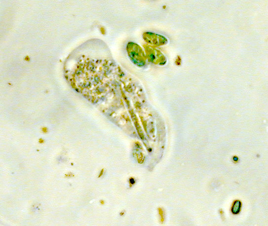 |
| Unsharpened
ccd image of Small Soil Amoeba x800.
(Wild x40 0.75 na fluorite plus x100 phase annulus) Note high contrast and colour saturation in particulates of protoplasm and inside algal cells , and also modest phase refractile boundaries around periphery of the cell walls. Visual appearance was superior to this image. |
NotesI must emphasise the fact that initial trials may prove disappointing because this illumination works best on certain subjects far better than others. I find it ideal for pond life and related subjects, were brightfield tends to flood delicate structures etc. It is undoubtedly however, a useful illumination alternative to brightfield, and deserves serious consideration, especially experimentation regarding working conditions.
In the next article ( Part 2 ) I shall cover annuli, condensers and also the very interesting variation of this COL lighting which uses a mixture of COL and DF.
| All comments welcomed to Paul James. |
Please report any Web problems or offer general comments to the Micscape Editor.
Micscape is the on-line monthly magazine of the Microscopy UK web site at Microscopy-UK