An ANNULUS or STOP ?
Strictly speaking an annulus
allows a coherent ring of light to pass through the condenser to produce
phase contrast or COL effects. My experiments
have led me to circumvent the making of a traditional annulus, because
it is highly desirable that the outer regions of the light cone should
be variable. Therefore an annulus not having variable outer
proportions is less versatile. An ordinary circular stop as used for darkfield,
though smaller, will yield the COL phase reversal effects, and if placed
near the iris diaphragm, the latter can be closed down to control the outer
proportion of light input to the specimen. Thus the overall effects can
be radically changed by simply altering the size of the iris diaphragm.
In practice this works extremely well, providing that the iris is concentric
with the stop. Complete concentricity in the microscope's optics makes
for better COL effects.
These initial trials are set out to introduce those who have no phase equipment, and have not experienced COL before, in a quick and uncomplicated manner.
Initial trials
Set up
your 'scope for brightfield with the x40 objective using a slide of diatoms
containing fine detail such as Actinoptychus heliopelta etc.. Optimise
the condenser's focus position and then open up the iris to maximum aperture.
Now place a disc of opaque material such as a 5p coin, or one cut from
silver foil of about 10-15 mm on the field lens of your microscope. Centre
it as carefully as you can by making sure that the back lens of the objective
shows a ring of peripheral light when viewed without the eyepiece in place
( a Phase telescope makes this much easier to accomplish ). Alter the size
of the stop if necessary until you see this down the draw tube :-
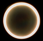 |
Then look at the diatom whilst altering the position of the condenser until you see a dark central zone, which will confirm the field stop's centrality, if you are in doubt about that. Now rack up the condenser until the dark zone just disappears. You will, if the condenser is of the simple Abbe form, see some splendid colour backgrounds as you move the condenser about its usual position. This is perfectly normal in these circumstances, and the final position can be determined which provides the most pleasing effects regarding background and contrast etc..
This
initial view contains a proportion of COL and darkfield components, but
the phase reversal contrast enhancing effects will still be seen.
 |
|
|
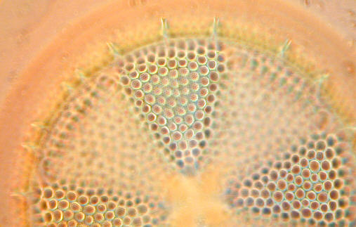
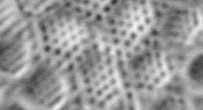
Wild x40 Achromat using simple field lens stop. Colour profusion from Abbe condenser's peripheral aberrations helps differentiate detail of phase reversals. Adjusting of condenser dictates colour backcloth. DETAIL SHOWN BELOW Unsharpened, unsized, grayscaled detail of the central ccd image of the same above but using the Zeiss Optovar with total amplification of around x2000 illustrates quite clearly the potential image enhancing properties of COL using the humble x40 achromat !
|
Optimising this simple set-up can be accomplished by adjusting the position of the condenser, and/or trying smaller or slightly large stops on the field lens.
If you cannot gain success so far, try another diatom slide, as the COL phenomenon is quite sensitive to the optical properties of the diatom and its mountant medium. If still no change, then it could well be that your objective has flaws of one kind or another around the edges of the elements such as delamination, or fungal growth.
NB.......The images you will have seen like those above are not purely COL, because there is the darkfield component overlaying the COL. However I have mentioned this because there can be a distinct advantage in the perceived resolution and general presentation of the diatom etc.. By using an annulus which blocks the DF component, pure COL will result, which might suit certain subjects better, and may appeal to some observers.
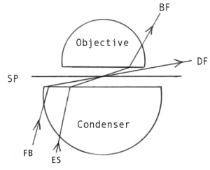
The diagram
below shows how this stop on the field lens works. Only one one side of
the condenser's output is shown of course. ES represents the light from
the edge of the stop, and FB represents the light at the maximum field
boundary, which in most microscopes with condensers of at least 0.9 NA
will project light beyond the x 40 objective's front element....This is
the darkfield component mentioned above.
 |
Optimizing COL for a favourite objective/specimen
Having experienced some success, and observing the benefits of a simple stop, it is likely that those who are impressed will want to 'tweak' the COL to maximum advantage. We will consider these in turn :-
1) The stop.
2) The position of the stop.
3) The suitability of the various forms of condenser.
The Stop
Thickish aluminium foil is the easiest material to cut into a disc for the stop, since its outer edge should be sharp and thin like the iris. For most observations the disc does not have to be perfectly circular, but this should be the case if possible because the iris can be used to greater effect when closed down to reduce the DF component. Blackening it is not essential but does reduce stray light from reflections.
Its diameter should be such that your objective's back element displays a circle of peripheral light of thinnish proportions as shown above. It will probably, in most microscope's, need to be a little narrower than you have used on the field lens, because it should ideally take its position near the iris diaphragm.
Siting the Stop
The ideal site for the stop
is near the plane of the condenser iris, i.e.. near the anterior focal
plane of the condenser. The filter tray is good enough to start with, but
suffers from the disadvantage that it should really be able to be centred
at times. Stops placed in the spare apertures of phase condenser Zernicke
discs work very well, provided the iris diaphragm is independent of this,
which is not the case with some Zeiss phase condensers.
 |
Above is shown the principal of a dedicated condenser for COL using a preferred objective, which has a stop attached to the underside of the condenser's bottom element, with the iris controlling the outer DF portion of light. What really matters is that the stop is concentric with the condenser's axis and iris. This is vital and the effort to put this into practice will pay dividends.
There are many ways of mounting stops near the iris in either a temporary or permanent way. This will ultimately depend on the observer's needs regarding COL/DF and their imaginative DIY skills. Adapting an old iris/lens mounting from an obsolete camera etc. incorporating a stop, or series of removable stops to suit different objectives mounted just beneath the condenser will appeal to those requiring speedier illumination changes. A case could be made for the acquisition of a phase condenser substage unit for your 'scope, if fixed annuli will suit your needs.
Those who have a second microscope may adapt it for COL?
The Condenser
Fortunately, the Abbe or achromatic condenser are both well suited to COL up to and including x40 objectives. The achromat has the edge regarding the use of oil immersion objectives for it provides a more accurately coherent cone, as does the aplanat over the Abbe. However, the intrinsic chromatic aberrations of the Abbe can provide some advantage, in that by its careful focussing we can bathe the specimen in apple green light, the preferred colour for achromatic corrections, which I find elicits the best imagery regarding contrast enhancement and general viewing comfort.
Tail Ends
One very important issue regarding the comparison of brightfield and COL, is that the former is rarely used at full aperture, prejudicing maximum resolution from the objective because of iris closure requirements. COL can utilise the max NA of the objective every time.
COL works
best with the higher NA objectives, but will work also with x10 and x20
too. Each objective will require a different diameter stop. If the iris
is opened up to allow some DF component to reach the specimen, it will
add a degree of highlighting too. Decentering the condenser can also add
depth and make for a very interesting image :-
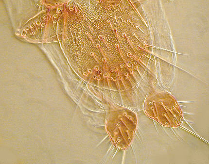 |
| Note almost 3D effect of COL/DF on this insect part |
The variations of annulus/stops are endless, and the experimenter might consider the effects of 2 concentric annuli, one just inside the NA of the objective (COL) and one just outside (DF), thereby bypassing the peripheral edge of the objective's elements where presumably some light might go astray etc............It's a thought.
Some Personal thoughts
For me COL, and especially when in use with the iris controlling the DF component, is the preferred illumination set-up to brightfield for a number of specimen types. More experimentation is on the way for me, and I hope some readers will have had their appetites whetted accordingly.
What truly amazes me is that COL has been known in principal and practised by a few workers for over 50 years, yet the microscope manufacturers, as far as I know, have never incorporated its great advantages into the standard microscope? The cost would be trivial compared to phase contrast, but the benefits would be so appreciated by many observers. ...............Pity.
| All comments welcomed to Paul James. |
Read Part I and Part III of the series.
Microscopy
UK Front Page
Micscape
Magazine
Article
Library
© Microscopy UK or their contributors.
Published in January 2003 Micscape Magazine.Please report any Web problems or offer general comments to the Micscape Editor.
Micscape is the on-line monthly magazine of the Microscopy UK web site at Microscopy-UK