Structures from Nature
Image Gallery by David Walker, UK
Nature's ability to make intricate microscopical structures out of inorganic materials such as silica and calcium carbonate is perhaps one of the most wonderful feats of nature that the microscopy enthusiast can appreciate. Diatom frustules, of course need no introduction, but sponge spicules, radiolaria 'shells' and echinoderm spines are other examples. This is a small selection of 'Nature's structures' under the microscope other than diatoms from the author's modest slide collection. The images were taken with a Nikon D50 DSLR on a Zeiss microscope using a Zeiss 10x Kpl W eyepiece as projection eyepiece. Helicon Focus image stacking software was used for some images to increase the depth of field and image stitching on others to increase the field of view.
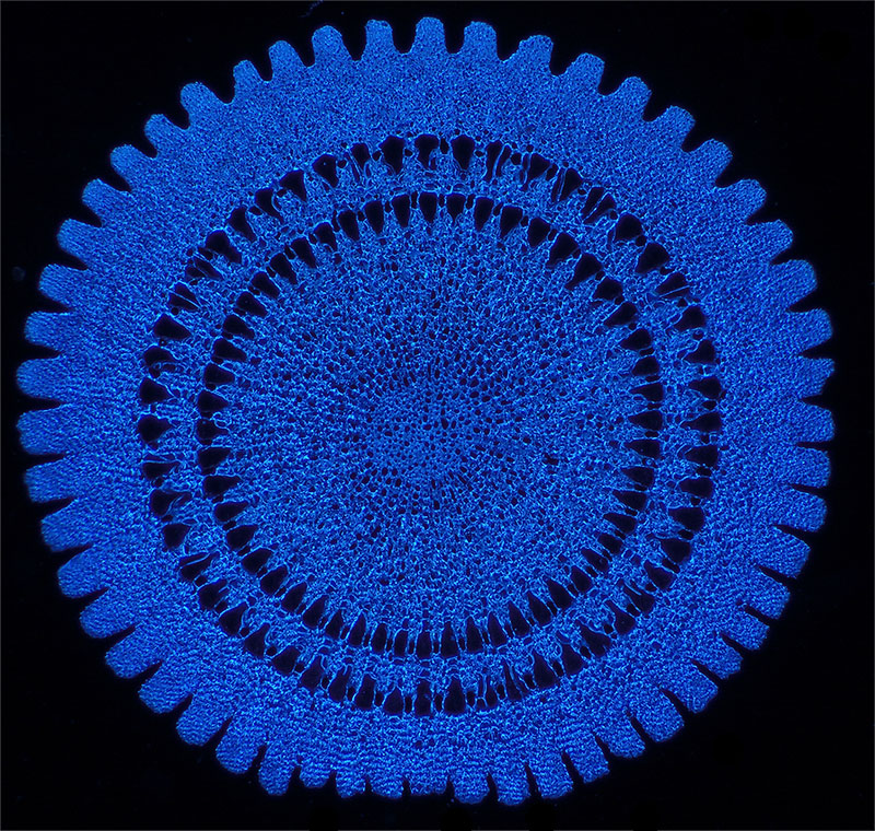
The image below is a cross section through an 'Echinus spine' labeled for 'Spot lens'. Echinus is a genus of sea urchin and is one of my favourite slides. It's a Victorian papered slide by an unnamed mounter. The mount of this specimen is still very clear and the section thickness is barely noticeable in side view; sectioning such a potentially brittle material (calcium carbonate) and keeping it intact must have been quite an accomplishment.
'Echinus spine' cross section 2.8 mm diameter. Zeiss 2.5x planachro, darkfield with blue filter, two images stitched. The darkfield 'D' setting on the Zeiss achromatic-aplanatic phase condenser works well with top lens removed for low power darkfield; not completely structureless darkfield but good enough. The Zeiss manual suggests that the D' setting is primarily intended for immersed condenser darkfield work.
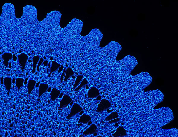
Detail
of above subject, Zeiss 4x planapo.

Further
detail, Zeiss 10x planapo. The apertures have silica in which are not obvious
from the lower mag images.
Modern biology books often seem to present a more matter of fact view of nature, but much prefer reading the accounts of the Victorian naturalists who had an infectious enthusiasm for nature's wonders on the microscopic scale. One of my favourite books is Richard Kerr's 'Hidden beauties of nature' which is particularly good at presenting such structures and has separate chapters on diatoms, radiolaria both fossil and present and sponges. As well as including a selection of plates from the Report of the famous Challenger Expedition it has some beautiful white on black drawings giving a darkfield effect. I possess a copy of Kerr's undated 4th edition but an 'Abe' book search (where copies can often be bought for $10 or so) suggests that the 1st edition was ca. 1895.
Radiolaria have particularly delicate structures, many are made of silica although apparently some have strontium sulphate skeletons (see for example the 'Miracle' website, link below). I've often thought that if the sci-fi film makers run out of ideas for exotic looking spaceships that many of these would look great on the much larger scale zipping through the cosmos! The selection below are all from an arranged slide kindly made by Wim van Egmond. The slide label states that they are from the Barbados tertiary deposit, a famous location for them.
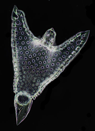
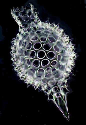
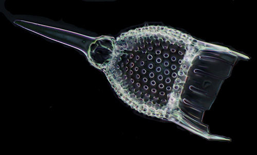
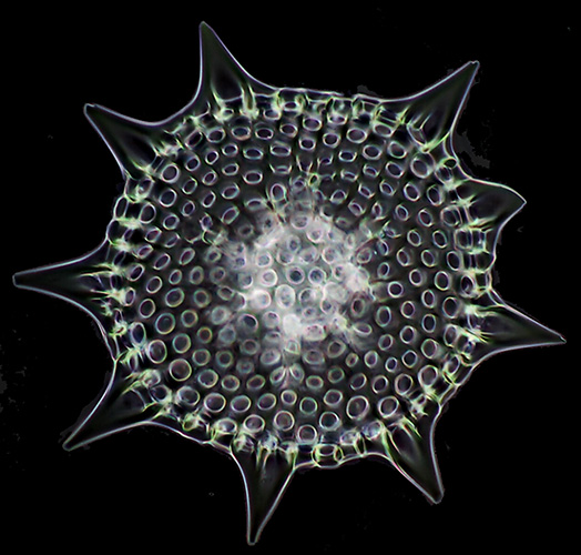
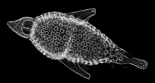
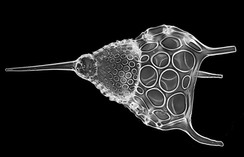
Typically five images were stacked to create each of these using a Zeiss 16x Neofluar objective. I preferred using just a few stacked images to capture the major planes of focus rather than tens of images as found the latter stacks were more prone to artifacts, often misinterpreting the spines. I'm a beginner at image stacking so further image parameter tweaking may remove multi-image stack artifacts. They are brightfield images with 'invert' to create an artificial darkfield effect.
As
well as enjoying selected radiolaria in arranged slides, the strews often found
in old collections are very attractive, especially under darkfield where the
feeling is of navigating through glass jewels on a velvety black background.
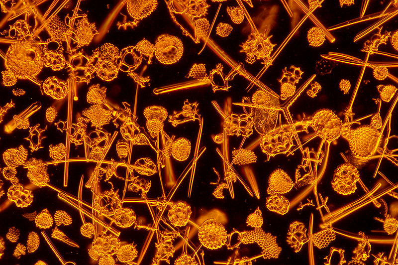
'Radiolarians, Jeremie, Haiti', 'Collection Albert Renaud'. I have three slides labeled 'Albert Renaud'; the mountant is uniformly brown so suspect may be an exotic mountant rather than mountant oxidation. They are very cleanly prepared mounts with high contrast. I believe sponge spicules are also present. A Google search gives a selection of interesting resources on the radiolaria and diatoms collected and studied from this locality. Zeiss 4x planapo, darkfield, orange filter.
I've always been intrigued by what single-celled organisms like diatoms and radiolaria are using as the 'architectural template' (for want of a better phrase) with such efficiency on the microscopic scale. It's this aspect that I really marvel at as a non-biologist and would be interested to hear from readers who can pass on some references to accessible reviews of these processes.
Comments to the author David Walker are welcomed.
Related Micscape links: A number of Micscape contributors have also shared their enthusiasm for nature's inorganic structures.
'Calcareous flowers: tests and spines of sea urchins' by Richard Howey
'Those who live in glass houses' by Bill Amos, illustrated by Wim van Egmond (marine diatoms and silicoflagellates)
'Cultural debris' by Richard Howey where he illustrates the amazing Venus' flower basket sponge, Euplectella.
Micscape Article Library Marine category has a large selection of illustrated articles on diatoms, radiolaria, foraminifera.
Micropolitan Museum has a wide selection of organisms illustrated by Wim van Egmond.
External websites:
www.radiolaria.org is devoted to radiolaria and a mine of info and attractive images.
University College London website 'Miracle' (Microfossil Image Recovery and Circulation for Learning and Education) is very informative with many images and gets my vote for the cleverest title!
Published in the February 2007 edition of Micscape.
Please report any Web problems or offer general comments to the Micscape Editor .
Micscape is the on-line monthly magazine of the Microscopy UK web site at Microscopy-UK
© Onview.net Ltd, Microscopy-UK, and all contributors 1995
onwards. All rights reserved.
Main site is
at www.microscopy-uk.org.uk
with full mirror
at www.microscopy-uk.net
.