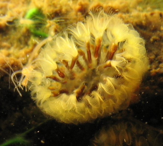
Cristatella mucedo
photographed in situ
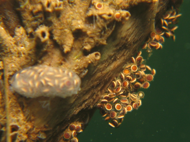
Cristatella mucedo statoblasts
the zooids have died
by Michiel van der Waaij, The Netherlands
There is a group of smallish aquatic invertebrates that live a well hidden life in lakes, ponds and small streams called bryozo (bryozoans or bryozoa). Read more about them in my earlier Micscape article. Freshwater bryozoa produce survival capsules called statoblasts to survive adverse conditions like the cold of winter, even freezing and desiccation. In this article I describe how I used now uncommon lighting methods to see structures I've never seen reported before and possibly discover something new to science.
One of the freshwater bryozoans that is quite common in North-West Europe is called Cristatella mucedo and looks a bit like a caterpillar that lives underwater. Below to the left is a young colony that has not yet developed the typical elongated shape. Byrozoa individuals are called zooids and are quite advanced with a complete mouth-stomach-gut-anus system. Bryozoa can also retract their polyps in case of a threat.
The rightmost picture below shows what happens at the end of the short life of a bryozoa colony. The colony has developed survival capsules called statoblasts and all zooids have died. Soon the remaining colony capsule will rupture (as shown on the right) and the statoblasts will disperse.
The righthand picture is an in-situ (underwater) photo that has captured an unique moment in time. Typically this situation lasts only for hours.
|
|
|
|
Cristatella mucedo |
Cristatella mucedo statoblasts |
 For an idea of the size of a statoblast - one polyp in the picture on the right, including the brownish 'body' is about 6 mm, so a statoblast is in the 1 mm range.
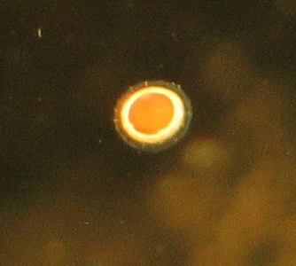
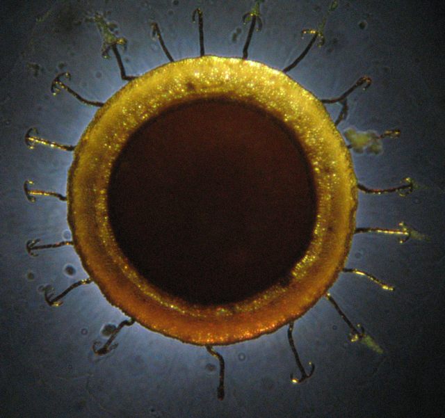
To the right you can see a free floating statoblast I managed to photograph while under water. There are several aspects that should be noted:
The smooth center is the part where a new bryozo lays dormant. I hesitate to call it an embryo as this is asexual reproduction. The bright ring is unique for a few species of fresh-water bryozoa and is filled with a gas that makes the statoblast float to the surface. The side with the ring is called the back or upper side. It is thought that the surface currents will enhance dispersal of the statoblasts enabling the species to colonize new locations more rapidly. The fuzzy outer ring is described in the literature
I know on this subject as 'barbed spines' that are thought to enable the statoblast to aquatic plants and even aquatic bird feathers.
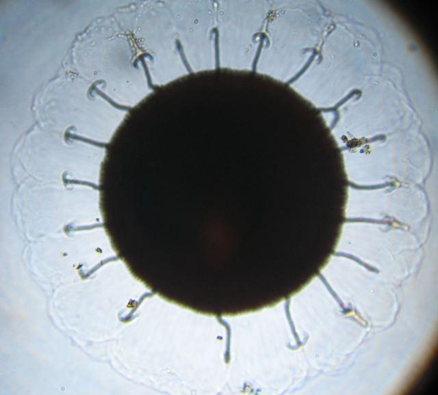
The typical representation in literature is shown below. This photo was made of a fresh statoblast floating just below the water surface in a small container with a normal (Zeiss standard) microscope with a Zeiss 1,6-5 zoom objective (using light from below only). I did not use a slide to carry/fix the statoblast on purpose, for reasons indicated below.
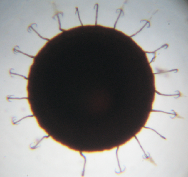 This representation clearly shows the 'barbed spines' but not much more. I was dissatisfied with the result and wanted to enhance the image by showing the floatation ring. I did this by using an old microscope lamp converted with a high-power white LED, which I used to add light from above. This required quite a bit of fiddling with the microscope lamp until the light beam entered sideways and from above between the objective and the object. Also I reduced light intensity
from below. The result was more satisfying.
This representation clearly shows the 'barbed spines' but not much more. I was dissatisfied with the result and wanted to enhance the image by showing the floatation ring. I did this by using an old microscope lamp converted with a high-power white LED, which I used to add light from above. This required quite a bit of fiddling with the microscope lamp until the light beam entered sideways and from above between the objective and the object. Also I reduced light intensity
from below. The result was more satisfying.
 Given the small space between the underside of the objective and the object I did not manage to light the inner part of the statoblast leaving it dark, but the flotation ring can be clearly seen and there is a nice shine on some of the spines, not unlike the shine you get on a beetle shield. This is in line with literature stating that significant parts of the statoblast is made of chitin, as are beetle shields.
However I was still dissatisfied. Close inspection of the in-situ image shown above shows a fuzzy outer ring that could not to my mind be fully explained by the barbed spikes. Something was missing I felt. Then I remembered an illumination technique described in Micscape. A quick search resulted in two articles by David Walker (links below) on (Asymmetric) Oblique Illumination. This is an old technique that uses some form of patch stop that blocks most of the light and lets a one-sided light stream through. This
is a simple and cheap way to bring contrast to very low contrast objects like thin living cells.
A patch stop was easily improvised and the result was beyond my expectation! There is a thin membrane extending sideways from the statoblast. (With apologies for the vignetting in the picture.)
The patch stop I used was a dustcap that was big enough to cover the opening above the field diaphragm in my microscope's base, adjusted in such a way that a small crescent shaped split remained for the light to shine through. It was a bit fiddly but it gave good results.
|
|
|
|
Cristatella mucedo statoblast |
Cristatella mucedo statoblast |
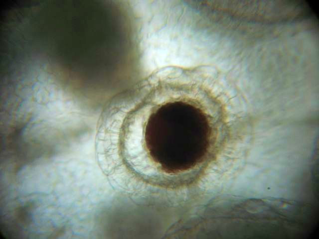
I found an earlier photograph of a developing statoblast inside a colony that also seems to show a membrane (shown above to the right). The dark shapes are the bases of zooids (individual bryozo animals) partly blocking the light.
So the barbed spines have a very different purpose as described in literature. They seem to support the membrane to keep it extended, more than that they act as hooks to attach to aquatic plants or aquatic bird feathers (although they could do that once the membrane has decayed). It seems to me that the membrane could act as a sort of adhesive to the water surface (using surface tension) and extend the time the statoblasts floats at the surface beyond the time the gas filled ring keeps it afloat.
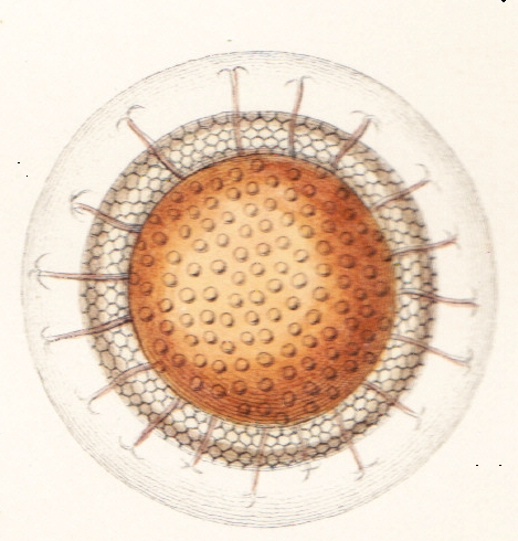
A quick scan through my quite extensive library on freshwater bryozoa showed no references at all to this membrane - I was on to something new! However I remained uneasy, thinking I had seen something somewhere. Weeks later I was scanning a famous work on fresh-water bryozoa by Mr Allman dating 1856 and thought to check. No reference in the text at all. However in the drawings there it wasa membrane supported by barbed spikes. So something seen by a 1856 artist with the microscopes of that day had been ignored and forgotten until now.
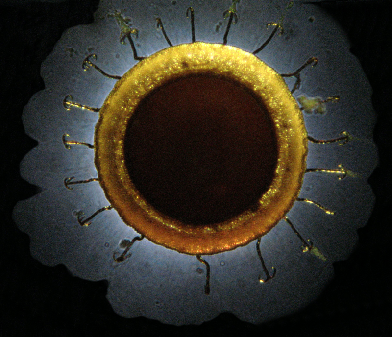
The final photo combines the above ones, showing both the membrane using asymmetric oblique lighting and one sided lighting from above. As the contrast between the background and the membrane was even worse than in the photo above I used a photo manipulation program to artificially turn the background to black, taking care not to introduce artificial structures. This is the only use of manipulation, other than resizing for the web, that I did.
|
|
|
|
Cristatella mucedo statoblast |
Cristatella mucedo statoblast |
One thing that puzzles me is why this has happened. My theory is that most researchers store specimens for later research and then make slides. This can easily damage the thin membrane beyond recognition. Also unless asymmetric oblique lighting is used (or possibly phase contrast, or DIC, - I did not try that and it was certainly not available around 1865) the membrane will remain invisible.
Relevant Micscape articles and other literature are:
Comments are welcomed by the author!
Published in the February
2010 edition of Micscape.
Please report any Web problems or
offer general comments to the Micscape
Editor.
Micscape is the on-line monthly magazine
of the Microscopy UK web
site at Microscopy-UK