|
An 1830s Cary-Gould style microscope by Carpenter and Westley:
Exploring the versatility and optical performance of a popular single lens / compound microscope.
by David Walker, UK
|
Antique microscopes can have an appeal on a number of levels—they are attractive for display and can prompt research into the model design and/or maker's history. To the amateur microscopist, one of their greatest appeals is that they are tools that can still be used. It's fascinating to explore their use and capabilities and to assess their pros and cons in a modern context by comparing with instruments of today.
My brother Ian and I have only a modest budget for antique microscopes (we have six collected over a decade) so we tend to pick good examples of a design that we can afford that are in a position to be brought back to good working order if not already so. The Cary-Gould style model from ca. 1830s described below with chromatic optics is an example of a very popular microscope that predated and overlapped somewhat the development of achromatic optics for the microscope.
There are superb resources on the history of the Cary-Gould design, that of the parent company and its place in the history of microscopy. Detailed studies of the optical performance of many antique microscopes have also been made and a selection of references are offered below. This article just presents a hands-on study of an example of this model.
The Microscope
|
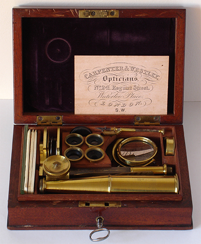
I've only been fortunate enough to handle and study this example, but from published illustrations, Cary-Gould style microscopes can vary in their box size and included components. The quality of the woodwork also varies.
This example sold by Carpenter and Westley features one of the larger mahogany boxes, making it portable rather than truly pocketable. Typical of this design, the components are neatly compartmentalised in the box and has a pad of purple velvet in the lid. The carpentry is to a high standard in this example with pinned dovetail corners.
Box size - 170 x 117 x 48 mm.
Microscope height (not including box height) - 124 and 250 mm (with and without compound head).
The height of the microscope remains fixed because it has the typical moving stage focus.
It has a lock rather than the more commonly seen two clasps. The only other example have seen to date with a lock is another Carpenter and Westley shown in the Golub Collection (which has both lock and clasps and differs on other features, e.g. a pivot at base of pillar).
The tray lifts out which allows access to all components while the microscope is mounted on the box. The space below the tray allows some storage of paperwork, in this case the leaflet and trade card.
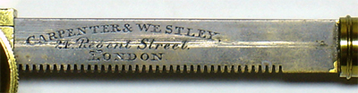
The rack is marked 'CARPENTER AND WESTLEY, 24 Regent Street. LONDON.' They are a well known company and dates the microscope from ca. mid 1830s. It may have been made for them (see Resources below for a history of the company and the link above).
|
|
|
Above, shown with the compound head and eyepiece in place. The head can screw in to the supplied lenses. The tubes on this age and style of microscope frequently show concentric circles of the varnish ageing, but this example is in excellent condition. The mirror is a single sided concave design which is reported to act as a condenser for NAs up to about 0.2.
|
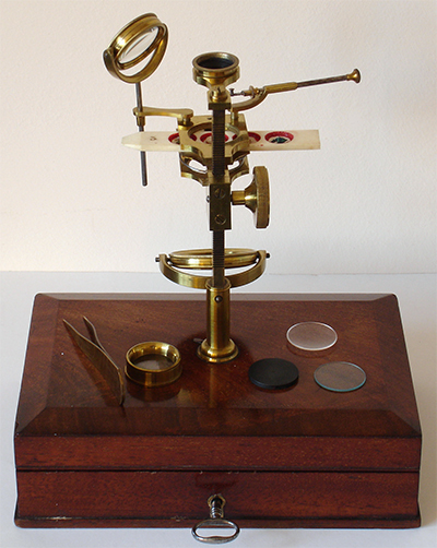
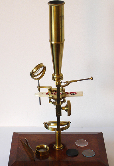
Above, shown in single lens mode, lenses can be used alone or stacked. Included accessories shown are a pair of tweezers and a live box with controllable depth to immobilise specimens. The three discs can each fit into a stage recess. A black / white disc for epi studies, a flat glass plate and a concave plate, the latter for e.g. aqueous studies.
|
|
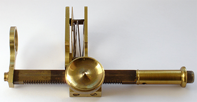
Right. The pillar has the typical straight cut rack of the period. The lenses screw into the fixed support left and the typical sliders of the time sat in the sprung loaded stage.
The microscope has a well engineered feel but the focus is rather coarse for the high power button lens. At higher powers this example also has a slight lateral movement when focussing.
The bright brasswork to the left side of the rack suggests that it has spent much of its life stored in the box rather than assembled, either for display and/or use. This observation was supported with the solid block of dirt in the pillar recess on top of the box and the extremely dirty lenses on receipt.
Antique microscopes being rarely used seems all too common from my brother's and my joint buying experiences over some years. In this case a pity, as it is a design that is very easy to service and clean without special skills and the owner misses out on rediscovering the experiences of the early 19th century microscopist.
|
|
|
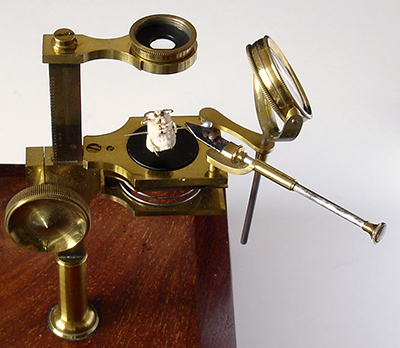
This style of microscope, as were many later 19th century compound microscopes, were particularly versatile for top lit studies because they had valuable features not seen on modern models.
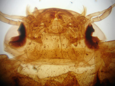
The stage forceps with sprung steel tips allows a subject to be supported and manipulated in all dimensions with accuracy while under study. An adult silk moth head is being studied here with the supplied black/white disc in stage recess. There's no reason why stage forceps couldn't be a feature of a modern compound with low power objectives or on a stereo. The model also has a stage condenser to focus incident light. This has been replaced nowadays with accessories such as the
flexible cold light sources.
The lens is shown in focus illustrating the good working distance for subject manipulation. The stage is a little small for dissecting work.
Some models of similar design had rotational and/or linear movements of the lens mount to allow scanning of samples and useful to avoid disturbance of live aquatic creatures.
|
|
|
|
|
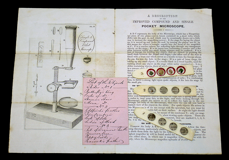
The set came with the original maker's leaflet and a trade card, the latter with the subjects on the three supplied sliders handwritten on the back. All subjects were still present apart from the classic diamond beetle elytra. Fortunately I had a piece of this from a broken deep cell Victorian slide so could be replaced. Slider 3 were uncovered subjects for top light. Sliders 1 and 2 had mica covers for transmitted and / or top lighting.
The microscope optics
|
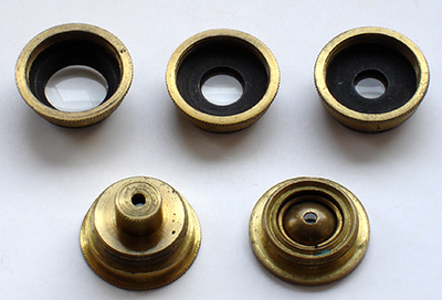
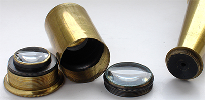
Five lenses were included, shown right, in increasing order of magnification. The top row numbered 3, 2, 1 had bi-convex lenses for lower powered work, were still intact and optically clear and were in excellent condition after a thorough clean.
Sadly the high power lens element in the screw fit brass cell (2nd row on left, shown inverted) was missing which was for use with the compound head only. An optical diagram in Savile Bradbury's 'The Evolution of the Microscope' (page 173) of a typical Cary model ca. 1830 suggested this was likely a small plano-convex lens, so I may replace with a modern
equivalent. The accompanying maker's leaflet stated it had an (area) magnification of 8,000 times, i.e. 89.4 linear.
There was included a button style high power spherical lens ('B', 2nd row, shown inverted right) for use without the compound head. This lens may have been added later because the thread didn't fully match the lens mount.
The compound head had a ca. 2 mm stop at its very base and tube well blackened inside with the optics solely in the slide-in three element eyepiece (both shown right). These elements were all slim bi-convex lenses, two in a spaced doublet ca. 11 mm apart and the third element ca. 30 mm below this with an intermediate ca. 13.5 mm field stop in the eyepiece housing.
All the optics were extremely dirty on receipt. As they were uncoated single glass elements, they were soaked in their housings in deionised water with a trace of detergent and left for 15 minutes. All dirt then readily floated off with the light action of a fine brush while inspecting each lens under the stereo while still immersed. This procedure avoided the potential danger of any gritty dirt scratching the glass surfaces.
|
|
Assessing optical performance
The magnifications of the lenses in single and/or compound mode were measured using a standard micrometer slide projected 250 mm away from lens top. It was more practical to lay the microscope supported on its side for these measurements to form a simple optical bench. Micrometer slides both 0.1 mm and 0.01 mm are useful for assessing field of view, aberrations and field flatness. A black scale on a bright background is a rather demanding image and would readily show chromatic aberration. This is a demanding test even for some modern achromatic compound microscope objectives.
All the test images of micrometer scales shown below are in colour so would show any marked chromatic aberration.
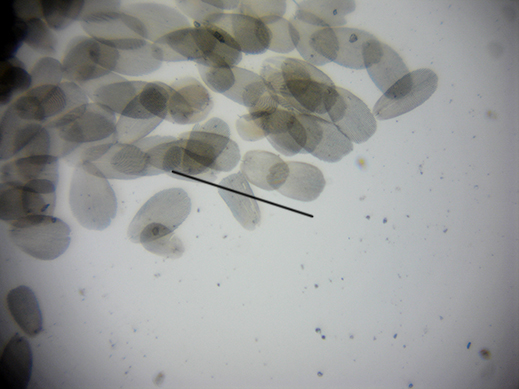
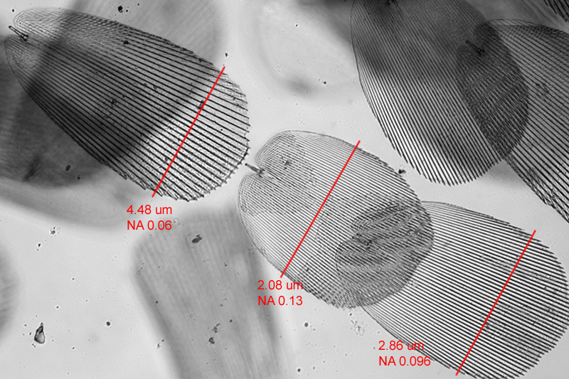
Estimating numerical aperture For the low NAs of these type of lenses, the classic test objects such as diatoms are unsuited and I don't own a rulings type test plate. A 0.01 mm micrometer slide is a useful start to show if the NA is greater or less than 0.028 (using R=λ/2NA at 550 nm). After browsing around my prepared slides, a strew of Lepisma saccharina (silverfish) scales proved useful as the coarse ridges vary quite widely in separation and also are non-parallel. This allowed three scales to be selected that required NAs of ca. 0.06, 0.096 and 0.13 to resolve the ridges. For the lowest power lens no.3, the finer ridges on the 'Scale of Sole' subject on slider included was useful which required an NA of 0.013. (See Appendix.)
The table below summarises the lens properties measured. The optical properties of a Cary microscope's optics in the Utrecht University Museum collection were reported by van Cittert in 1934 and recently recalculated as part of a larger study by Deiman (see Refs.). Deiman used a Grayson ruling slide and the NAs are compared in the table above. The mags and focal lengths of my example differ to the likely earlier Utrecht Cary so cannot be directly compared but the NAs are broadly similar. A thorough study of the optical properties of an 'early nineteenth century' Cary microscope was also reported by White (see Refs.)
|
Lens
|
Working distance / cm
|
Field of view / mm
|
Magnification
|
Estimated NA
bold indicates nearer to this value
|
Properties of Cary microscope optics*
Lens no. - Focal length/mm - NA (Deiman)
|
|
1
|
1.8 (2.1)
|
7 (4.3)
|
12 (42)
|
>0.028 and <0.06
|
III - 18.6 - 0.031
|
|
2
|
2.5
|
14 (6)
|
9
|
>0.028 and <0.06
|
II - 29.7 - 0.016
|
|
3
|
3 (5)
|
15 (8.7)
|
6.5
|
>0.013 and <0.028
|
I - 38.7 - 0.011
|
|
'B'
|
0.4
|
1.4
|
45
|
>0.096 and <0.13
|
L - 9.6 - 0.072
|
|
1 + 2
|
|
|
20
|
|
|
|
1 + 2 + 3
|
|
|
22.5
|
|
|
Figures in brackets are when the lens was used with the compound head and eyepiece.
*Values when used as a single lens. Cary microscope described in van Cittert pp.54-55. Table data with thanks to
Deiman (see refs.), Appendix 2, page 155, entries UM73.01 to UM73.04. Cary lenses numbered in opposite mag order to current example.
|
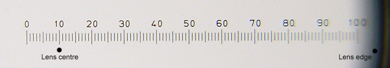
Above. Micrometer slide 0.1 mm scale. Lens 3 (6.5X) showing that for the lowest mag, chromatic aberration was minimal across most of field and a good flat field of view. A useful scanning lens akin to a modern hand lens for studying larger subjects.
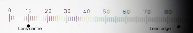
Above. Micrometer slide 0.1 mm scale. Lens 2 (9X) chromatic aberration remained modest but image quality fell off from centre more rapidly.
Above. Micrometer slide 0.1 mm scale. Lens 3 (12X) chromatic aberration remained modest but image quality fell off from centre more rapidly.
|
|
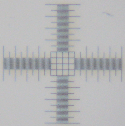 
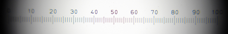
Above. A 0.01 mm grid using the lens number 1 (12X) and number 2 (9X). Both comfortably resolved this for an NA >>0.028 and gave good aberration free imagery. The grittiness is the limitation of the noisy sensor at the 1:1 imaging level on the rather aging Sony P200 camera used.
|
|
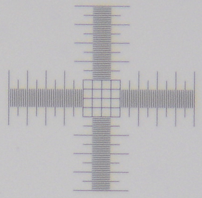
Above. A similar 0.01 mm scale taken using a modern Leica S8 fully apochromatic zoom (1-8X) stereo microscope set at an optical mag of 10X (see this Micscape article). The striking difference is, of course, because at 10X on the stereo, the primary optical objective is 1X with a lower NA used with a 10X eyepiece to gain the advantages of a wider field of view and greater working distance. The models with wider zoom lenses also inevitably further compromises resolution at the lowest mag of this type of stereo (and the latest wide zoom flagship macroscopes). So the humble Cary-Gould style microscope with number 2 lens of the 1830s can significantly outperform a typical modern stereo macro / microscope if resolution is the key at 10X!
|
|
Above. Micrometer slide 0.01 mm scale. Button lens 'B' (45X). The scale covers almost the entire field with near undistorted field with little chromatic aberration and shows a useful performance for smaller subjects.
|
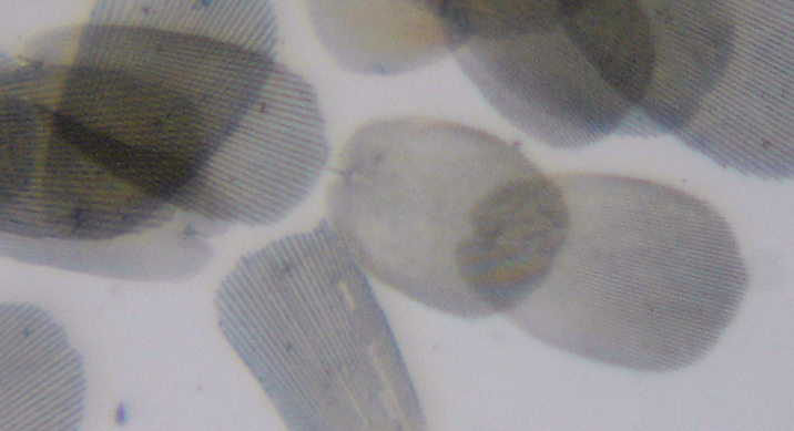
Above: Full field of a strew of Lepisma scales(Biosil slide) using the lens 'B' (45X). The bar shows the three scales selected for resolution studies.
|
|
Right. Detail from the same image right above using the button lens 'B'.
From the calibration image in the Appendix, the lefthand and righthand scale ridges are resolved but not quite the centre (confirmed visually, the camera was not the limiting factor nor the tricky focussing.)
This suggested that the NA of lens 'B' was >0.096 and <0.13 and likely closer to the former.
|
|
Approaches tried for taking photographs
|
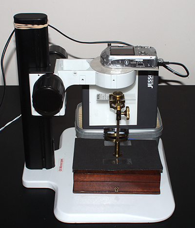
Ideally, direct projection onto the sensor of either my Sony NEX 5N or Canon 600D body (with APS sensors) would have been preferred, but making a light-tight flexible shroud and a versatile support for these larger cameras proved tricky.
The most practical approach was to use a lightweight, smaller Sony P200 7 Mp fixed Carl Zeiss lens camera—the 'small bore' zoom lens is eminently suitable for close coupling to either the single lens mount or the compound eyepiece, giving a good light baffle. The zoom allowing either a full field or zoomed-in image to be captured.
Camera focus was set to infinity and relying on the live rear LCD screen image to set the microscope focus. ISO 100 was used and the manual mode 'M' to set the largest aperture at a given zoom.
The flat camera face sat on the top of my stereo microscope stand with a support, providing a useful vertical height adjustment. (Camera shown raised in the image.) Used on mains power to avoid camera auto shutting off which moves the lens further forward, causing possible damage to camera or microscope.
The lower powered lenses require a large illumination source, ideally a good large window for daylight but was not practical in my hobby room which has small framed windows (winter doesn't help either!). A 35 mm slide viewing panel was adopted instead. A comparison of images taken with this and daylight on an occasional bright day did not reveal any noticeable differences for the lenses and subjects used.
|
|
|
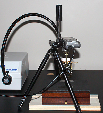
The above set-up was unsuited for top lighting studies because the stand collar blocked light access. The set-up right was used instead allowing a cold light source (30W) to be used, with some modelling using a small crescent of paper on the stage.
The disadvantages of this set-up was that setting the vertical height of the camera was awkward and not readily altered. Access was also limited by the tripod legs.
The rubber mat placed on the box for both set-ups was a safety precaution because standard 3 x 1 inch slides when in use did not sit in the sprung clips and had to sit untethered and more precariously on top of the stage.
For transmitted work above the supplied mirror was somewhat undersized for evenly filling the field, so a Russian LOMO convex mirror was temporarily placed on top (it had a similar focal length to the original).
|
|
A selection of images
|
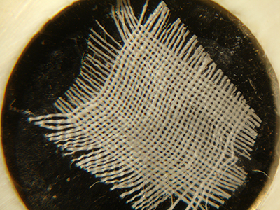
'Fine Cambric'. Lens 3.
|
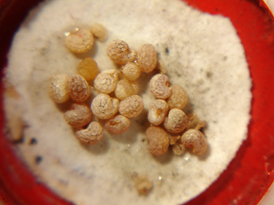
'Piece of Cane'. Lens 1.
|
|
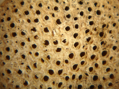
'Poppy Seeds'. Lens 3. A classic subject for the microscope described and illustrated as 'corn poppy seeds' by Dr. Henry Power in his book of 1663/4 Experimental Philosophy in Three Books which just predates Hooke's Micrographia (September 1665) as the first English book devoted to microscopy. Power's book had very limited basic illustrations and simple commentaries compared with those of Hooke. (Power lived at Elland, near Halifax. The house, now 'New Hall Farm', still stands and is a couple of miles from my house.)
|
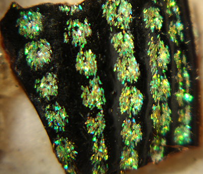
'Bit of Diamond Beetle'. The iridescent scales on the elytra of the S. American diamond beetle (Entimus imperialis) was a popular prepared slide subject throughout the 19th century (see this Micscape article). The scales are crisper visually.
|
|
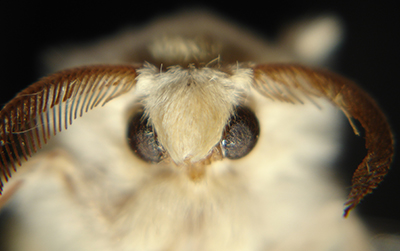
Head of the cultivated silk moth, Bombyx mori (own sample). The camera does not accurately reflect the crisp visual image seen.
|
Left. The stage forceps proves a valuable manipulator for subjects such as this insect or small flowers etc. The cultivated silk moth was a subject studied intensely by Leeuwenhoek, see 'Exploring the cultivated silk moth Bombyx mori' a three part series in Micscape 2012 repeating some of Leeuwenhoek's studies using both a Leeuwenhoek replica microscope and modern compound microscope.
|
|
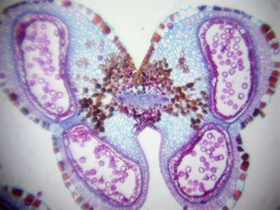
Lens 1 as single lens. Madagascan flamboyant tree, Delonix, flower-bud, T/S (Biosil slide). Although a modern stained plant thin section, it's a useful subject to compare the merits of using the compound head.
|
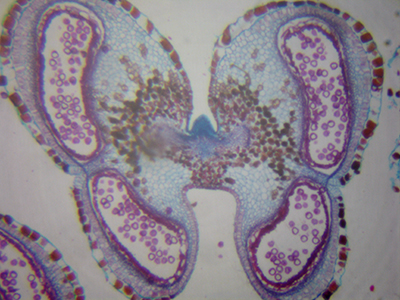
Lens 1 with compound head which gave a ca. 3.5X mag increase, field of view approx. halved. Image colour, contrast or detail is not improved as expected by adding a pre-achromatic three element eyepiece.
|
|
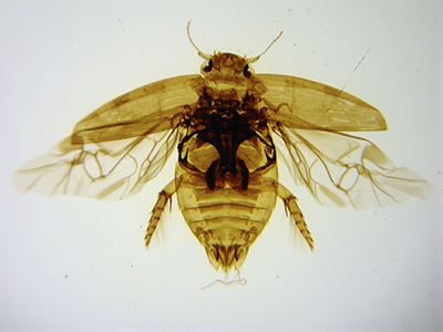
Lens 1: Slight optical zoom to fill field. Victorian water beetle slide Laccophilus minutus. Very competent aberration free images are provided to the eye and camera.
|
Lens 3. Same subject which fills the field of view of the lens.
|
Concluding remarks. I greatly enjoyed exploring the performance of this modest looking microscope and shows its versatility and usefulness well for a range of subjects. The compound head does not really add useful optical magnification and much preferred the simplicity and crisp imagery which the single lenses alone offer.
Comments to the author David Walker are welcomed.
Appendix:
A strew of Lepisma saccharina (silverfish) scales (Biosil slide) showed that the coarse ridges on the scales varied quite widely in separation and also were sometimes non-parallel. These proved useful for assessing lens resolution. A grouping was chosen on the strew that was both easy to relocate under the Cary-Gould microscope and which provided a selection of ridge separations / NAs.
A photomicrograph of the selected area of the strew was taken in brightfield using a modern compound microscope (Zeiss Photomicroscope III, Neofluar 16X NA 0.4 objective with Canon 600D camera body). An A4 print was made and the ridge separation measured along the lines shown in the image below (horizontal field width 461 µm). This gave areas on the test scales requiring NAs of ca. 0.06, 0.096 and 0.13 to resolve the ridges (using R=λ/2NA at 550 nm), i.e quite useful steps of 0.03 NA. Plus the 0.01 mm micrometer slide providing an NA test for 0.028.
To assess the NA of the lowest power lens number 3, the 'Scale of Sole' subject supplied on the slider proved useful because of its ridges were coarser than the Lepisma scales.
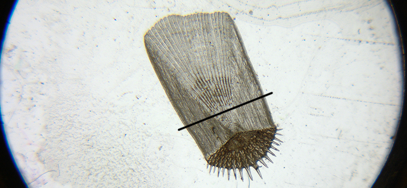
Above: Full field of view of the 'Scale of Sole' using lens 3 (6.5X), the lowest power. The fine ridges on left of scale along line were measured using the photomicroscope as being ca. 21.5 µm apart.
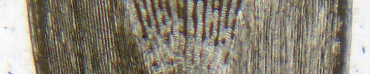
Above, detail from image of scale of sole along the line shown. The finer ridges left of the coarser centre are comfortably resolved, suggesting the lens has an NA of at least 0.013.
Selection of resources and references
Brian Stevenson, The Cary-Gould-Porter-Optical businesses, Micscape Jan 2014. An in-depth study of the company who instigated the design.
Brian J. Ford, 'The Single Lens. The Story of the Simple Microscope', William Heinemann Ltd., London, 1985. A fascinating account of the often underestimated role of the simple microscope in the history of science.
Savile Bradbury, 'The Evolution of the Microscope', Pergamon Press, London, 1967. A classic work which discusses the Cary-Gould design and its development.
P. H. van Cittert. 'Descriptive Catalogue of the Collection of Microscopes in Charge of the Utrecht University Museum with an Introductory Historical Survey of the Resolving Power of the Microscope', P. Noordhoff NV, Groningen, 1934. A widely cited and invaluable work which includes the optical performance where possible of extant lenses of each model, including an example of a Cary microscope.
Brian Bracegirdle, 'Notes on Modern Microscope Manufacturers', Quekett Microscopical Club, London, 1995. Entry 'CARPENTER. P (& WESTLEY)' p.20.
R. H. Nuttall, 'Philip Carpenter and the 'Microcosm' exhibition; with a note on Carpenter & Westley's microscopes', J. Quekett Microscopical Club, 1976, 33(2), pp.62-65.
J. C. Deiman, 'Microscope optics and J. J. Lister's influence on the development of the achromatic objective 1750 - 1850.' Ph.D. thesis Imperial college, London, 1992. CD version 2008 - Little Imp Publications available from Savona Books (Catalogue no. A15 via the 'Archival Series CDs' option in menu). An indispensable work for those interested in the optical performance of a wide range of antique microscopes and extant lenses. Includes new measurements of the Cary microscope optics described in the van Cittert work.
G. W. White, 'A Cary Microscope', J. Quekett Microscopical Club, 1966, 30(5), pp.113-116, 156.
Revision history. Uploaded January 13th 2015.
Some additions and minor revisions Jan 14th, Jan. 24th 2015
©
Microscopy UK or their contributors.
Published in the January - February 2015 edition of Micscape.
Please
report any Web problems or offer general comments to
the
Micscape
Editor
.
Micscape
is the on-line monthly magazine of the Microscopy UK web
site at
Microscopy-UK
©
Onview.net Ltd, Microscopy-UK, and all contributors 1995
onwards. All rights reserved.
Main site is at
www.microscopy-uk.org.uk.