JELLYFISHES 
by Jean-Marie Cavanihac, France
 
The same organisms often have various names in different languages or cultures. An example are jellyfishes. Literally the English word seems to suggest a sort of fish with a translucent and light body,.... like gooseberry jelly on our plates!

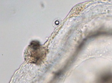
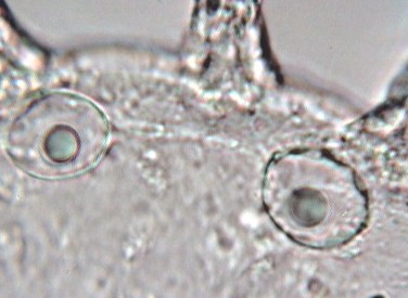
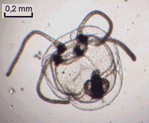
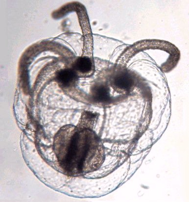
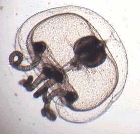
In French, jellyfishes are called: Meduse (Medusa in Latin). Why??? Remember in mythological times, there was the three gorgons living near the Hesperides Garden and one of them was called Medusa: she had snakes in her hair and the poor humans who met her gaze were turned into stone. She was defeated by Perseus, and he gave Medusa's head to the goddess Athena who used it to protect the town of Athens. But these legends are not so far from the reality; indeed jellyfishes have long pendant tentacles which seem to be hairs, and when a fish, for example, touches one of them, poisonous stings inject paralyzing toxin into its flesh ... (the fish don't become a stone but they can't move though!)


Above: hydromedusa (see below for description). Picture taken with x2,5 objective. Right hand side: taken with 6,3x objective. 


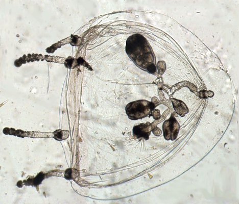
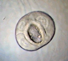
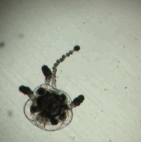
Side view of hydromedusa

 
When summer comes along, the sea often contains plenty of jellyfishes and the larger ones (20 or 30 cm diameter!) can cause serious 'burns' and then scars on swimmer's skin especially in young children.

It's safer and very interesting to study some species of marine organisms with microscopic 'jellyfish' stages in their life cycle, some of them you already know like the medusa stage of obelia. (See the past articles in Micscape e.g. Obelia). How do these jellyfish stages form? In many species they are produced by a polyp fixed on rocks or algae which forms buds. Buds grow on the stem one after the other and when they are mature, their umbrella begins to move and young jellyfish leave the stem.


Below is an unusual picture which shows a mature jellyfish with its umbrella still connected to the polyp. (It looks like an inflatable toy!) Above and below the transparent umbrella you can see the stem and other buds on it at various stages of development.  
  Sometimes, when you catch plankton in the middle of spring or summer you can capture very tiny jellyfish stages called hydromedusae. An interesting subject is the obelia 'jellyfish' (medusa) stage because its umbrella is flat and can be easily observed. (Shown below with a polypoid form in the right hand picture. See Micscape article on cnidaria.)  Hydromedusae have a bell shaped umbrella and the internal organs are a little more difficult to see in the larger species.  
  Other individuals are shown below: you can see the four tentacles, the manubrium (mouth open) and 4 rounded gonads located near its base. (The right hand picture shows detail of an obelia's medusa stage gonad.) The digestive system is constituted by a central stomach (last picture) with 4 channels crossing gonads and connected to a ring near the base of the tentacles. Jellyfish adopt sexual reproduction and its sometimes possible to see spermatozoids.  
 
 
  Frequently you can find a single specimen which regularly contracts its umbrella to eject water contained inside: in fact it's a tiny 'jet propulsion' mechanism! Click here for an MPEG animation.  
The left hand picture is (I must confess it!) a photo-montage: it's otherwise impossible to see clearly both the mouth and the bottom of the umbrella. It was created by merging two images at two levels of focusing: the first on the umbrella and the second on the mouth (some software like CombineZ can do this automatically). 
In the right hand picture you can see the tail of a larvacean (arrowed) body inside the jellyfish mouth !
  Another interesting subject for the microscopist are the tentacles which are covered with cnidocysts which produce nematocysts. A nematocyst is a sort of chitinous capsule containing a coiled thread. Cnidocysts often feature a hair (cnidocil) which act as a trigger when prey touch it. At this time a nematocyst projects a sort of little harpoon which pierces the skin or exoskeleton of the prey and injects venom. It's difficult to see the detail of a nematocyst (except with an electron microscope) but easy to make them discharge by adding a little vinegar to the slide. The animated gif below shows a small hydromedusa deploying one tentacle with detail shown in the right hand picture.  
 
  Upper right hand picture above shows detail of tentacles. Lower left hand picture above shows detail of cnidocysts. Note the cnidocil (trigger) and the folded structure inside cnidocyst. The lower right hand picture is a discharged cnidocyst showing thread.  
One final detail you can observe in some species are the equilibrium organs i.e. the statocysts which are located in an ampulla near the side of the umbrella (see the left hand picture below). The right hand picture below shows the obelia's jellyfish stage statoliths. Statoliths are little calcareous stones contained within a statocyst. (See the author's Micscape article on Statoliths.) 
 
 
  Don't be frightened if you see a large jellyfish near your favorite beach: avoid touching it but observe the graceful movements of the umbrella!  
Comments to the author Jean-Marie Cavanihac are welcomed.

Microscopy UK Front Page
Micscape Magazine
Article Library

All drawings and photographs © Jean-Marie Cavanihac 2002
Published in the June 2003 edition of Micscape Magazine.

Please report any Web problems or offer general comments to the Micscape Editor


© Onview.net Ltd, Microscopy-UK, and all contributors 1995 onwards. All rights reserved. Main site is at www.microscopy-uk.org.uk with full mirror at www.microscopy-uk.net.