|
|
The second voyage text by Richard L.
Howey
|
 |
|
|
The second voyage text by Richard L.
Howey
|
|
As you may recall, on our previous visit to Professor Moriarty's laboratory, you put on scuba gear, entered his new inventionthe resizerand emerged the size of a Colpidium, about 60 microns long. You were then removed from the tank of water in which you were immersed and by means of a Pasteur pipet, placed into a culture dish filled with all sorts of wonderful microscopic delights and monsters. The second voyage will involve encounters with equally wondrous micro-aliens and Professor Moriarty and I will keep track of your encounters by observing you with a stereo microscope while you are in the culture dish.
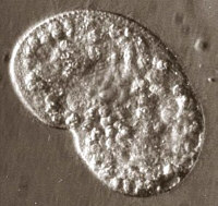
ColpidiumThe resizing goes smoothly with no difficulties and the tiny video camera in your suit will record your adventures. As you descend, you notice a small island on the bottom, off to your left. As you approach its vicinity, you see that it has what appear to be trunks rather like the stalks of the Vorticella you encountered on your previous journey, except, in this case, there are no bells on the stalks. Then as a current sweeps you back away from these clear stalks, you get an altogether different perspective and realize that these are extending out of a large branching structure almost like a tree with a number of branches. The overall height is nearly 2,500 microns! As you watch, a Paramecium aurelia, one of the smaller species, brushes against the tip of one of the stalks and then begins to struggle to get free. You observe a small pad at the end of the stalk which is apparently sticky and has trapped the Paramecium. In rapt horror, you watch as the protoplasm of the Paramecium is sucked out of its body through the stalk, like a straw emptying a glass of soda.
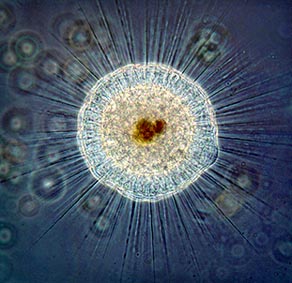
ActinosphaeriumThis fascinatingly grotesque creature is Dendrosoma radians, a suctorian. They have complex reproductive cycles and in only one stage are they ciliated, so seeing an adult wouldn't lead you to realize that these are classified as ciliates. Sometimes a Dendrosoma will be feeding on half a dozen or more Paramecia at once. You decide you've seen enough of this bizarre monster and swim away. Ahead you see a cloud of ultramicro plankton and in its center what appears to be a transparent sea urchin with long spines. It looks like a glass sphere with smaller spheres (vacuoles) packed around the inside edge. The spines are flexible and sticky as well as a consequence of a streaming flow of protoplasm up and down the axis. Bacteria and tiny flagellates are pulled down toward the vacuoles by this flow. Slightly larger flagellates and small ciliates prompt the spines to bend and curl so as to entangle the prey more thoroughly. This magnificent sunburst creature is a heliozoan (or sun animalcule) called Actinosphaerium. Another genus, Nuclearia, secretes a gelatinous or mucous dome around itself and is able to extend its thread-like pseudopodia through the dome and feed on the bacteria which seem mysteriously attracted to the area and swarm in great numbers. Yet another genus, Raphidophyrs, goes through an extraordinary process of partially fusing with 3, 5, 7 or even more of its fellow heliozoans to form a formidable feeding trap for small organisms and then after the feeding process, they separate off again into individuals and go about their own business. This creates an occasion for some interesting speculations regarding the formation of colonies in eukaryotes, and, in fact, this topic could make a fascinating book.
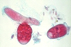
Two Didinium; one is eating a Paramecia.Ahead you see a large mound of detritus and a lot of actively feeding Paramecia, which you find comforting, since although they are larger than you, they don't pose any real threat But today seems to be your day for lessons about micro-predators and micro-prey. Whirling through the water like a barrel comes a rotund ciliate which appears to have a long, narrow snout or nose. This creature is a highly specialized and efficient predator called Didinium nasutum and it has an appetite almost exclusively for Paramecia. This narrow snout or proboscis has the cytosome or mouth-opening at the tip and in the length of the snout is a series of rods which can cause the mouth-opening to expand. What is astonishing is that this expansion is such that it allows a Didinium to engulf a Paramecium twice its size! Didinium is a very active and fast organism (unlike me) and as a consequence feeds frequently, multiplies rapidly, and can quickly deplete a Paramecium population. So, it appears that Didinium isn't very smart; it destroys its food source and then dies offright? Wrong! Didinium has developed a clever survival strategy which involves encystment. When they manage to eat up all of the Paramecia, the lack of food triggers a remarkable series of events. The Didinia dedifferentiate their organelles while secreting protective cysts, in other words, what's left is basically a blob of protoplasm, a nucleus, and a cyst wall. The Didinia reduce their metabolic activity to a minimum and wait for conditions to change or for them to be transported to another environment with abundant Paramecia. The cyst wall itself is extraordinary, for, on the one hand, it seals up and protects the delicate living packet of protoplasm, but, on the other hand, allows biophysical and biochemical "communication" with the environment in such a manner that when conditions are once again favorable, the whole process reverses and excystment occurs. The Didinium somehow gets the proper set of signals and begins to rebuild its organelles, thereafter it breaks through the cyst wall and begins hunting Paramecia again.
ParameciumAs you continue observing the Paramecia, you notice some much smaller organisms which are clustered around a Paramecium and seem to be feeding on it. You count them. There are seven of them and the Paramecium is helpless. These are Coleps, the jackals of the protozoan world. As you look more closely, you see that they too are barrel-shaped and armored with a series of plates that cover the body. In addition, this particular species has eight spines projecting from the posterior area. With so many predators, it is fortunate that Paramecia have such a high rate of reproduction given desirable conditions.
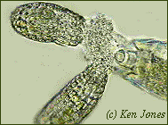
Two Coleps eating the
egg laid by a rotifer (right).After all this violence and predation, a Halteria grandinella bounces into your vicinity and offers a bit of comic relief. These entertaining little creatures can swim with dizzying rapidity and then suddenly stop on a dime, dive headfirst to the bottom and spin in one place. They are like miniature street sweepers and the oral ciliary movement is like the brushes sweeping up bacteria and detritus. But Halteria also has a series of long semi-rigid spines which project from around its midsection and when these come into action, Halteria becomes a pole vaulter with a plethora of poles and goes springing off through the water.
As you swim further toward the center of the culture dish, you hesitate, for once again you see stalks and your previous experiences with stalks have been somewhat disconcerting, especially with the suctorians. This time there are two distinctly different kinds of stalks, one solitary and clear, the other dark brown and branched toward the top. You allow yourself to drift slowly upwards, 25 microns, 50, 100, 150, 200, 300 and there you are gazing at a superb specimen of Clathrulina elegans, a stalked heliozoan. The central envelope is about 90 microns in diameter or about half again your size and there are many pseudopodia projecting from the central sphere on all sides through circular or polygonal openings in the central envelope which make Clathrulina an elegant and impressive creature indeed. As you continue to ascend, you can begin to see the top of the brown, branching stalks, and at the top of each stalk, there is what appears to be a cluster of flowers, each tiny bud of which is extending two tendrils which lash the water creating minute currents. This deceptively plant-like creature is Anthophysa (Anthophysis) vegetans. The flower-like bundles at the top of each stalk are colonies of flagellates and as you watch, one of the colonies breaks off from its stalk and swims tumblingly through the water.
These proceedings are interrupted by what in the protozoan world is the equivalent of hyperactive, athletic, showoff teenagersa half dozen or so Urocentrum turbo. They almost always show up in small groups, I suspect, because they cannot endure the idea of not having an audience for their antics. Urocentrum turbo lives up to its name and is a micro-dynamo. In appearance it is an elongated oblate spheroid pinched slightly in the middle, as though it were wearing a belt that was too tight with the result that the upper and lower halves are each slightly puffed out. Each half has a thick band of cilia and at the posterior end, to one side of center, is a fairly long tuft of fused cilia. They use this tuft to anchor themselves to a substrate and then spin around that point like a toy top gone mad. Just when you get one nicely aligned and in focuszipit's gone. When the adhesive loosens, Urocentrum tears off through the water like a turbo-charged hotrod.
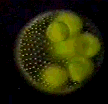
Volvox colony with daughter colonies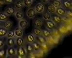
Close-up of Volvox
Note: These animated gifs of 50kB and 125kB need to fully download before the animations begin. Fortunately, you were still clinging to the Anthophysa stalk when the Urocentrum took off, so their wake didn't sweep you away. While you are holding on for dear life, the light overhead dims sharply. When you look up, you see an enormous green sphere with hundreds and hundreds of cells rotating through the water driven by flagella, each cell having a pair. This giant orb reminds you of the Death Star from Star Wars, but, on this occasion, you are quite safe. Volvox feeds photosynthetically. You are mesmerized by the sight of this giant transparent globe with smaller globes inside which are "subcolonies" having to do with the complex reproductive cycle of Volvox. Some colonies produce only female gametes, some only male gametes, and some produce both male and female gametes. Nature's strategies to achieve high levels of diversity are astonishing and to most people, the idea of protists having complex patterns of sexual development seems amazing. So, while your prurient impulses are absorbed with protist perversity, your attention regarding your surroundings fades until suddenly you are aware of powerful currents coming at you from all directions and sweeping you from side to side as you desperately cling to the Anthophysa stalk.
Another enormous protozoan, this time a ciliate, looms on the horizon and this one is clearly no vegetarian. Equipped with a huge V-shaped cytostome, it swoops through the water devouring virtually everything in its path; small flagellates and ciliates, medium-sized flagellates and ciliates, Paramecia, Colpidium (your size), Tetrahymena, nearly anything and it's headed in your direction, engulfing one Paramecium after another. Suddenly through the microphone, Professor Moriarty and I hear you shriek: "GET ME OUT OF HERE NOW!"
Clearly your survival instincts have caused you to lose your aesthetic sensibilities. From our vantage point, Professor Moriarty and I are admiring the translucent elegance of this graceful creature which looks rather like a handblown Steuben lead crystal vase slowly sweeping through the water towards you.
"GET ME THE HELL OUT OF HERE!" crackles through the microphone. "Very well, very well," Professor Moriarty grumbles and inserts a pipet into the culture dish, extracts you and puts you into the expansion chamber. When you have recovered your size and your strength, you begin yelling at the Professor that you would never go into a culture again. "Then you won't get paid," says Moriarty tersely. "You agreed to three journeys."
I remember that later after you and I reviewed the magnificent video tape which you brought back, you did, after all, agree to one more adventure.
Comments to the author Richard Howey are welcomed.
Click links below to read parts one and three:
Hunting micro-aliens: The first voyage
Hunting micro-aliens: The third and final voyage
| Picture credits(with
links to the Micscape article they were sourced from, for further information.)
Wim van Egmond: image of Actinosphaerium, page background, animation of ciliate and Spirillum bacteria. Lizzie Harper: painting of Paramecium. Mike Dingley: image of Didinium from the article Didinium - the master feeder. James Evarts: Colpidium(oblique illumination). Ken Jones: Volvox animations (gif versions by the Editor of original video clip), from the article 'Volvox - jewel of the pond' by Ken Jones and Maurice Smith. Coleps image from video still by Ken Jones from the article 'The good, the bad and the ugly' written by Dave Walker from material kindly supplied by Ken Jones and Eric Hollowday. |
Web page prepared by Dave Walker
Please report any Web problems or
offer general comments to the Micscape
Editor,
via the contact on current Micscape
Index.
Micscape is the on-line monthly magazine
of the Microscopy UK web
site at Microscopy-UK