|
|
The first voyage text
by Richard L. Howey
|
 |
|
|
The first voyage text
by Richard L. Howey
|
|
| If you have a few billion
dollars to spare, access to rockets, enormous electronic monitoring facilities,
the dedication of thousands of scientists and technicians, and lots of
time and patience, then you might be able to launch a space probe to Mars,
collect some very expensive samples, and perhaps, with a great deal
of luck find some hints of possible fossilized life forms of a very primitive
sorthowever, you'd also have to invest in a couple of electron microscopes
at this point, but that's just small change after all the money you've
already spent.
Fortunately, there's an easier way and one, that to my mind is more exciting, although granted, a lot of people would be very excited to find fossilized tuberculosis bacilli on Mars. For those of us with more modest means, a couple of microscopesa low power dissecting microscope and a standard compound microscope¹a fine mesh net or two, some jam jars, a few slides, coverglasses and culture dishes and we're ready to go hunting for some of the most amazing organisms on the planet and, best of all, the probabilities are very high that some of these creatures can be found within walking distance or a short bicycle ride.
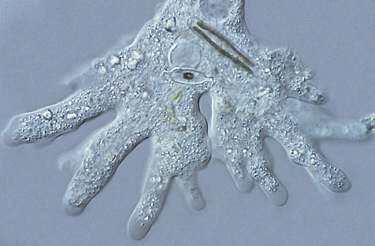
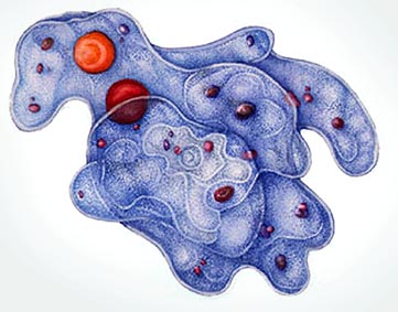
One of the wonders about microscopic organisms is that they can be found most anywhere, but they are often dormant or camouflaged and require some encouragement to make an appearance. A mud sample from the bottom of a pond or a gravel sample from the bottom of a lake will sometimes produce amoebas. A few grains of boiled wheat added to the sample will provide food and many organisms will appear over the next several days². It is not common to find large amoebas, but when one does it is well worth the trouble to isolate some and start several cultures. Every beginning biology student has seen drawings and photographs of amoeba, but there is no substitute for observing live specimens. Two years ago, I found some specimens of Amoeba proteus which averaged about 600 microns in size. I took a sample from an 8,000 foot lake and placed a bit of it in a watch glass and was examining the sample under my stereo dissecting microscope. I wanted to get a fairly close look, so I pushed the magnification up to about 60x. Over the years I have looked at a lot of small amoebas, but with a compound microscope. Looking at a large amoeba under a stereo microscope is a completely different experience.
We tend to think of amoebas as fundamentally two-dimensional, so that when you see a pseudopodium, then another, and another extend upwards at you, you have to rethink your whole image of an amoeba. Imagine yourself as a scuba-diving micronaut about the size of a Colpidium, a small ciliate protozoanand this came about one afternoon when you wandered into Professor Moriarty's laboratory and let him convince you to try out his new reduction changerin other words, you are only about 60 microns long or only 1/10 the length of the amoeba and its volume relative to yours is enormous. This is not a critter you want to get too close to. You have already observed that a pseudopodium can suddenly "erupt" and surge upward, outward and forward and if you're not careful you may suddenly find yourself enclosed in a bubble-like food vacuole with digestive juices beginning to attack your scuba gear. It's not only these pseudopodial surges that you need to watch out for, since you might be quick enough to pull back in the nick of time, but the surface envelope of the amoeba is covered with minute tentacle-like structures and is sticky as well, so you might initially escape a pseudopodial surge and then find yourself stuck to another part of the surface which has reacted by beginning inexorably to close around you. And this from an organism without a nervous system!
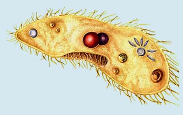
The micro-world is one which we sometimes tend to think of as rather simple, but only because we are so little familiar with it. Here we are surrounded by biochemical and biophysical reactions on a scale very different from our world. The micro-world is one in which a few molecules interacting or not interacting can make the difference between life and death. While your attention is caught up in avoiding the gigantic amorphous blob of the amoebawhich had to have been the inspiration for the two grade B sci-fi movies titled The Blobyou find yourself caught in a powerful current which suddenly releases you, leaving you with the impression that you have just been run through a blender at high speed. Being Colpidium-sized, you just escaped becoming lunch for a foraging Paramecium. Paramecia feed primarily on bacteria and very tiny flagellates and ciliates and often appear like hyperactive children in need of a dose of Ritalin.
Paramecia are so widespread and so abundant, that we tend to overlook them or take them for granted. They are so easy to culture in large quantities that a staggering amount of research has been done on them and there is a tendency to regard them as not only ordinary, but typical of ciliate protozoans. Here we need to remind ourselves of a few basics: 1) There are no typical protozoans. 2) There are no typical ciliates. 3) Paramecia are weird and if there were a prototypical ciliate, Paramecium certainly would not be my first choice. There is a considerable variation in size both within a given species and between species. For example, Parameciumcaudatum, probably the most common species, varies in length from about 175 to 300 microns. P. trichium can be as small as 50 microns and P. multimicronucleatum can grow as large as 350 microns. Paramecia have one large macronucleus and 1 to 8 micronuclei, depending on the species. They also generally have two contractile vacuoles, but some can have as many as seven. The contractile vacuoles regulate the pressure and salt concentrations within the organism by expelling excess water taken in during feeding. They have a distinctive flower-like appearance with six petal-shaped canals feeding into a central spherical vacuole which has a pore that connects to the outer surface of the Paramecium. When the sphere reaches a certain pressure as it expands, the pore opens and the excess fluid is discharged into the surrounding water. P. bursaria, a fairly small species, is easy to pick out because it is bright green due to the presence of symbiotic algae of the genus Chlorella. This arrangement turns out to be a very nice benefit for the P. bursaria. The Chlorella produce food photosynthetically and this Paramecium gets to take advantage of the metabolic by-products as a food source for itself. The Chlorella benefit from P. bursaria's maintaining a proper balance of sunlight. P. bursaria, however, has the ultimate advantage and can become a very inhospitable host. If a culture is placed in the dark for an extended period, P. bursaria starts digesting the Chlorella if other food sources are not adequate.
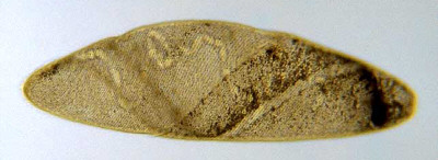
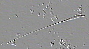
While you have been preoccupied with Paramecia, a cetacean of the micro-world has been approaching from behind, a Spirostomum ambiguum40 times your length!truly a micro-whale. This gigantic creature is narrow and band-shaped, and fortunately for you, a grazer on bacteria and very tiny protozoa. As it glides past you, you get a glimpse of an enormous posterior contractile vacuole which takes up nearly a quarter of this leviathan's length, when suddenly you find yourself once again being pushed and tumbled through the water. The Spirostomum has contracted and is now only about 1/3 its original length. As you thrash around, you find yourself pushed onto an island with some thin, transparent, but sturdy tree trunks and you grab hold of one and watch as the Spirostomum slowly stretches itself to its full magnificent size.
But, back to Vorticella. When the stalk has fully contracted, you have an opportunity to look inside the bell and observe the streaming protoplasm and get a glimpse of the enormous "C"-shaped nucleus that runs from almost the top down to the bottom of the bell. The bell of the Vorticella begins to move away and you notice the thick fibril that runs through the length of the stalk and which functions essentially in the fashion of a muscle. As you begin to examine the little island on which you landed, you observe that it consists of a thick tangled mat of strands of algae, both living and dead, and large clumps of bacteria, both living and dead. All around the edges of this "island" and from above as well, countless organisms are feeding and breeding and what is disconcerting is that many of them are bigger than your Colpidium-size, including some Spirillum bacteria that seem to bore through the water like long, thin, black corkscrews. However, the vast majority of the beasties here are much smaller than you are and the water seems thick, so densely populated is it with tiny flagellates, ciliates, algae, amoebae, and bacteriaa living soup! As you gradually accustom yourself to this profusion of life, from your perch on a mound of filamentous algae, you see a large ciliate swimming rapidly toward the island and it is an astonishing rich pink color. Along one side of the anterior end are longer cilia and a distinct oral membrane which extends about 1/3 of the length of the body. This amazing creature is Blepharisma and as you watch it swim toward you, there appears, apparently out of nowhere, another Blepharisma, three times the size of the first one which it draws up to and slowly engulfs in its enormous cytostome (mouth). This creature is a cannibal giant and poses a real threat to you. No one is certain of the exact mechanisms involved, but under certain conditions, Blepharisma will form giants and feed on their own kind. The pigment is unique and thus has the name blepharismin. It is a photoactive pigment, which means that the organisms use it to maintain a proper position in the water column so as to avoid strong sunlight which can activate the pigment in such a fashion as to make it toxic and if the exposure to intense light is prolonged, even fatal.
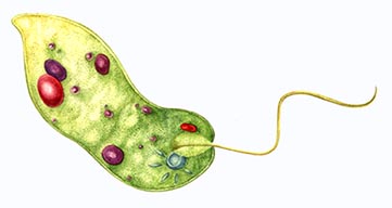
Fortunately, the Blepharisma is looking for larger, more nutritious prey and ignores you. In the distance you see a long, narrow, green tubelike creature approaching rather lethargically, slowly rotating through the water, drawn forward by a short whip-like structure lashing through the watera flagellum. This creature is about 7 times as long as you are and is a Euglena oxyuris. It is filled with green disc-shaped chromatophores which contain chlorophyll, by means of which it photosynthetically manufactures food and the fat and oil are stored in two larger ovoid bodies called paramylum bodies. But the feature which captures your attention is a deep rich red eye spot or stigma located very near the front of the organism. A bit above the Euglena, a smaller, close relative comes twisting through the wateran elegant creature which looks like a green leaf with a flagellum about two-thirds its body length and a single bright red cyclopsian eye, glistening like a miniature ruby. This lovely creature belongs to the genus Phacus and is well worth looking for in the cold water of early spring.
Another bright pink Blepharisma comes churning past and while you are admiring all the contrasting colors, it suddenly begins to thrash around and a chunk is missing out of one side and the area is leaking protoplasm. You caught the first strike only out of the corner of your eye, but as you watch, a head with a gaping mouth, on a long snake-like neck strikes again and tears another chunk out of the Blepharisma which swims off as rapidly as possible. The creature that has been feeding in such a grisly manner now comes out from its hiding place in the debris and you feel like you have been dropped into Loch Ness. The body is long and teardrop-shaped and the anterior end has a long neck and a head with a cluster of cilia 2 or 3 times longer than the body cilia. This monster is Lacrymaria olor, the tear of a swan. As it swims off, you notice other Lacrymaria still anchored in the neighboring debris and as you watch, one of them extends its head and the neck stretches out 2, then 5, then 7, then 10 times the length of the body! This remarkable creature is one of the most fascinating denizens of the micro-world.
There are a number of protozoa which produce monsters, including our old friend, Paramecium; but, from my observations, S. coeruleus holds the record. Sometimes there is little else than a pinch of blue protoplasm with a few cilia and about 1/10th the size of an ordinary Stentor. Only a bit of a nucleus, no cytostome, no vacuoles. This little blob is going to have to undergo some extensive regeneration or die. The odds are almost certainly death. These monsters assume all sorts of weird shapes and have varying degrees of deficiency in terms of organelles. Here is a fascinating project for someone who is intrigued by complex challenges. In the midst of these ruminations, your oxygen tank scuba alarm goes off, signalling that it is time for you to return to the surface and the macro-world. You swim up to the top of the water in the culture dish and there Professor Moriarty plucks you out of the water with a Pasteur pipet and places you into the resizing chamber. You are exhausted, but elated and are already anticipating another journey into the micro-world. Comments to the author Richard Howey are welcomed. Click links below to read parts two
and three:
Footnotes
² There are two reasons for boiling the wheat grains: 1) it makes the nutrients more readily available and 2) it helps kill fungal spores and bacterial and protozoan cysts that might otherwise take over one's culture. Picture credits (with
links to the Micscape article they were sourced from, for further information.)
|
Web page prepared by Dave Walker
Please report any Web problems or
offer general comments to the Micscape
Editor,
via the contact on current Micscape
Index.
Micscape is the on-line monthly magazine
of the Microscopy UK web
site at Microscopy-UK
 Just
as you are ready to take a rest, the "tree trunk" to which you have been
clinging starts to vibrate and shake and then coils like a snake. When
you look up you see what appears to be an upside down transparent bell
being pulled down by the coil. It is a Vorticella contracting. This
is a world in which contraction can mean survival. The first dedicated
microscopist, a Dutch draper and lens grinder, Antony van Leeuwenhoek,
was the first to describe these wonderful organisms. By means of his ingenious
single-lens microscopes, he was able to open up worlds that no one had
ever ventured into before. The avid curiosity of this man changed the world
in ways that he could never have dreamt. He was not a scholar, but a man
whose passionate desire to explore the micro-world created that noble beingthe
amateur microscopist. Today, with all the high-tech imaging techniques,
it is fashionable to dismiss the amateur microscopist as a dabbler, a mere
hobbyist, a dilettante. Up through the first half of the 19th Century,
biology made many of its advances as a consequence of the contributions
of amateurs. Even today the amateur can still make significant contributions,
since so many aspects of the vast micro-world have still only been sporadically
explored and are even now poorly understood.
Just
as you are ready to take a rest, the "tree trunk" to which you have been
clinging starts to vibrate and shake and then coils like a snake. When
you look up you see what appears to be an upside down transparent bell
being pulled down by the coil. It is a Vorticella contracting. This
is a world in which contraction can mean survival. The first dedicated
microscopist, a Dutch draper and lens grinder, Antony van Leeuwenhoek,
was the first to describe these wonderful organisms. By means of his ingenious
single-lens microscopes, he was able to open up worlds that no one had
ever ventured into before. The avid curiosity of this man changed the world
in ways that he could never have dreamt. He was not a scholar, but a man
whose passionate desire to explore the micro-world created that noble beingthe
amateur microscopist. Today, with all the high-tech imaging techniques,
it is fashionable to dismiss the amateur microscopist as a dabbler, a mere
hobbyist, a dilettante. Up through the first half of the 19th Century,
biology made many of its advances as a consequence of the contributions
of amateurs. Even today the amateur can still make significant contributions,
since so many aspects of the vast micro-world have still only been sporadically
explored and are even now poorly understood.
 Just
when you thought that your A.Q. (Astonishment Quotient) had peaked, out
from under the same clump of debris, there arises an enormously long (2000
microns!) pale blue trumpet-shaped creature, which as the light shifts,
becomes a lovely purplish-rose color and then shifts back to pale blue.
This organism possesses a pigment which is dichroic; that is, which of
the two colors you see, depends upon the angle of the light. This protozoan
giant has the apt generic name of Stentor and its species name is
coeruleus.
This genus is remarkable in so many respects that Professor Vance Tartar
was able to write an entire book about Stentor containing many suggestions
for further investigations beyond his own extensive researches. At its
posterior tip, S. coeruleus has a special organelle called a holdfast
designed for attaching itself to a substrate. It has a chain nucleus, like
a string of beads, running almost two-thirds of the length of the body.
It is contractile and when disturbed, frequently forms into an almost ball
shape, releases its holdfast, and goes swimming off to feed or find a new
location to anchor. But perhaps the most bizarre facet of this curious
creature is its quirky ability to regenerate in strangely deformed ways.
In this respect, S. coeruleus is rather like a spatially dyslexic
architect. When conditions start to become less than desirable, for example,
rather limited food supplies, lots of waste products from other organisms,
especially bacteria, then S. coeruleus begins to produce significant
numbers of "monster formations." On a number of occasions, I have run across
samples that I had left sitting for weeks or months which were full of
monster formations. Some of these odd, truncated fragments, you would hardly
recognize as anything derived from S. coeruleus were it not for
the telltale blue pigment, stentorin, which is unique. But, life's never
easy and there are other species of Stentor that also produce monster
formations and they don't have stentorin as an identifier.
Just
when you thought that your A.Q. (Astonishment Quotient) had peaked, out
from under the same clump of debris, there arises an enormously long (2000
microns!) pale blue trumpet-shaped creature, which as the light shifts,
becomes a lovely purplish-rose color and then shifts back to pale blue.
This organism possesses a pigment which is dichroic; that is, which of
the two colors you see, depends upon the angle of the light. This protozoan
giant has the apt generic name of Stentor and its species name is
coeruleus.
This genus is remarkable in so many respects that Professor Vance Tartar
was able to write an entire book about Stentor containing many suggestions
for further investigations beyond his own extensive researches. At its
posterior tip, S. coeruleus has a special organelle called a holdfast
designed for attaching itself to a substrate. It has a chain nucleus, like
a string of beads, running almost two-thirds of the length of the body.
It is contractile and when disturbed, frequently forms into an almost ball
shape, releases its holdfast, and goes swimming off to feed or find a new
location to anchor. But perhaps the most bizarre facet of this curious
creature is its quirky ability to regenerate in strangely deformed ways.
In this respect, S. coeruleus is rather like a spatially dyslexic
architect. When conditions start to become less than desirable, for example,
rather limited food supplies, lots of waste products from other organisms,
especially bacteria, then S. coeruleus begins to produce significant
numbers of "monster formations." On a number of occasions, I have run across
samples that I had left sitting for weeks or months which were full of
monster formations. Some of these odd, truncated fragments, you would hardly
recognize as anything derived from S. coeruleus were it not for
the telltale blue pigment, stentorin, which is unique. But, life's never
easy and there are other species of Stentor that also produce monster
formations and they don't have stentorin as an identifier.