|
Winter 2002 - 2003 By E.M. Kinsman, USA |
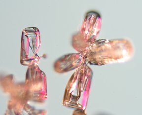
A rare doublet 2.9 mm wide. |
|
Winter 2002 - 2003 By E.M. Kinsman, USA |
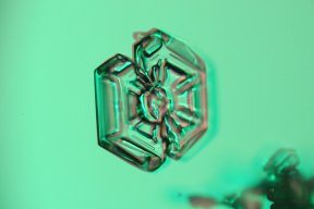
A rare doublet 2.9 mm wide. |
For the past ten years I have been an avid snowflake photographer. I had developed a number of illumination systems to try out in the winter of 2001-2002, but the winter was one of the
warmest on record and did not supply even one snow storm suitable for photography. This winter (2002-2003) has been one of the coolest on record and supplied some 20 storms suitable for photography. With the cooperation of this great winter, I was able to test out some of my illumination ideas, and I would like to share some of the resulting images below.There are several procedures that must be followed to collect good snowflake images. Technically snowflakes are called snow crystals.
The equipment has to be stored in a place out of the snow, but within easy reach of the snow crystals and be ready to go at a moments notice. The equipment must also be kept at the same temperature as the outside temperature. I keep my equipment in the garage and leave the door open when a storm is approaching. I will not collect images unless the temperature is below 25F.
The snow crystals are allowed to fall on a black sheet of paper then picked up with a pin and transferred to a very clean microscope slide. Since a snowstorm is really a storm or particles, it is not uncommon for little bits of dust and organic material to fall on the slide while you are collecting snow crystals. Another problem is the magnification, I use three main objectives on a microscope to collect the images - a 2x, 4x and a 10x. Depending on the size of the snow crystal the objective can be switched - this has problems with changing the illumination to a new technique. An ideal system would have three microscopes with three cameras and three illumination systems.
This winter I have used two cameras, a OM2N Olympus 35 mm camera for film collection and a digital Canon D60 with a 6.3 megapixel array. The digital camera has the advantage of being able to preview the shots much quicker and verifying if the shot is correct. Without going into the advantage of each system, most of my photography this year has been with the Cannon D60.
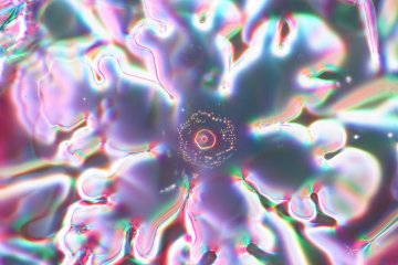
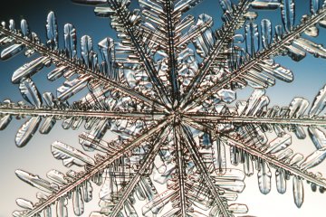
This is a 2.9 mm wide view of a spatial dendrite illuminated by a 5 color LED substage illuminator. This technique allows each LED to be on a variable resistor so that the color can be changed, but the LED generates too much heat so close to the crystal and will melt the crystal in only a few seconds. The LEDs were replaced by fiber optics for a much cooler light source.
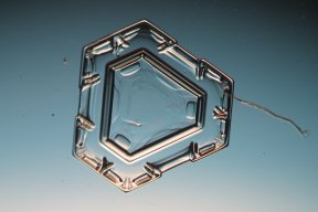
This night (Jan 27, 2003) we had a storm of 7 mm snowflakes and my equipment was set to up photograph a 2.9 mm field of view - this got me thinking about building a system for lower magnification shots.A 4 mm field of view. The illumination is a graduated blue filter. This image, like several below show the problems of photographing snow crystals. The crystals can range in size from 1 mm (or smaller) to huge crystals like this 4 mm crystal. It would be good to have an even lower power objective for these huge crystals.
The center of a stellar dendrite with a 10X objective. This image is 1.4 mm wide.
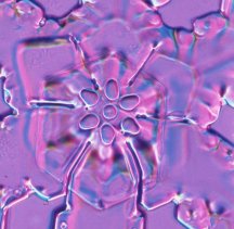
A 1.4 mm wide field of view - this is the center of a spatial dendrite, the illumination is a fiber optic multi-colored sub-stage source. I particularly like these close ups that show the six sided symmetry of the bubbles that form in the crystals.
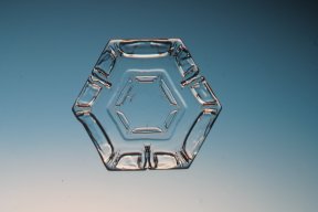
Both of these images are a 1.4 mm field of view. These platelets are quite flat and allows the whole crystal to be in view. Note the organic material on the crystal on the right. Snowflakes have an electrical charge and will attract dust, other snowflakes, and in this case what looks like a fragment of a plant.
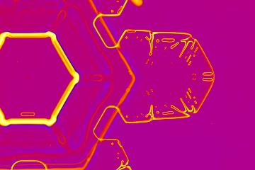
A huge platelet is photographed with a 1.4 mm field of view.
The image has false color applied to it.
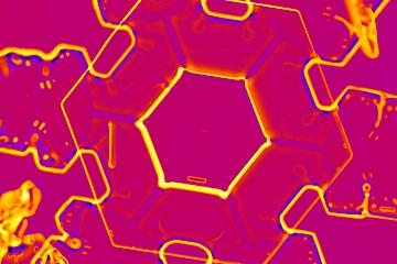
Same snowflake as above, but the central portion. This image is also false color.
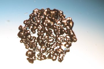
A 2.9 mm field of view of a large platelet that has fallen through a layer of rime ice. The super-cooled droplets of water instantly freeze to any crystal that comes into contact with them. These types of crystals are often associated with heavy accumulations.
A group of columns with mixed illumination, 1.4 mm wide image.
A single platelet (side view) image is 1.4 mm wide.

A 2.9 wide field of view of a stellar dendrite crystal with a fiber optic colored illuminator.

The author observing a few stellar dendrites while waiting for conditions to cool
to a point where photography can start.
Please note the above images are all copyrighted by Kinsman Physics Productions.
All comments to the author Ted Kinsman are welcomed.
Editor's note: The author's previous Micscape articles on snow crystals are:
Photographing snowflakes
Snow crystal photography
Please report any Web problems or offer general comments to the Micscape Editor.
Micscape is the on-line monthly magazine of the Microscopy UK website at Microscopy-UK