SUCTORIA PART II 
by Jean-Marie Cavanihac, France
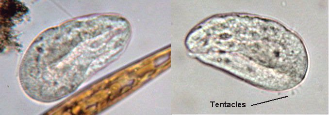
Young suctorians have cilia but they lose them when they become sessile. For this reason they are classified as CILIOPHORA / PHYLLOPHARYNGEA. These keywords are useful for searches on the Web. Unfortunately they are difficult to identify in this early state. 
Indeed, one of the most intriguing things about them is their reproductive process. If suctorians are classified as ciliates, it's because they can produce several ciliated offspring, for example by budding and also because they possess both a macronucleus and a micronucleus like ciliates.
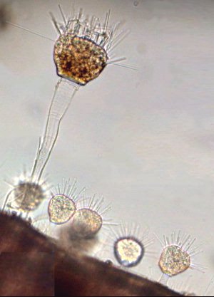
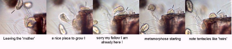
Another sub-species which I had collected, but without a lorica this time, illustrates perfectly the reproduction process. Some individuals were attached to a Bugula and one of them was budding. Click HERE to see an animation (time lapse sequence original duration 15 min) which shows budding and release of the 'youngsters'. 
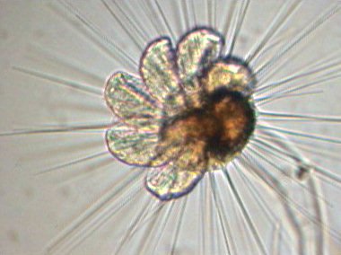
This sort of flower is the 'mother' suctoria and the 'petals' are the young larvae, which are ready to leave the mother. In real time, it was possible to see cilia beginning to move on each larva.
The young shown below look like ciliated protozoa (but move more slowly) and have a short existence in this form. (The picture was taken with a x15 objective; when they were examined with a x40 objective the detail of two individuals seem to have a 'folded' structure). 

After finding a suitable substratum, they lose the cilia and metamorphose immediately into adults.

But it was time to eat and I left the slide in place and the young to live their life (a microscopist too, must eat from time to time...!) Half an hour later, I observed the image below: now the 'mother' was surrounded by young adults growing rapidly! 
I searched for another specimen still in the budding stage, waited for larvae to be released and I took some pictures assembled below like a cartoon. 

Then I took a second lapse time sequence showing metamorphosis of one of the ciliated larvae which is arrowed in the 'comic strip' below. An hour later, the young looked like adults. (See picture at left-hand side below.)

Note the speed of their growth. They behave almost like seeds falling from a tree, sprouting immediately and making bushes under it!

Time lapse sequence: original duration 20 min showing growth of the individual arrowed in the 'comic strip' above.
It was earlier in this winter when I sampled these specimens in sea water. The sea temperature was around 14-15°C. Maybe it's the thermal shock caused by rapid change of sample temperature (when I came home) which prompted this extensive budding; the bottom of the Petri dish was covered by dozen of young. I think such a long observation (more than one hour on the same individual) has been possible because I have used white LED lighting. (For further info' on LED's, see this Micscape article). An incandescent bulb would have warmed too much the sample contained within a 3 mm high rectangular well slide (For further info' on well slides, read this Micscape article)

 
RETURN PAGE 1
Comments to the author Jean-Marie Cavanihac are welcomed.

Microscopy UK Front Page
Micscape Magazine
Article Library


All drawings and photographs © Jean-Marie Cavanihac 2002

Published in the March 2003 edition of Micscape Magazine.

Please report any Web problems or offer general comments to the Micscape Editor,
via the contact on current Micscape Index. 

Micscape is the on-line monthly magazine of the Microscopy UK web
site at Microscopy-UK

WIDTH=1


© Onview.net Ltd, Microscopy-UK, and all contributors 1995 onwards. All rights reserved. Main site is at www.microscopy-uk.org.uk with full mirror at www.microscopy-uk.net.