Topical Tip: The versatile 'light panel'
compiled by David Walker
The
small light boxes designed for viewing 35mm slides can prove very useful to the
microscopy enthusiast and amateur naturalist and both my brother Ian and I
use
them a lot. The older designs are quite deep
but a more
recent variant of it, the 'Light panel' is a mains or battery operated version
that is only 1.4 cm high and this is proving even more versatile; a selection
of uses from Ian's and my own experiences are shared below. It costs about Ł25
from suppliers like Jessops
(UK).
Update January 2017. With the demise of film, Jessops no longer stock the fluorescent tube style. LED versions e.g. the Medalight LP-100N are now available e.g. ProcessUK currently sell one for ca. Ł34 and are also sold on eBay UK. This may still be a more cost effective option than a dedicated microscope maker's base lighting for e.g. stereo microscopes.
Advantages of cold fluorescent light boxes as a light source:
Low voltage, either from 6xAAA batteries or suitable mains low voltage power adapter (the dealers will also usually offer a suitable power supply for about Ł12). This is safer than direct mains voltage driven lamps where water may be around e.g studying pond life and for youngsters. (There is usually an inverter internally to generate a higher voltage for the fluorescent lamp but is well away from the user.)
White light - as it is designed for viewing 35 mm slides it provides an excellent daylight balanced light, both for glare free viewing or for photo work.
Cold light source - useful for viewing live organisms.
Large structureless light for pleasing background - useful in low magnification settings.
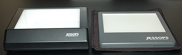
Features
The typical current design is shown left below and provides a 13 x 10 cm viewing area but is 4 cm deep. This is useful when the light source needs to be put on its side as a self supporting source but less useful for other applications. The more recent 'light panel', in this case the Jessops' LP45 shown right below, offers the same viewing area but is only 1.4 cm deep (overall size 15.8 x 15 mm). A slip-on case is also supplied; handy if using as a portable light source in the field. Larger models are also available in the slimline design.
Typical uses
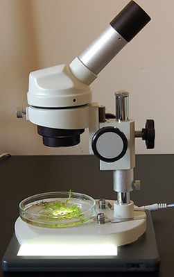
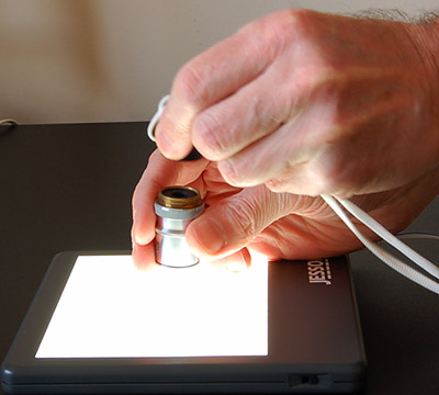
Base light for low power microscopes: Many of the cheaper and more expensive designs don't have a transmitted light source feature, but they usually have a removable disc to reveal a hole in the base. The smaller microscope models sit very well on the light panel to give a superb light source, as shown below for this 20x fixed mag microscope. Incidentally I think this set-up is close to ideal as a first microscope for a youngster, either in its battery powered form or using a mains low voltage supply as it's a safer system than using direct mains voltage. If required the illuminated area can be decreased to just beyond the objective's field of view with a card disc with hole cut out placed in the base aperture.
Superior cold light source for stereo microscopes with built-in light source: Many stereo microscopes do have a transmitted light source built in, but although a lot of care is often given to the mechanics and optics, the transmitted light source can often be quite poor. For example, the popular Meiji EMZ stereo series shown here with the PBH base has a small quartz halogen light that requires a lot of diffusing to give an acceptably even off-white light. The light panel sits nicely on the stereo scope base with not much increase in thickness to give a superior light. A few pieces of Blu-Tak stop the panel from slipping. (The intensity knob top right below has been taken off for the light panel to fit the stand exactly but there's only a small overhang at the front without doing this.)
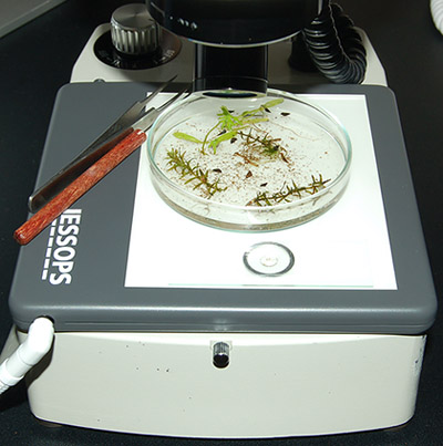
The larger backlit area provides an extra working area for transferring selected critters from e.g. a pond water sample onto a microscope slide for study with a compound microscope.
The light is also cold, unlike the halogen bulb, which allows extended viewing of live critters without distressing them.
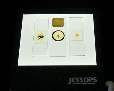
Photographing subjects requiring transmitted light: Such subjects require a structureless good quality white light as background and the light box can be very suitable for this. Large subjects on microscope slides for example may require a camera with a macro attachment rather than a microscope such as the whole insect slides below. (I believe light boxes may not provide a true white light spectrum but I've never noticed any colour balance problems with it; the photo below was photographed on the Auto White Balance setting of a DSLR although many cameras do have a fluorescent setting.)
Inspecting optics for damage or delamination: A light box can be useful for checking optics for major faults like delamination, haziness or cracks off the microscope (first image below). Studying the optical planes carefully from the rear over a light box with a 5x or 10x hand lens or low power microscope will often reveal most major faults (this can be more effective in fact than using a phase telescope on a microscope as some telescope designs don't focus fully through all the optical planes). Illuminated white paper as a light source is another way.
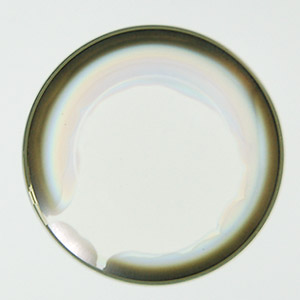
Left
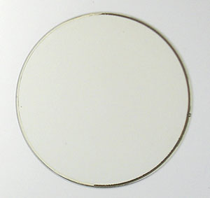
above: doublet of delaminated Zeiss 10x Kpl W eyepiece on light panel. Right
above: same doublet, two months later after the 'accidental fix' below.
(The
overall very slight yellow cast of glass is seen on good objectives as
well.)
The
doublet from a Zeiss 10x Kpl widefield eyepiece above left shows extensive delamination
of the doublet cement when placed on the light box giving a marked brown darkening
over 30% of the field edge when in use. (As
an aside as these are very large elements, unlike in an objective, I thought
I'd try fixing it. I tried curing this by letting immersion oil seep into the
side by capillary action, this didn't work, it only penetrated a millimetre
or so. Then decided to try an extended acetone soak for a few days to split the
elements to reglue with Canada Balsam. The acetone didn't seem to be touching the cement,
but when completely dried the delamination had completely gone, not the faintest
trace.
I'm not sure how; my two theories are either that residual immersion
oil may have been driven deep into the doublet by the acetone, or the acetone has
slightly dissolved the original cement and healed it. I'm not sure if it's
a permanent fix but works a treat at present.)
Update:
After two months in a warm room the doublet of the eyepiece still seems fine,
above right. This was carried out on a 'nothing to lose' $20 eyepiece but would
be wary of trying this on anything that is irreplaceable!
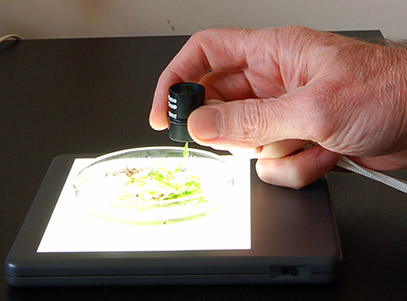
Studying subjects at low power if a stereo microscope not available e.g. in the field: If a stereo microscope isn't owned or it's needed to study pond life samples for example in the field, the light box (battery powered if required) with a hand lens can give excellent views of, for example, a sample in a Petri dish.
Other uses of a light box: Light source for compound microscope and mimic of the old edge of flame effect. My brother Ian has used a light box a lot as a light source for an old Victorian microscope with mirror. The glare free light and daylight balanced white is more than bright enough up to medium powers and particularly useful for providing a large structureless light source in critical illumination mode for the very low power obectives often supplied with Victorian microscopes. See Ian's article Experiments with a simple light source. Revisiting an old illumination technique for details where he also recreates the modern analogue of the paraffin flame microscope lamp by making a cardboard slit mask for the box. The light box models thick enough to place on their side may be more useful in this instance.
Comments to the compiler David Walker are welcomed.
Published in the March 2007 edition of Micscape.
Please report any Web problems or offer general comments to the Micscape Editor .
Micscape is the on-line monthly magazine of the Microscopy UK web site at Microscopy-UK
© Onview.net Ltd, Microscopy-UK, and all contributors 1995
onwards. All rights reserved.
Main site is
at www.microscopy-uk.org.uk
with full mirror
at www.microscopy-uk.net
.