Stereoscopic imaging with a normal compound microscope 3D microscopy by Wim van Egmond, the Netherlands |
It is already a long time ago since I started experimenting with stereoscopic imaging through the microscope. In one of my first Micscape articles I described some methods that can be used to create three dimensional images with an ordinary microscope. Stereomicroscopes do exist but they have a limited magnification. I published some examples of 3D images through the microscope. Since there was not much purpose for these images I only made a small series and put my attention to other techniques. However, recent technical developments and the introduction of the digital camera persuaded me to make a new dive into 3D microscopy. I discovered some promising methods to create 3D images with high magnifications. In this little article I want to show some of my recent results. The images on this page are presented as red and blue anaglyphs and they have to be seen with special glasses. But in case you don't have these please go to the cross-eyed stereo version of this page. | 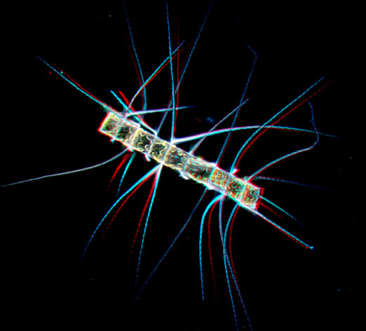 |
Digital imaging makes it much easier to use the method of 'stacking'; combining images with different focus layers. One of the problems with high magnification photography is that the stronger the magnification, the more shallow the depth of field becomes. With special stacking software like Helicon focus, and CombineZ and orthers it is possible to combine different focus layers and create an image with every part of the subject in focus. Some of these applications, like Helicon Focus, already have a function to create 3D images from these stacks but unfortunately for me that is only available in the PC version. Since I use a Mac computer I had to find my own way of creating stereo images from stacks. |
|
 |
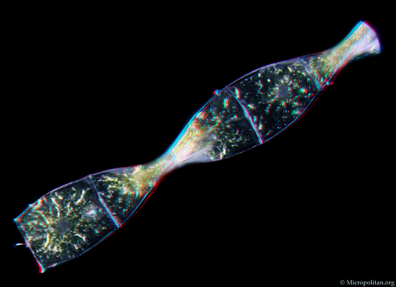
This
image of radiolaria was my first try to make a 3D image using
stacking software. My method was simple. Similar to my original
method I used oblique illumination. Using Helicon focus I processed
the stack twice. Once
with
the horizontal
position adjustment set to 0 percent and a second stack with
the adjustment set to one percent. This resulted in two images
that could
be used
for
a stereo
image.
|
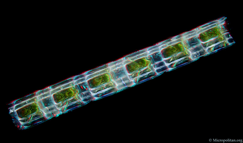
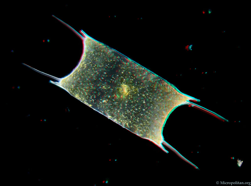
The following series of images shows diatoms, single celled organisms that can be found in marine plankton. These are all living organisms. From top to bottom we see the colonial Lithodesmium intricatum, photographed in darkfield with a 25X objective. The second image is Odontella sinensis (16X objective), the third image is of a helical colony of Helicotheca tamesis. The bottom image shows the strangely curved frustule of the diatom Actinoptychus senarius, photographed with a 40X objective. |
|
|
|
|
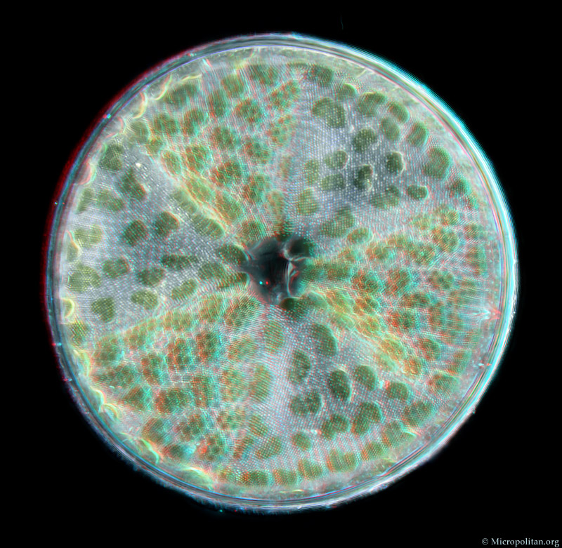
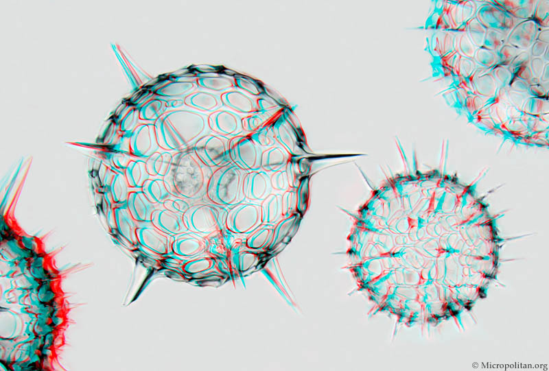
Diatoms are single celled algae that have a shell (the so-called frustule) that is made of silica, so they literally have glass houses. When the living tissues are removed from the cells (with chemicals) the structure and texture of the frustule can be observed in high detail. |
The images on this page are presented as red and blue anaglyphs and they have to be seen with special glasses. But in case you don't have these please go to the cross-eyed stereo version of this page. |
Comments to the author Wim van Egmond are welcomed.
Visit the Micropolitan Museum
Microscopy UK Front Page
Micscape Magazine
Article Library
all material © Wim van Egmond