Stereoscopic imaging with a normal compound microscope Page 2 by Wim van Egmond, the Netherlands |
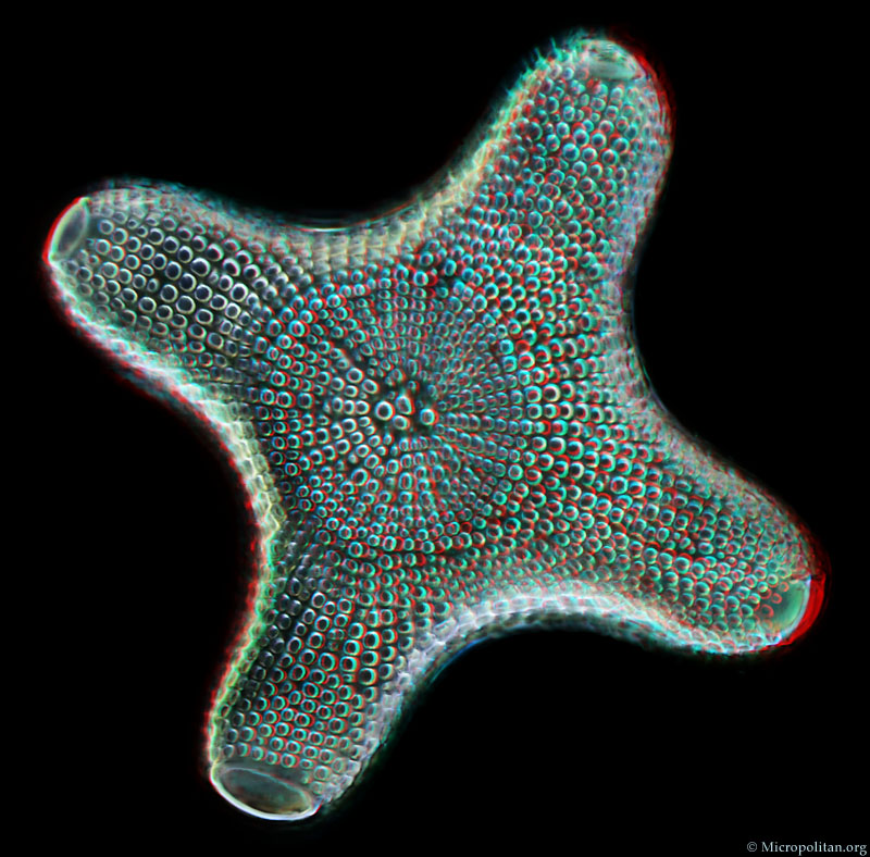
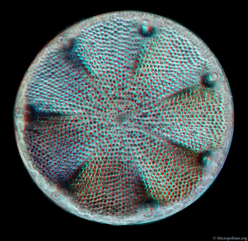
Cleaned diatoms, from a mounted slide made by diatom specialist Klaus Kemp, photographed with a 40X objective. Diatoms are single celled algae that have a shell (the so called frustule) made of silica, they literally have glass houses. When the living tissues are removed from the cells (with chemicals) the structure and texture of the frustule can be observed in high detail. |
|
|
|
|
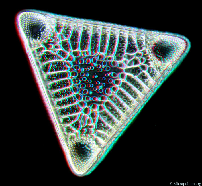
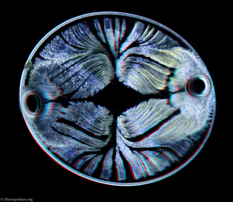
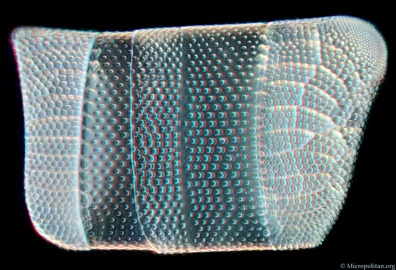
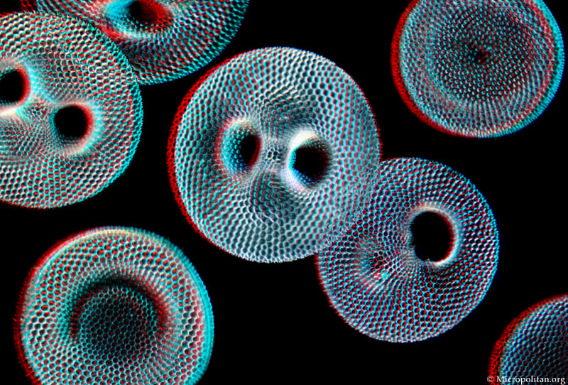
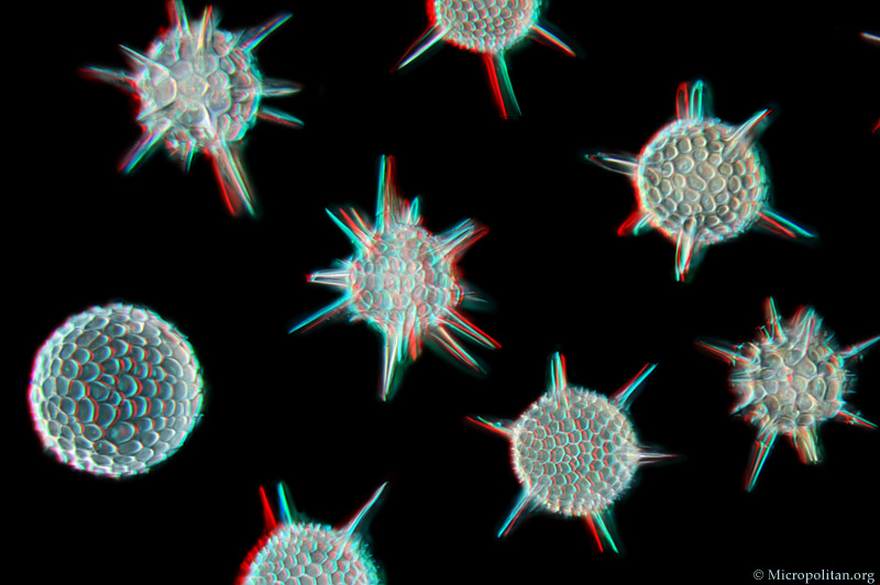
The following images were made from mounted slides I made of fossil diatoms from Barbados, and radiolarians from the same location. The objective used was 16X.The images on this page are presented as red and blue anaglyphs and they have to be seen with special glasses. But in case you don't have these please go to the cross-eyed stereo version of this page. |
|
|
The images on this page are presented as red and blue anaglyphs and they have to be seen with special glasses. But in case you don't have these please go to the cross-eyed stereo version of this page. |
Comments to the author Wim van Egmond are welcomed.
Visit the Micropolitan Museum
Microscopy UK Front Page
Micscape Magazine
Article Library
all material © Wim van Egmond