|
A "palette of palates". by David Walker, UK |
Please excuse the main title but I couldn't resist, although a Google search reveals that it's not original, it has been normally used in a food or drink context.
Prepared slides of the feeding organ of certain molluscs are quite common and were a popular Victorian subject because of their attractive features under the microscope, especially with polarised lighting. A variety of names have been used for this structure in the past which include 'tongue', 'lingual band', 'odontophore', 'palate' (1). Some writers have remarked that some of these names aren't strictly correct biologically (eg ref. 5) and 'radula' from the Latin radere 'to scrape' (2) now seems the most commonly accepted description. 'Odontophore' more correctly refers to the supporting structure of the radula (ref. 2a). Wikipedia entries (3, 3a) note that Alexander von Middendorff is generally credited with establishing the term 'radula' in his monograph on molluscs (1848-9).
My own interest as a hobbyist in this type of slide is exploring their structures under different lighting conditions and their potential as attractive microscope subjects rather than a particular interest in molluscs or their anatomy. There are some splendid online illustrated resources describing how the radula is used by different molluscs (e.g. 3) and its importance in their classification. Many of the classic Victorian microscopy texts have extended entries on this feeding organ (commonly referring to it as the 'tongue'), these include Carpenter (6), Hogg (7), Gosse (8) and Kerr 1905 (1). Hogg has a splendid plate of coloured line drawings of 16 different radulas. Protocols for preparing permanent slides of mollusc radula include those by Hogg (7) and Quekett (9).
A selection of other interesting references found on aspects of radulas, their structure and composition are given in refs. 11-14. (All are available online to freely download.)
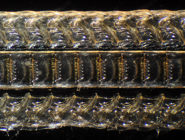
The slides: The author's complete collection is shown below and present a selection of images of some of these slides using different lighting techniques. Using the standard 3x1 inch slide size as a guide, the large size of the radula in some species is apparent.
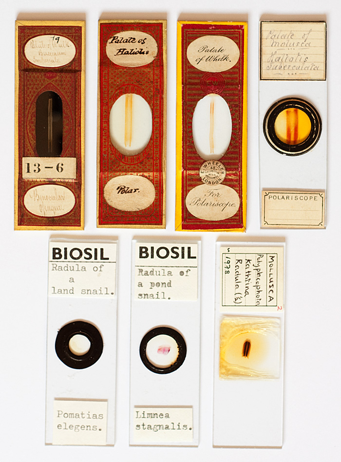
Top row: The author's earliest examples; all use the label 'palate'. The slide for incident lighting (top left) is my favourite, by mounter 'J&TJ' (see ref. 10 for a paper on the identity of this mounter). The second is by an unnamed mounter and the 3rd left by Wheeler. The three for transmitted work suggest the use of polarising light.
Second row: Two modern examples by Biosil (John Wells). The far right slide was prepared by my brother in law John Atkinson during a biology teaching course.
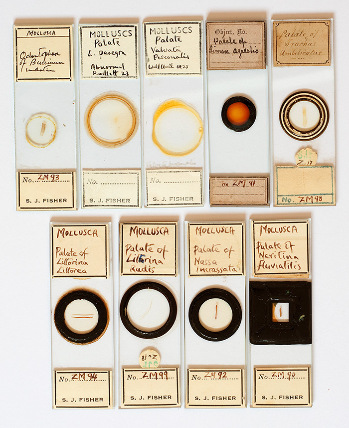
These slides were bought as a set from Brunel Microscopes and are all labelled 'S J Fisher' and/or 'S.J.F', except 4th slide top row. Bracegirdle has an entry for this mounter in his invaluable book on slide mounters which states that Fisher lived from 1927-1984 (4). This 20th century mounter preferred the label 'palate' or 'odontophore'. Each slide has a 'ZM' coding which I suspect stands for Zoology - Molluscs' as a slide labelled 'S J Fisher' and a 'ZI' code showing an Insect mount is illustrated in Bracegirdle's book (4). The two slides top right seem to have a different handwriting style to the others, so unclear which, if any, S J Fisher made himself. Especially as Bracegirdle's example slide shows S J Fisher's own labelling of a slide prepared by 'Flatters and Garnet' (which Bracegirdle notes in the slide caption).
Image details:
Microscope: Zeiss Photomicroscope III using Zeiss 10X Kpl eyepiece on short collar for projection.
Camera: Nikon D5000 DSLR. The author is currently assessing this 'entry level' model from Nikontheir first DSLR combining an articulated screen, Live View and recordable video including HD video. A review of this camera's suitability for still and video photomicroscopy will appear in Micscape at a later date; it is reported to have essentially the same sensor as the mid-range D300 for a third of the cost.
Images: All at ISO200 unless stated. Typical exposures for transmitted light techniques 1/5th sec. Auto white balance. Full image frame used, resized, minor levels adjust, no colour or sharpness adjust.
Images and comments on lighting techniques
(HFW = horizontal field width in mm)
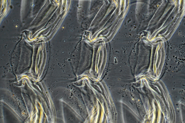
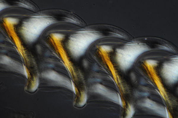
Palate of haliotis (unnamed mounter) - crossed polars with lambda plate: Simple structure towards the centre are well presented but can be hard to interpret the complex edge structures. Suppliers like Knight Optical (UK) sell lambda tint plates over a small range of wavelengths, this plate with blue tint I often prefer to the more traditional rose red tint. Unlike autofluorescence, see later, any detritus in the subject is clearly seen if artistic imagery is being sought. Zeiss 2.5/0.08 planachro' objective. HFW=3.1 mm.
Palate of haliotis (unnamed mounter) - phase: A closer look at the simple structures towards the centre where phase worked better. Zeiss 10/0.22 achromatic phase objective. Again detritus is emphasised. HFW=0.82 mm.
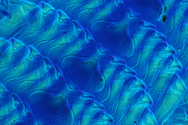
Palate of haliotis (unnamed mounter) - darkfield and crossed polars: Darkfield with complex radula gave busy images, adding crossed polars defined the structures to some extent but not very successful at this low power. Zeiss 2.5x planachro. The 'D' setting on the Zeiss long working distance condenser with top element off to give NA0.32 gives good darkfield with the lowest power objectives. HFW=3.1 mm.
Pond snail. Limnea stagnalis (Biosil) phase + crossed polars: I sometimes preferred this mixed technique imagery to phase or polars alone, this one with a lambda tint plate. Note the large difference in number of 'teeth' on the radula compared with e.g. the whelk shown below. The older books especially state the teeth number for various species and remark on the orders of magnitude variation in number. Zeiss 10/0.22 achromatic phase objective. HFW=0.82 mm.
'Teeth' on selected mollusc radula (estimates by Kerr. ref. 1):
Common whelk (Buccinum undatum) - 250
Common periwinkle (Littorina littorea) - 3500
English limpet (Patella vulgata) - '1920 glassy hooks in 160 rows of 12 teeth each', Kerr also notes the radula is 'longer than the shell itself'.
Helix pomatia (Roman or edible snail) - 21 000
Large garden slug (Limax maximus) - 26 000, Kerr notes that 'A single tooth measures only the 10,000th of an inch!'.
Umbrella mediterranea and Umbrella indica, Kerr notes 'They seem to baffle calculation. Possibly 750,000 may be somewhere near the truth.'
Polyplacophora, Kathrina - brightfield: Kathrina is a Chiton mollusc. In many radula slides which the author owns, the 'teeth' seem to be visibly of similar composition to the supporting structure but not in this example where they are opaque but uncertain of their composition. Zeiss 2.5/0.08 planachro' objective with 2x on Optovar. HFW=2.0 mm.
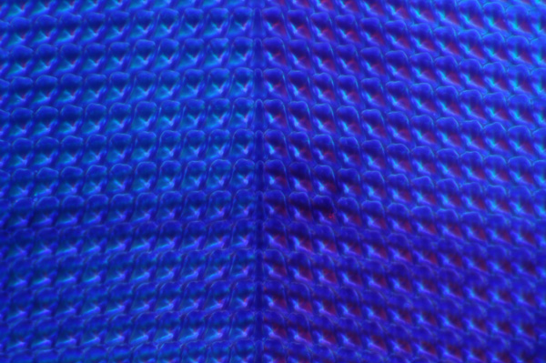
Polyplacophora, Kathrina - crossed polars: The polars with lambda tint plate offer attractive imagery but don't think it aids the study of the support structures. Zeiss 2.5/0.08 planachro' objective with 2x on Optovar. HFW=2.0 mm.
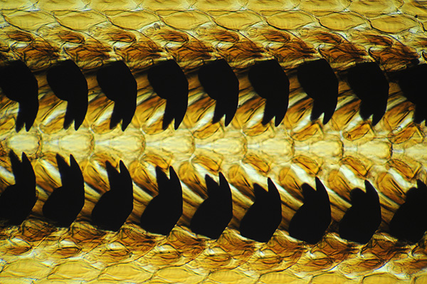
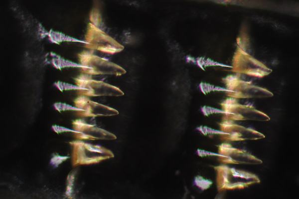
Trochus umbilicatus (labelled 'SJF' for S J Fisher) - darkfield: Some radula had very complex structures, visually some idea of their construction can be gleaned by repeated focussing through the specimen using a range of lighting techniques but single shot, single focus plane imagery was not very satifactory. Zeiss 2.5/0.08 planachro' objective with 2x on Optovar. HFW=2.0 mm.
Trochus umbilicatus (labelled 'SJF' for S J Fisher) - crossed polars: Polars with lambda tint plate helped to delineate some of the structure which differed in their birefringence, particularly the central detail cf darkfield above. Zeiss 2.5/0.08 planachro' objective. HFW=2.0 mm.
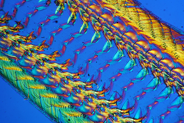
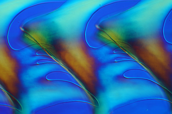
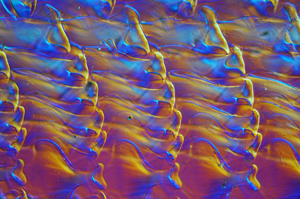
Palate of whelk (Wheeler) - Nomarski interference contrast: The birefringency of a radula coupled with DIC's ability to reveal low contrast structure can give attractive effects. A lambda plate was also used. Zeiss 16/0.35 planachro objective. HFW=0.51 mm.
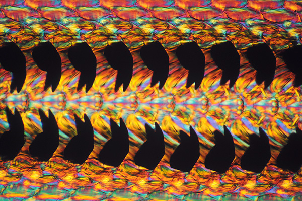
Palate of whelk (Wheeler) - Phase and crossed polars: Phase alone often gave busy hard to interpret images with more complex radula. Here combined with crossed polars shows fine structure along teeth edges and modelling with the birefringent effects. Zeiss 10/0.22 achromatic objective. HFW=0.82 mm.(On the camera front the top notch sensor on the D5000 copes well with this tricky range of shades from almost total black to textured off white.)
Trochus umbilicatus (labelled 'SJF' for S J Fisher) - Nomarski interference contrast: With lambda tint plate. Close ups of aspects of the radula structure can give interesting imagery. Zeiss 16/0.35 planachro objective. HFW=0.51 mm.
Trochus umbilicatus (labelled 'SJF' for S J Fisher) - phase and crossed polars: With lambda tint plate. Interesting artistic imagery can be explored but again detritus spoils the effect. Zeiss 16/0.40 Neofluar phase objective. HFW=0.51 mm.
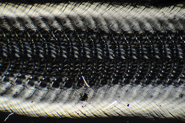
'Palate of whelk'. Victorian dry mounted slide by 'J&TJ' - off-axis incident light: The paper label states 'Binocular Opaque'. A LOMO 6V 15W lamp was used at full intensity at a near right angle to long axis of radula. The high contrast and often specular reflections didn't seem very effective for studying the structure. Zeiss 2.5/0.08 planachro' objective. HFW=2.0 mm.
Axial brightfield epi with the III RS head hasn't been successful to date because of the glare from the coverslip, even with polars to reduce this. Dry mounts of uncovered radulas may be interesting subjects to study with brightfield epi both axial and off axis.
'Palate of whelk'. Victorian dry mounted slide with opaque background by 'J&TJ' - off-axis incident light: The observer needs to be wary of potential artefacts from covered objects such as this in strong incident lighting. When the slide was rotated so that the light fell along the long axis, an apparently new set of 'teeth' emerged. They may be a secondary image of the true teeth on the coverslip underside as they disappear as rotate the slide (and are severely suppressed with polarised incident light). Zeiss 6.3/0.16 planachro' objective. HFW=1.3 mm.
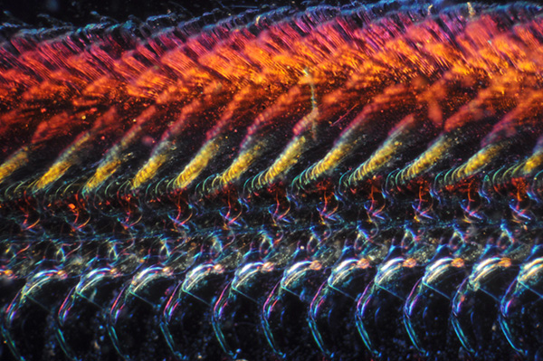
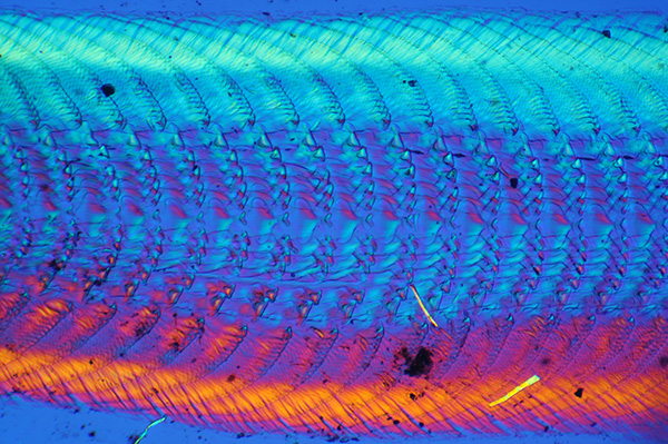
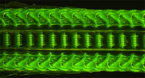
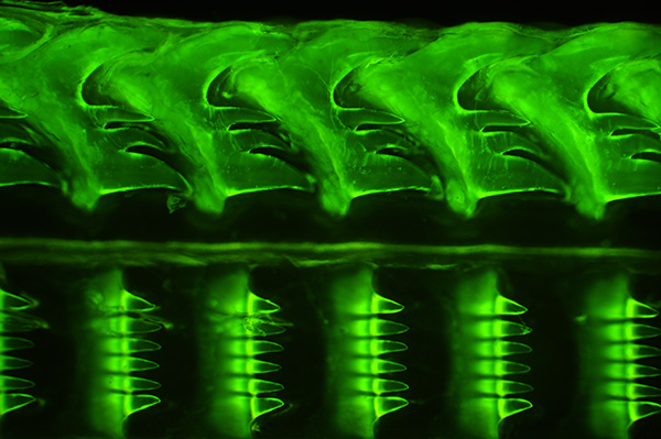
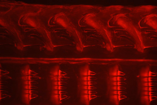
Autofluorescence: In an earlier 'Forays into Fluorescence' article I explored my prepared slide collection to see what sort of subjects autofluoresced. Many subjects could not be studied because of the fluorescence of the mountant, usually Canada Balsam. The radula slides shown here also suffered from this, except the dry mounted slide by 'J&TJ" for incident light. This gave a broad band fluorescence with green, blue or violet / UV light and gave an attractive three dimensional effect with surface detail of the radula. Radulae are generally described as being a 'horny' and/or 'toothed chitinous ribbon' (3,6) but have found very little data on the excitation and emission spectra of chitin structures (any information welcomed). Whether this broad band emission was due to the radula and/or how it had been treated, I'm uncertain but is now one of my reference slides for autofluorescence.
'Palate of whelk'. Victorian dry mounted slide by 'J&TJ' - autofluorescence: Compared with the images from brightfield incident light above, these are much more successful images. Images of this radula were first presented in 'Forays into fluorescence 2' taken using a Leitz Diaplan, with mercury arc 100W lamp and Ploemopak head (now sold) and Nikon D300 DSLR. They have been retaken here on the Zeiss PMIII stand with renovated Zeiss III RS epi head.
A mercury lamp isn't vital for blue and green excitation if subject autofluoresces brightly, a 100W quartz halogen lamp gave exposures about 3-4 stops slower and the visual imagery from blue excitation although not as bright can easily studied by eye in normal room lighting with a 6.3x objective.
Zeiss 2.5x NA0.08 objective. Blue excitation. Exposure 6 secs ISO800. HFW=3.1 mm.
Leitz 6.3x NA0.20 objective. Blue excitation. Exposure 3 sec ISO400. HFW=0.13 mm.
(As brightness of epi fluorescence is to the fourth power of NA, using an NA0.20 Leitz objective rather than the Zeiss 6.3X NA0.16 gives a 1 stop shorter exposure.
Zeiss 6.3x NA0.30 objective. Green excitation. Exposure 16 secs ISO400. HFW=0.13 mm.
Comments to date: Prepared slides of the radulas of molluscs, both older and more modern examples, are widely available and can offer rewarding studies under the microscope both visually and for imaging. Because of their often three dimensionality, complex structure, semi-transparency and birefringence they offer a challenge to explore with single or mixed lighting techniques. I've yet to make Rheinberg discs for my microscope so this technique wasn't tried in the above examples or image stacking but either could well be of benefit.
The enthusiast interested in preparing their own slides could try some of the published protocols as some of the classic species are common molluscs and the organ is often quite large, often tens of millimeters in length and/or width.
It's a pity that I only possessed one dry mount as the autofluorescence seemed to give the most convincing imagery on the whelk's radula three dimensionality and was interested in exploring further the nature of the autofluorescence between species/mounts. This technique also suppresses detritus in the mounta disadvantage for artistic imagery in many of the author's examples.
Comments to David Walker welcome.
Acknowledgments: Thank you to Brian Stevenson for informing me of reference 10 and for providing a copy.
References
1) R. Kerr, 'Nature Through Microscope and Camera', London, 1905, pp. 57-63. Two photomicrograph plates.
2) 'The New Oxford Dictionary of English', Editor J Pearsall, Clarendon Press, 1998, 'radula' entry. The entry also notes that its 'origin' was 'late 19th cent.'.
2a) as above, 'odontophore' entry.
3) Wikipedia entry 'radula' and 3a) 'Alexander von Middendorff'.
4) B. Bracegirdle, 'Microscopical Mounts and Mounters', Quekett Microscopical Club, 1998, London, entry 'FISHER, S. J.' p. 38, and Plate 16, slide N.
5) E W Bowell, 'Radulae of Mollusca', Journal of the Quekett Microscopical Club, 1924, vol. XV (series 2), pp. 57-64 and Plate 1.
6) W B Carpenter, 'The Microscope and its Revelations', 1857, 2nd edition, pp. 559-563. Includes five figures. Also other editions.
7) J Hogg, 'The Microscope. Its history, construction, and application', 1898, 15th edition, p. 554-558. Plate V. Also other editions. (Plate V is less well reproduced in some earlier editions.)
8) P H Gosse, 'Evenings at the Microscope ...', 1859, pp. 55-60.
9) J Quekett, 'A Practical Treatise on the Use of the Microscope', 1848, p. 418-420. (Fascimile edition published by the Science Heritage Library.)10) S Warren, 'Letters to G A Walker Arnott from J & T Jones', Quekett Journal of Microscopy, 2002, vol 39, 379 - 390.
11) I B J Sollas, 'The Molluscan Radula: its Chemical Composition and Some Points in its development', Quarterly Journal of Microscopical Science, 1907, vol. 51, pp. 115-136 and Plate 9.
12) J E Gray, 'On the Tongues of Mollusca', Transactions of the Microscopical Society, 1853, Vol. I New Series, pp. 170-177.
13) J Hogg, 'The Lingual Membrane of Mollusca, and its Value in Classification', Transactions of the Royal Microscopical Society, 1868, Vol. XVI New Series, pp. 93-104, Plates VIII-XI.
14) S P Woodward, 'A Manual of the Mollusca', 1880, 4th edition. 'Digestive system' pp. 20-23.
Microscopy UK Front Page
Micscape Magazine
Article Library
© Microscopy UK or their contributors.
Published in the March 2010 edition of Micscape Magazine.
Please report any Web problems or offer general comments to the Micscape Editor,
via the contact on current Micscape Index.Micscape is the on-line monthly magazine of the Microscopy UK web
site at Microscopy-UK
© Onview.net Ltd, Microscopy-UK, and all contributors 1995 onwards. All rights reserved. Main site is at www.microscopy-uk.org.uk with full mirror at www.microscopy-uk.net.