|
Exploring classic insect test objects for the optical microscope:
III - The wing scales of Morpho butterfly species - one of the oldest test objects but subject to intense modern research.
.. and revealing an, as yet unsolved, historical puzzle.
by David Walker,
UK
with prepared slide images by Brian Stevenson and Howard Lynk;
SEM images by Andrew Syred.
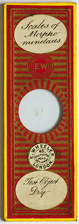
Right: A test slide by Wheeler ca. 1866-84. Author's slide.
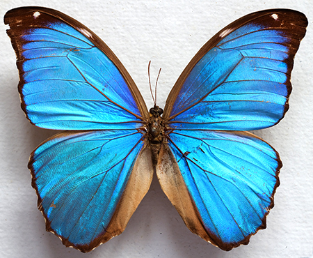
Morpho menelaus, a mounted and boxed early 20thC display item owned by the author.
In the Explorer Browser, setting Print to 60% prints out on 6 x A4 sheets.
|
|
Series
Introduction: Using
certain subjects as tests for microscope optics has a long history
and had a vital role in the optical development of the achromatic compound microscope in the early 19th century. Some species of diatoms are well documented as
tests and still very popular with enthusiasts, but using other subjects including animal hairs and various insect parts are
also long established.
A selection of test objects in the author's slide collection will be shared in this
occasional series in hope they are of interest. As others have
noted, given the potential variability of a subject between mounts
and the sometimes vague specification of the species, it's arguable
if insect parts can be regarded as reliable test objects. But it's
both great fun and instructive to inspect these slides while
consulting the old books and to repeat the studies they
recommended. The enthusiast nowadays can also try techniques
developed since the Victorians such as phase and interference
contrast if available.
Note on the butterfly specimens. All recent specimens were sourced from an accredited European Lepidoptera dealer, imported under EU regulations from captive breeding programmes in S. America, run by and for the local people.
Introduction
The Morpho genus contains some of the most striking species of large blue butterflies that would have no doubt attracted the attention of early naturalists visiting their habitats such as those in South America. The wing scales of the species Morpho menelaus had a particularly important role in the optical development of the microscope. It was one of the oldest (if not the first) 'test object' to be specifically described
by species in English by Dr. Goring in his 1827 paper (1). In this same work there is a key series of experiments described and illustrated using these wing scales showing how the angle of aperture of the light cone reaching the objective had a profound effect on the resolution of scale fine structure. These empirical observations were important in optics development with the underlying theory being formulated by Ernst Abbe in the 1870s (3).
As optics developed rapidly with the help of this and other test objects (4), the Morpho scales became a less demanding test and was largely superceded later in the 19th century by diatom frustules where the fine structure of some species can stretch the optical microscope to this day. However, there is an interesting modern twist to the study of Morpho genus scales
under the microscope. A cursory search with Google will find a large number of modern papers in the field of 'photonics' reporting studies of the scale ultrastructure of Morpho and other genera as revealed by the electron microscope. In 2014 there was a feature on some modern studies of Morpho on the BBC News website 'Butterfly wings inspire cosmetics and bomb detectors.'
Aspects of the study of the
scales from the 1820s to 21st century are presented below and how the historical puzzle arose.
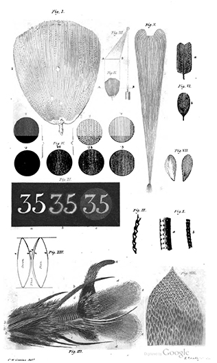
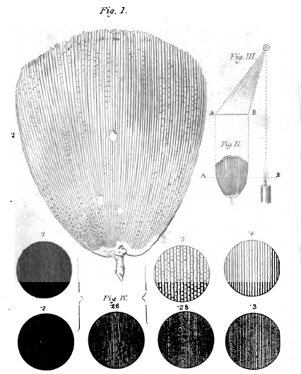
Left - Full plate accompanying part 1 and part 2 of Goring's 1827 papers showing a selection of test objects - insect scales, a moss leaf, watch face, fly ('blue bottle') foot and the improved achromatic objective (triplet) made by Mr. Tulley for Lister are shown. Lister drew the large scale. Part 1 only discussed the 'Menelaus' series of images, part 2 discussed the remaining subjects.
Right - Detail of same plate, showing a single 'Menelaus' scale and the effect of 'aperture' on 'penetrating power' (resolution in modern terms) of the scale structure. The discs read anticlockwise with aperture of .1, .2, .26, .28, .3, .4 and .5, the last being the maximum clear aperture (diameter of lens) used. Increasing aperture revealed first the coarser longitudinal structures but required the maximum aperture to show the cross structures. (Objective aperture nowadays
uses numerical aperture as defined by Abbe.)
A diagram (Fig. III) illustrating the off-axis transmitted light which enhanced observation of the finest scale structure is also shown.
|
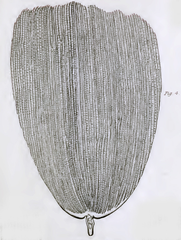
Right: A scale of Morpho Menelaus from Plate 4 in John Quekett's 'Practical Treatise on the Use of the Microscope', 1848.
(Surrounding parts of other subjects removed for clarity. Some fine detail lost because of the file compression of the copy on www.archive.org).
Quekett describes the object as a test in the extensive section on 'Test Objects'. But he notes:
"In former times it required a good quarter to exhibit the transverse striae ; but the half-inch, as now constructed, will show them readily."
(Objective focal lengths are being referred to, for a 10 inch tube length microscope, these would have a primary mag. of ca. 40X and 20X respectively.)
|
|
Examples of old slides, kindly supplied by Howard Lynk and Brian Stevenson
The Morpho butterfly was a popular permanent microscope slide subject from the 1830s through to the 20th century presenting either a fragment of the upper wing or strew of the scales. My own late 19th century example of a strew by Wheeler is shown in the header. I am indebted to Brian Stevenson and Howard Lynk for the images of their own examples shown below. Only the earliest example of a slide pair owned by Howard are shown here,
his other splendid examples
and imagery are presented in the accompanying article discussing the 'historical puzzle'.
By comparing the scale strews with their likely production date from Howard's, Brian's and my own example, there's some evidence for a trend of later strews using the lower wing scales (i.e. with their distinct toothed edges). Howard Lynk offers a plausible explanation: this potentially allows the maximum number of both wing and strew scales slides to be made from a possibly limited
supply of wings by a major slide maker. Using lower wing scales for the strews would allow the same part of a wing to be used for the upper wing fragment slide (although only the blue upper wing scales were described as tests by the early workers).
|
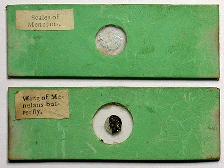
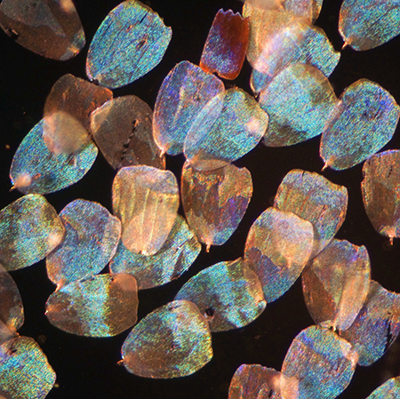
Above: Papered slides of both a scale strew and wing fragment. 'Tentatively identified as being from Cary/Gould ca. early 1830s, ... with mica slips'.
Right: Image of the above scales skillfully photographed using a mix of incident and transmitted light.
Images and notes in quotes courtesy of Howard Lynk.
|
|
|
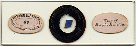
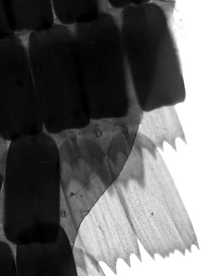
Above: A later 19th century slide by Samuel Stevens of a wing fragment.
Right: Detail of the wing edge. These scales do concur with the modern M. menelaus but not those of the early scale strews. This shows nicely how the scales on the upper surface are of a different shape to those on the lower, the latter being markedly toothed and dark brown.
Images courtesy of Brian Stevenson.
See Brian Stevenson's Micscape article on this mounter.
|
|
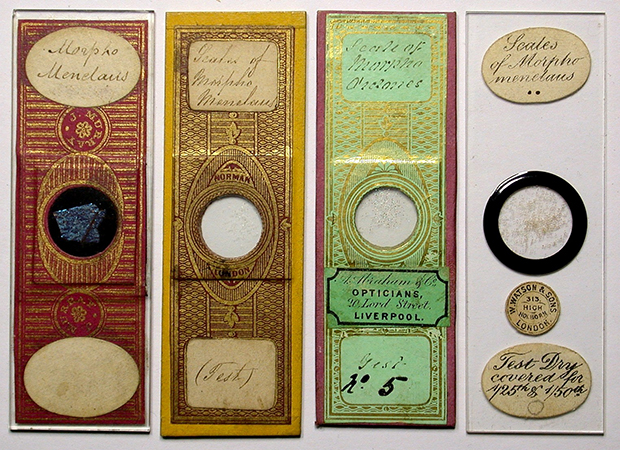
Above (left to right). A slide by J. Murray (wing fragment) 'guessing ca. 1860s' and two strew slides by J T Norman ('ca. 1850s-60s') - the third from left with 'secondary label of A. Abraham' and Watson (1892-1908 address label).
Images and date estimates courtesy of Howard Lynk.
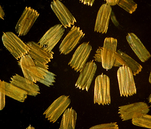
Right - scales from the Watson slide, lower wing scales only. Described as a 'test' slide on label despite the lower wing scales never being described as test scales by the early workers such as Goring, Pritchard or Quekett.
Images courtesy of Howard Lynk.
|
|
Wing scale structure across selected Morpho species and the effect on their perceived colour
The Wikipedia entry for the Morpho genus notes that there are 'over 29 accepted species and 147 accepted subspecies'. In addition to M. menelaus some other species are attracting particular attention in photophysics research, two, M. didius and M. rhetenor are compared with M. menelaus below. The whole wing photographs were taken at the same time with the same natural light from the side
although they don't show the subtleties as well as by examining real examples.
Some species have two distinct type of scales on the upper wing: the iridescent 'ground' scales responsible for the colour and the translucent 'cover' or 'glass' scales, The photo-physics of how the cover scales modify the interaction of light with the ground scales has been studied in depth in recent years by scientists.
The respective scale images below were all taken on a Zeiss Photomicroscope III using a Zeiss 16X NA0.4 Neofluar objective and Optovar set at 1.6X, with incident off-axis lighting from a 3 mm LED. The wings were held in a compressorium to reduce gross wing folding and permit an unstacked single shot image. The light source was
from the NE with respect to each scale image.
|
|
|
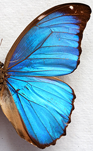
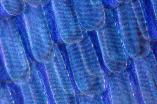
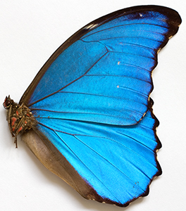
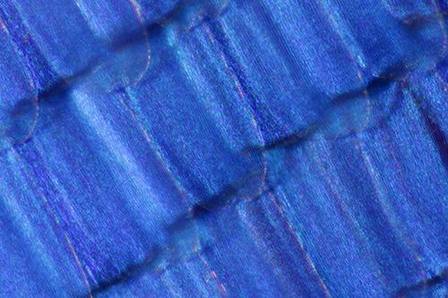
Morpho menelaus upper wing and scale detail.
This species has colourless cover scales which do not completely overlay the blue iridescent ground scales (cf. didius).
The translucent cover scales are longer and narrower than the ground scales. Under the microscope their modifying effect on the colour and intensity of the blue ground scales can be compared with uncovered areas.
|
|
|
|
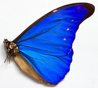
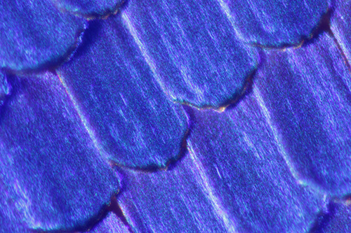
Morpho didius upper wing and scale detail.
This species has both ground scales and broader cover scales, the latter which completely overlay the ground scales when the scales are undisturbed (cf. menelaus).
|
|
|
|
Morpho rhetenor upper wing and scale detail.
This species has ground scales only to give the more intense blue.
|
|
|
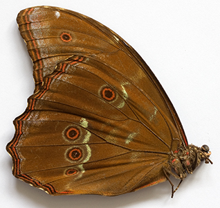
|
Above. Underside of M. menelaus and scales near a roundel (Zeiss 16X NA 0.35 objective with incident off-axis lighting from a 3 mm LED). These scales are pigmented scales and show no structural colour by iridescence except a few near the roundel centre. Unlike the upper wing scales, they have toothed edges and can be readily distinguished from the iridescent scales when studying strews of mixed scales from the upper and lower wing (see Watson slide above of
underwing scales).
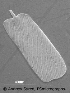
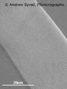
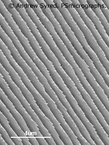
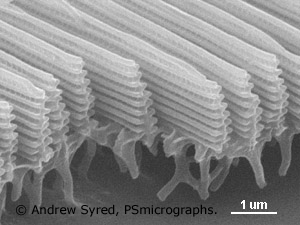
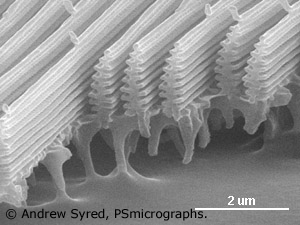
Modern studies and their value to the advancement of photonics
The series of SEM images below are kindly supplied by Andrew Syred of PSmicrographs (UK). As the magnification increases the ultrastructure of the ribbing is revealed. It is this ultrastructure found in e.g. some butterfly wing scales, beetle elytra and how they interact with light that attracts so much research interest, forming part of a branch of science called photonics. The potentially wide applications that can arise from this type of research
include colour displays, cosmetics, paints, fabrics
and opto-electronics. The BBC News item mentioned earlier highlighted the work of Professor Peter Vukusic of Exeter University who is one of the leading workers in this field. A selection of reviews in this field and research papers covering aspects of the Morpho genus are as follows (links are to free access downloadable pdfs).
'Photonic structures in biology' by Pete Vukusic and J Roy Shambles, Insight Review Article, Nature 2003 (August 14th), vol. 24, 852-680.
'Investigating the Use of Replica Morpho Butterfly Scales for Colour Displays', by Rebecca E Coath, Univ. of Southampton School of Electronics and Computer Science, May 2007.
'Quantified interference and diffraction in single Morpho butterfly scales', by P Vukusic, J R Shambles, C R Lawrence and R J Wootton, Proc. Roy. Soc. Lond. B, 1999, 266, 1403-1411.
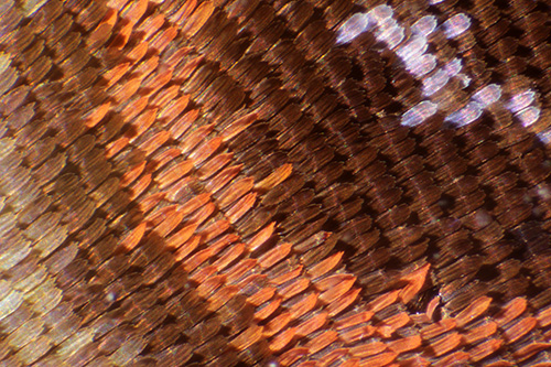
Wing scale from male Brazilian Morpho (Morpho aega) by SEM
Images (top four): 'Contextual shots of increasing magnification of a single scale showing a rib structure running along the length of the scale'.
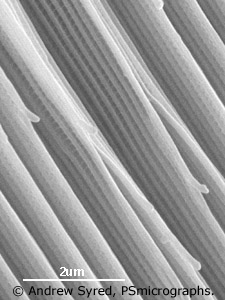
Images (second row): 'Fractured cross-sections show the ribs to be 'laminate ridges'. These act as interference mirrors responsible for metallic interference colours when viewed at certain angles.'
Images and caption text by and © Andrew Syred of PSmicrographs and shared with permission. Not to be further used without the permission of PSmicrographs.
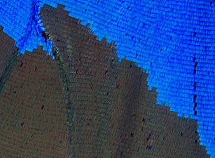
Effect of changing the scale/air interface on scale colour: The image lower right above shows the effect of dropping some solvent on the wing—in this case methanol seen in the brown patch. Replacing the interface between the scale ultrastructure from air to a solvent changes how light interacts—dramatically changing the colours seen. As the solvent dries back the interference blue colour is restored. Image taken under a stereo with
10X total optical mag.
Modern compound microscope studies of M. menelaus scales - were ground or cover scales the 'test object' - neither?
During my own studies of whole specimens of Morpho menelaus and its wing scales and following discussions with Howard Lynk of his own studies, who owned examples of both early scale strews and wing sections, it became clear that the early scale strews are very different to those of the wing scales of the currently accepted Morpho menelaus. Neither the ground scales nor the cover scales of the modern accepted species
match those used as the test objects.
Essentially, prepared slides of the scale strews labelled 'Menelaus' or 'Morpho menelaus' inspected, dated ca. 1830s - ca. 1860, do match the contemporary descriptions and illustrations of the test object scales but do not match those of the modern M. menelaus. However, slides of sections of the upper wing prepared at the same
time by the
same maker do have the very different scales typical of the modern M. menelaus. This suggests that this mismatch was something more than an early 19th century understanding of the Morpho genus differing from our current classification. (Another famous test object, the scales of the springtail 'Podura' identified at a similar date to 'Menelaus' were the subject of taxonomic uncertainties for some decades as to which species was being referred to, see the Micscape
article by David Walker.)
As we have not seen this historical puzzle reported before, it is presented separately and co-authored with Howard Lynk in the accompanying Micscape article: 'An historical puzzle - the wing scales of 'Morpho menelaus' butterfly species - one of the oldest test objects for the optical
microscope. Which species
was being used for the earliest scale strews and related curiosities?'
Morpho menelaus upper wing scales - a variant of the first test subject as a challenging modern test object for the optical microscope?
From the ultrastructure shown above, the ribs are ca. 1.6 µm high and the rib 'branches' ca. 0.17 µm apart, i.e. just below the traditional cited limit of the optical microscope of ca. 0.2 - 0.25 µm. But with suitable scale preparation, can these structures be resolved with 'pushing the envelope' techniques such as using near UV perhaps with annular lighting with crossed polars as successfully used e.g. by Osamu
Oku with the toughest diatom frustule fine structure? Using near UV of 390 µm with an NA 1.4 objective can theoretically resolve ca. 0.14 µm (from R=λ/2NA).
My own admittedly crude attempts have just tried to physically crush scales under a slip on the chance that pieces may break off presenting some of the ribs at right angles to the microscope optical axis, but these attempts to prepare suitable slides have been unsuccessful to date. An enthusiast familiar with microtome work and wax embedding may be able to take cross sections and mount for study. I would be interested
to hear from readers who try this or other techniques and who successfully show some evidence of the fine structure.
The author David Walker is an amateur microscopy enthusiast not a historian and welcomes any comments / corrections.
Acknowledgements:
Thank you to Brian Stevenson and Howard Lynk for photographing their respective slides, providing information on their provenance and also for the photomicrography of the strew scales under their microscopes.
Howard Lynk maintains the splendid resource 'A Cabinet of Curiosities' at www.victorianmicroscopeslides.com.
Brain Stevenson maintains the splendid resource 'Historical makers of microscopes and microscope slides' at microscopist.net.
Thank you to Andrew Syred for the SEM images of the wing scale.
References:
Many of the older journals and books are available on www.archive.org.
1.
C R Goring, 'On
Mr. Tulley's thick Aplanatic Object Glasses, for Diverging Rays; with an Account of a few Microscopic Test Objects', Quarterly Journal
of Science, Literature and the Arts, 1827, XXII, 265-284. (Link
is to Google Books beginning of paper.)
2. C R Goring, 'On
Achromatic Microscopes, with a description of certain Objects for
trying their Defining and Penetrating Power', Quarterly Journal
of Science, Literature and Art, 1827, Jan-Jun (XXIII), 410-434. (Link
is to Google Books beginning of paper.)
3. S. Bradbury, 'The Evolution of the Microscope', Pergamon Press, 1967. This book provides a detailed but very accessible discussion; chapter 5, 'The Development of the Achromatic Microscope'.
4. G. L'E. Turner, 'The Microscope as a Technical Frontier in Science', in ''Historical Aspects of Microscopy, Eds. S. Bradbury and G. L'E. Turner, The Royal Microscopical Society, 1962, pp.175-199. Figs. 9-11 plot the increase of numerical aperture and resolution of test objects with year showing the particularly rapid improvement from 1830s to ca. 1860.
General resources on microscope test objects
W B Carpenter, 'The Microscope and its Revelations', London, 1857,
second edition. Section 108, 'Test-Objects', 183-189.
Cheese
mites and other delicacies: the introduction of test objects into
microscopy, Endeavour, Volume 27, Issue 3, September 2003,
134-138, Jutta Schickore. (Link
is to an Abstract and full access to subscribers.)
J
W Griffith and A Henfrey, 'The
Micrographic Dictionary', third edition, 1875, p.773 - 776,
('TEST-OBJECTS' entry). (Link
is to www.archive.org
full download of
the 1856 first edition.
A Pritchard, 'An Account of Test Objects for Microscopes', The London and Edinburgh Philosophical Magazine, 1833, 3rd series, vol. II, 335-342 and Plate III.
A Pritchard and C R Goring, 'The Microscopic Cabinet ...', 1832, London.
S Bradbury, 'Test Objects', Quekett Journal of Microscopy', 2002, 39, 215 - 224.
J Schickore, 'Test objects for microscopes', History of Science, 2009, 117 - 145.
©
Microscopy UK or their contributors.
Published
in the March 2015 edition of Micscape.
Please
report any Web problems or offer general comments to the
Micscape
Editor .
Micscape
is the on-line monthly magazine of the Microscopy UK web site at
Microscopy-UK
© Onview.net
Ltd, Microscopy-UK, and all contributors 1995 onwards. All rights
reserved.
Main site is at www.microscopy-uk.org.uk
with full mirror at
www.microscopy-uk.net
.