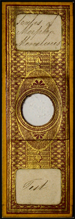
|
An historical puzzle - the wing scales of 'Morpho menelaus' butterfly species -
one of the oldest test objects for the optical microscope.
Which species was being used for the earliest scale strews and related curiosities?
by David Walker,
UK and Howard Lynk, USA.
(Text by David Walker compiled using a combination of his own studies and essential contributions from Howard Lynk, USA
including imagery of Howard's rare early microscope slides.)
In the Explorer Browser, setting Custom Print to 55% prints out on 8 x A4 sheets.
|
Some material presented below overlaps the main article to present a standalone commentary.
Introduction
In the main article published in Micscape March 2015, the history of the use of wing scales from the Morpho menelaus butterfly as optical microscope test objects was presented in the context of modern studies of the genus. As remarked, the wing scales of this species had a key role
in the optical development of the microscope. It was one of the oldest (if not the first) 'test object' to be specifically described
by species in English by Dr. Goring in his 1827 paper (1). In a fascinating historical twist, the genus has re-attracted intense scrutiny under the microscope in the field of photonics to study the ultrastructure of some Morpho species' wing scales, which is only revealed under the electron microscope.
While compiling this study with the essential contributions from Howard Lynk and Brian Stevenson, what appears to be an historical oddity surfaced which we haven't seen reported before. This is presented here separately from the main article in the hope that a reader may be able to offer an insight into the queries raised.
Essentially, prepared slides of the scale strews labelled 'Menelaus' or 'Morpho menelaus' inspected, dated ca. 1830s - ca. 1860, do match the contemporary descriptions and illustrations of the test object scales but do not match those of the modern M. menelaus. However, slides of sections of the upper wing prepared at the same time by the
same maker do have the very different scales typical of the modern M. menelaus. This suggests that this mismatch was something more than an early 19th century understanding of the Morpho genus differing from our current classification. (Another famous test object, the scales of the springtail 'Podura' identified at a similar date to 'Menelaus' were the subject of taxonomic uncertainties for some decades as to which species was being referred to, see the Micscape
article by David Walker.)
Illustrations of the test scales described as those of 'Menelaus' and later 'Morpho menelaus'
The detailed illustration of the test scales by leading experienced workers, from Goring's first reports in the late 1820's onwards, are all consistent on how they illustrate the test scales described initially as 'Menelaus' and later as Morpho menelaus and typical examples are shown below. The early achromatic optics of the time were being assessed for their ability to resolve the main longitudinal ridges on the scales and their cross ribbing. They are
shown as broad scales with expanding edges and barely toothed.
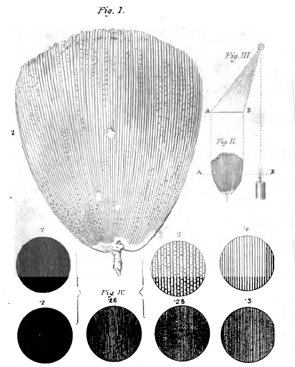
|
Right - Detail of a plate accompanying part 1 and part 2 of Goring's 1827 papers which showed a selection of test objects. This part of the plate shows a single 'Menelaus' scale and the effect of 'aperture' on 'penetrating power' (resolution in modern terms) of the scale structure. The discs read anticlockwise with aperture of .1, .2, .26, .28, .3, .4 and .5, the last being the maximum clear aperture (diameter of lens)
used. Increasing aperture revealed first the coarser longitudinal structures but required the maximum aperture to show the cross structures. (Objective aperture nowadays
uses numerical aperture as defined by Abbe.)
A diagram (Fig. III) illustrating the off-axis transmitted light which enhanced observation of the finest scale structure is also shown.
|
|
|
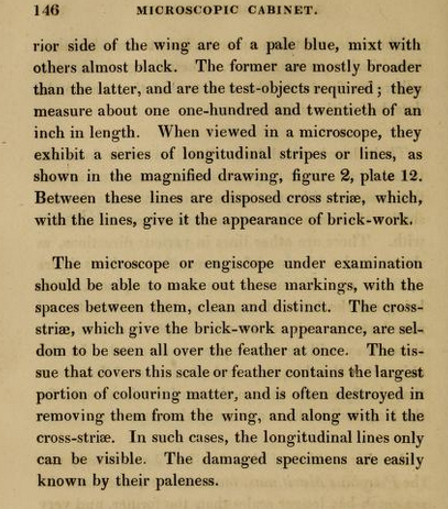
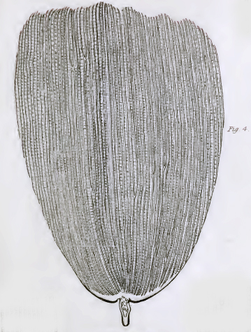
Right: A scale of Morpho Menelaus from Plate 4 in John Quekett's 'Practical Treatise on the Use of the Microscope', 1848.
(Surrounding parts of other subjects removed for clarity. Some fine detail lost because of the file compression of the copy on www.archive.org).
Quekett describes the object as a test in the extensive section on 'Test Objects'. But he notes:
"In former times it required a good quarter to exhibit the transverse striae ; but the half-inch, as now constructed, will show them readily."
(Objective focal lengths are being referred to, for a 10 inch tube length microscope, these would have a primary mag. of ca. 40X and 20X respectively.)
|
|
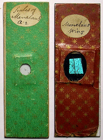
Early to mid 19th century slides showing the test scales as described by the workers at the time
A fellow microscopy enthusiast, Howard Lynk in the USA, is fortunate to own examples of the rare slides made at the time of the early test studies. This forms part of his collection of old Victorian microscope slides which are splendidly presented on his website with his own studies. A pair of ca. 1830s slides made by the same worker of a scale strew and part of wing of 'Menelaus' are shown below. It was common
at that time for the same worker to prepare both of butterfly subjects.
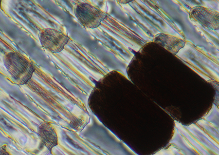
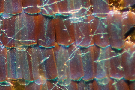
The 'Scales of Menelaus' or later 'Scales of Morpho menelaus' slides below dating from the 1830s to ca 1860s clearly shows scales that match the descriptions and illustrations of the time when described as a test object. The scales have distinctive non-parallel expanding sides with barely any toothing and are partly translucent to permit ridge structure studies in transmitted light.
One puzzle immediately arises—the test scale morphologies shown below are very different to those from the upper wing of the species now regarded as Morpho menelaus which have iridescent narrow parallel sided scales (the ground scales) underlying the slimmer translucent cover scales (presented later below).
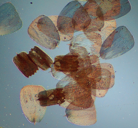
|
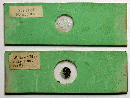
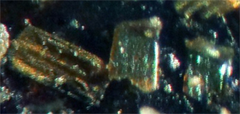
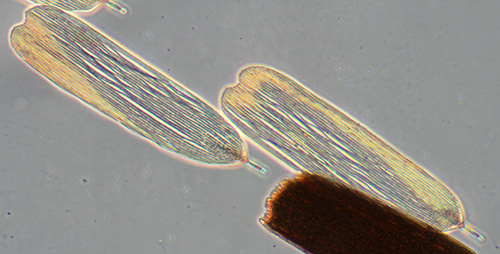
Above: Papered slides of both a scale strew and wing fragment. 'Tentatively identified as being from Cary/Gould ca. early 1830s, ... with mica slips'. Made at the time of intense development of achromatic optics when the strew scales slides were still valuable as test objects.
Right: Image of the above scales skillfully photographed using a mix of incident and transmitted light.
Images and notes in quotes courtesy of Howard Lynk.
|
|
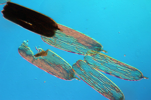
A later slide ca. 1840s shown below also shows a scale strew with the same scale morphology (with likely some toothed underwing scales as well).
|
Above. Two papered slides of both wing fragment and scale strew. 'Two early mounts by J W Bond ca. early to mid 1840s'.
Right: The strew has both upper and lower wing scales.
Images and notes in quotes courtesy of Howard Lynk.
|
|
A later slide ca. 1850s-60s by Norman shown below also shows a scale strew with the same morphology as those reported for the test scales (with likely some more opaque and toothed underwing scales as well).
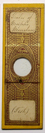 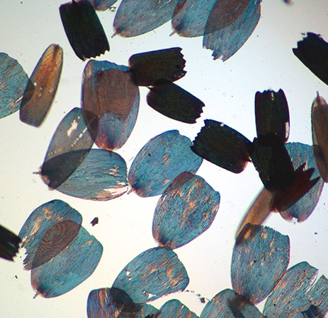
Above. A scale strew slide by Norman 'ca. 1850s-1860s' and their appearance under the microscope (right).
Images and date estimates courtesy of Howard Lynk.
|
|
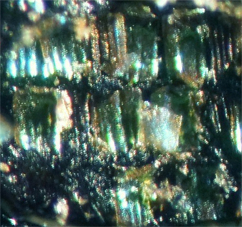
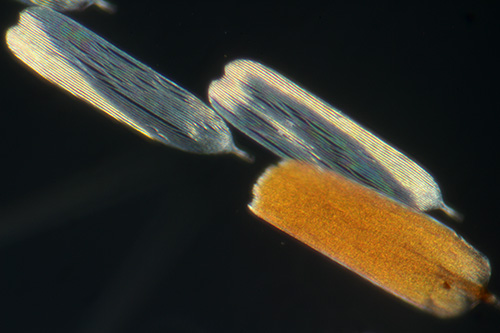
Above. A papered slide labelled 'Scales strew of Morpho menelaus' likely by Norman ca. 1850s - 1860s by comparing with known slides.
Right. Scales from above slide all to same scale, optical mag used 400X. This is the latest date slide handled showing the test scale morphology. This slide also shows scales from likely other parts of the wing.
No evidence of the almost transparent cover scales with very coarse ridges seen typical of the modern M. menelaus are present (see below).
This slide suggests continued accessibility to the source of the test scales up until ca. 1860—as also suggested by the illustration shown earlier by Quekett of the test scales in his treatise of 1848 and later editions.
Images and slide provenance courtesy of Howard Lynk.
|
|
Early 19th century slides showing sections of the upper wing of 'Menelaus' or 'Morpho menelaus'
It was common for a given maker to make both a scale strew slide and one of a section of the upper wing at the same time. The believed Cary-Gould and Bond pairings above are of this type. As the 19thC scale strew morphologies don't match those of the modern M. menelaus, what do the scales on the 19thC wing sections scales show?
|
Cary/Gould ca. early 1830s the green papered slide above. Detail from wing section. Unfortunately this slide is in poor condition and only a few scales were still present on the upper wing, shown above and left. The scales do look parallel and narrow typical of M. menelaus.
Detail from master image supplied by Howard Lynk.
|
|
Right. 'J W Bond ca. early to mid 1840s'. The slide with red paper with stars shown above. This slide is in good condition and the narrow parallel sided ground scales with partly overlapping cover scales typical of the modern M. menelaus are clearly seen. See other article for a modern view of a M. menelaus wing.
Detail from master image supplied by Howard Lynk.
|
|
|
Left above. A slide by J. Murray (wing fragment) 'guessing ca. 1860s'. Right above. As for the Bond slide above the ground and cover scales are clearly seen and typical of the modern M. menelaus. Detail from master image supplied by Howard Lynk.
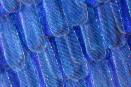
Above. The upper wing of a recently sourced whole specimen of M. menelaus. The iridescent ground scales and slimmer translucent cover scales are seen and match closely those of the Bond slide. Image by D. Walker. From main article.
Above. Image of the same wing under a ca. 1830s Cary-Gould style microscope made by Carpenter and Westley described in the January 2015 Micscape issue. A contemporary 'button' single lens of ca. 45X with a working distance of ca. 4 mm was used. This shows that low power microscopes of the time could clearly distinguish between the ground and cover scales if used prior to higher power work to examine scales in situ.
The very small working distance suggests selecting scales with low power microscopes of the time may have been unlikely. Rubbing the wing or as mentioned in some papers of the time breathing on the slide and touching the wing may have been used. This would lift in preference the cover scales if present.
|
The present authors are not experts on the Morpho genera—the taxonomy of the genus and that of M. menelaus in the 1820s could be different from today's but the scales in the earlier slides of parts of the upper wings do seem to match that of the modern species. Although there are many sub-species reported (see Wikipedia entry).
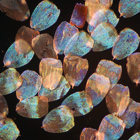
Modern compound microscope studies of M. menelaus ground or cover scales
Scales taken from a modern specimen of the adult M. menelaus are shown below using various sampling and lighting techniques. These were deferred from the main article.
|
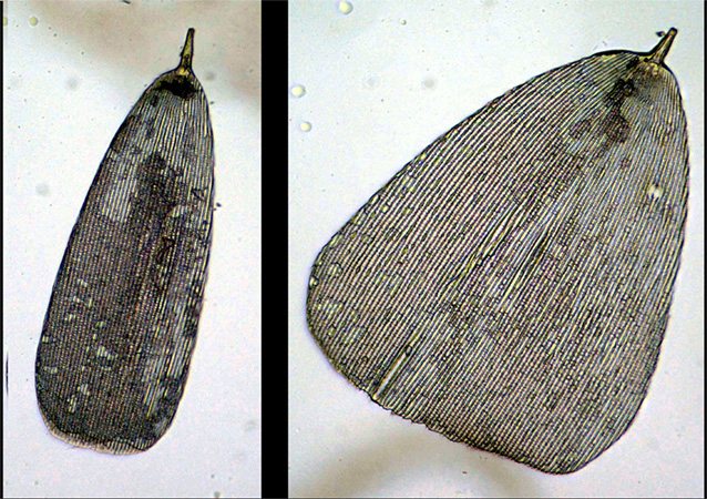
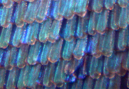
Right: Modern specimen of Morpho menelaus, upper wing scales lifted using Sellotape. The translucent cover scales are shown and the near opaque ground scales with their parallel sides.
Unlike the original test scales of the ca. 1830s, it was very difficult under a modern compound microscope using either transmitted brightfield or other techniques to study the ridge structure of the ground scales, suggesting that they did not use the ground scales as the test objects.
None of the 19th century slides seen to date show the distinctive cover scales of the current M. menelaus.
Objective 10X.
|
|
|
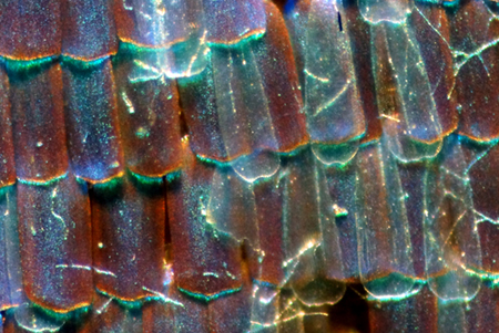
Above: Cover scales in brightfield. These are translucent, long, thin and coarsely ridged—very different from the traditional test objects scales. These scales can easily lose an upper layer.
Zeiss 16X NA0.4 Neofluar objective.
|
Above: Cover scales and a ground scale using phase contrast. Zeiss 16X NA0.4 Neofluar objective.
|
|
Above: Two cover scales and a ground scale under crossed polars. Zeiss 16X NA0.4 Neofluar objective.
|
Above: Cover scales and a ground scale under DIC with a λ tint plate. Zeiss 16X NA0.35 objective.
|
|
|
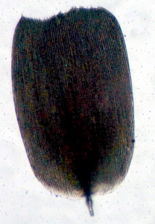
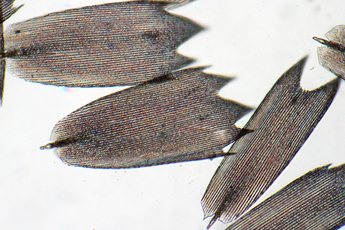
Left: Scales from the under wing are distinctly toothed. Prepared slides of scales from the second half of the 19th century often used these scales and described as a 'Test object'. Although not the traditional test from this species.
Zeiss 16X NA0.4 Neofluar objective.
|
Comments and queries
One potential explanation of the scales on the strews not matching those of the upper wing sections, we believe can be ruled out, i.e that the preparers were seeking a particular morphology of wider scale on M. menelaus for the test subjects. There is no evidence from my own or of Howard Lynk's careful studies of the whole upper wings under stereo microscopes of multiple modern specimens that these type of test object scales are present on M. menelaus. Neither do
the published modern research reports on this species seen to date show any.
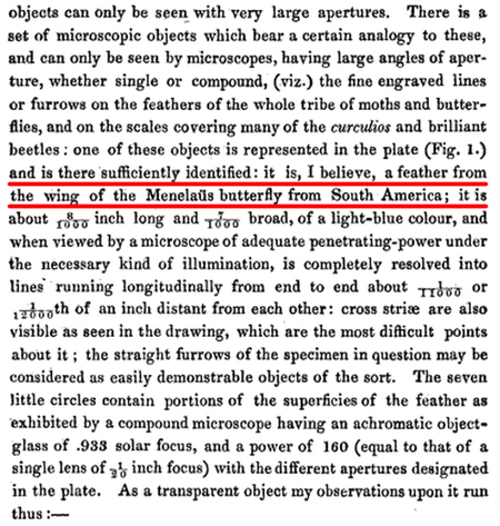
Early reports stated it was identified as a potential test object in the 1820s by Thomas Carpenter, London who was described as an 'experienced entomologist' and wrote extensively on entomological subjects for Gill's 'Technological Repository of the 1820s and 1830s.
Right. Dr C R Goring in the first of his two seminal papers of 1827 describing the value of test objects (ref. 1, p.268). This is the earliest mention seen to date of the use of 'Menelaus' as a test object.
Note the scale size and ridge separation also that the scales have 'cross striae'.
(From www.archive.org and in the public domain.)
|
|
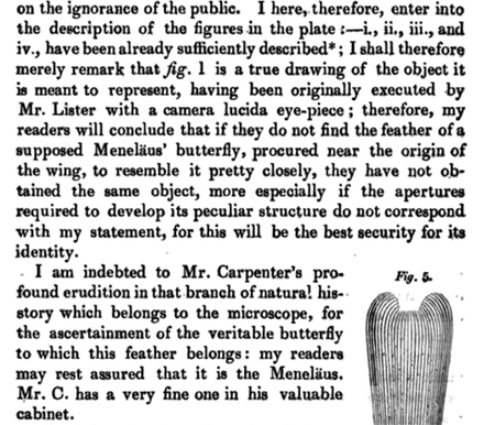
Right. Dr C R Goring in his second paper 1827 describing the value of test objects (ref. 2, p.423). This is the earliest mention seen to date of the use of 'Menelaus' as a test object.
'feather' .... 'procured near the origin of the wing'. Thomas Carpenter confirms the identification and 'has a very fine one in his valuable cabinet'.
(From www.archive.org and in the public domain.)
|
|
Further support that they were not seeking out special scales on samples of M. menelaus is the early descriptions of where the scales were sourced on the butterfly—see extracts below from the work of the leading workers Andrew Pritchard and John Quekett.
|
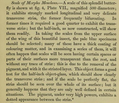
Above and right. 1832 A. Pritchard 'The Microscopic Cabinet', p.145-146 below. 'Centre of the superior side of the wing'.
Both this and citations above note that pale blue scales are the 'test' scales. Modern cover scales on their own are a pale blue in mixed light. The 'almost black' scales in the citation right are to be avoided. Modern ground scales are also very dark on their own in mixed light and almost opaque by transmitted.
(From www.archive.org and in the public domain.)
|
|
Summary of scale morphologies from historical and modern sources
The table below compares the scale parameters of historically reported 'test' scales and modern observations. In my own work in the table, photomicrographs of a range of scales were printed out on A4 paper together with a micrometer scale. Ridges were measured over at least 10 ridges and averaged to give a value for the separation.
| Observation |
Source |
Size x length in µm / length: width ratio |
Scale ridge separation in µm
|
Comment |
| Ground scales |
Modern M. menelaus. D. Walker. |
182 x 85 / 2.1:1 |
1.36 |
Almost opaque in transmitted by brightfield or any modern method, suggest unsuited as test scale. |
| Cover scales |
Modern M. menelaus. D. Walker. |
204 x 64 / 3.2:1 |
2.42 |
Translucent scales, pale blue in incident, suggests right sort of scales for a test. But scale shape very different to test scales. Also no cross striae noted by early workers for the 'test' scales.
|
| True test object scales, modern measurements |
From M. menelaus scale strew ca. 1830s. Cary/Gould? H. Lynk.
Three typical scales. |
240 x 171 / 1.41
229 x 182 / 1.26
229 x 175 / 1.31 |
1.87
1.86
1.83 |
Ridge separation consistent between scales.
Length : width of scale less square than Goring's measurements. |
| 'Test objects' 'Morpho menelaus' description in paper. |
Goring - Pritchard, 1832 (ref. 5). |
212 (1/120th inch) |
|
Within limits of measurement and scale variation, the length could apply to either modern cover or ground scales. |
| 'Test objects' 'Morpho menelaus' description in paper. |
Goring, 1827 (ref. 5). |
203 x 178 (8/1000 x 7/1000) / 1.14 |
2.1 (1/11000 - 1/12000th inch)
|
Scale length:width matches neither modern ground or cover scales |
|
'Test objects' 'Morpho menelaus', large drawing in book plate.
|
Quekett, 1848 (ref. 6, Plate 7 using declared mag of 500X.) |
ca. 196 x 136 / 1.44:1 |
2.1 |
Ratio more accurate than length estimation. |
| SEM of uncertain species but with apparent 'test scale' morphology |
H. Frederick Nijhout, SEM |
width 174 |
ca. 1.5 |
Estimate from SEM image suggest that the ridge separation is considerably finer than for 'test' scales. |
As the table attempts to summarise, there seems a baffling contradiction between the parameters of the original test scales (transparency, ridge separation, length : width ratio) and those for both cover and ground scales sourced from a modern M. menelaus. The transparency of a cover scale and the observation by Goring to select pale blue scales suggests that cover scales type scales rather than dark ground scales
were the test object. But the modern cover scales have a very different shape to those of the 'test', the ridges are rather coarse and there's no cross striae described by the early workers.
Modern ground scales also don't match the 'test' scales. Although Goring remarked to avoid the dark scales when selecting test scales. Even assuming that their ground scales were more transparent than for modern examples, the ridges are much finer and likely pushing the capabilities of the early achromatic optics.
Access to an 1830s slide of the true test scale strew allows modern measurements (by Howard Lynk) to be compared with Goring's. The modern average ridge separation of 1.9 µm is in quite good agreement with Goring's value of 2.1 µm considering the limitations of the microscopes and the likely extra difficulties of micrometric measurements of the time.
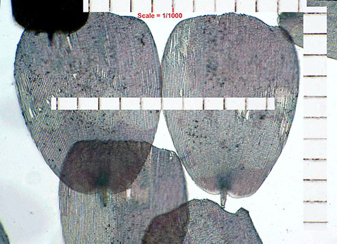
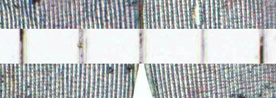
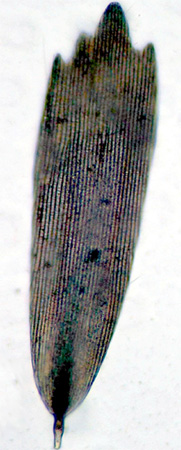
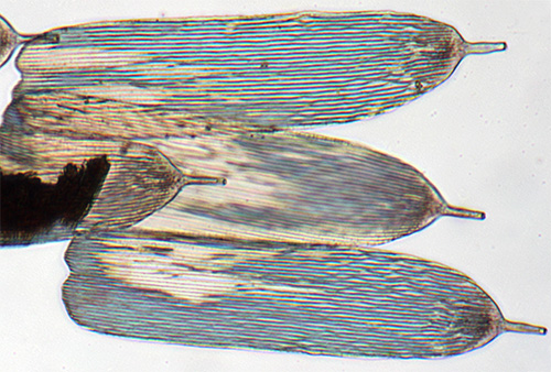
Above. Image and extracted detail used to measure the scale ridge separation and scale sizes of the ca. 1830s M. menelaus strew
which has the true 'test' scale morphology. There are typically 13 -14 ridge spaces to 1/1000th inch. Image by Howard Lynk.
Queries arising:
1) What species of butterfly was used for the test scale strews—although Howard's slide strews match the contemporary reports of the scales as test objects, they do not match those of the modern M. menelaus?
2) For the slide pairings of scale strews and wing sections made at the same time by the same maker, why do the scales on the wing section not match those of the scale strews if the same source of butterfly was used? Although the wing sections do match those of the typical modern M. menelaus suggesting that they had not misidentified the species or that at the time M. menelaus referred to a different species.
Do any of the many sub-species show this
scale form?
3) Does the understanding of the Morpho genera and that of M. menelaus of the time offer any insight.
A possible contender for the true identity of the test scales is that of the species' upper wing shown right—an image courtesy of Professor H. Frederick Nijhout. Although ascribed in its caption to M. menelaus, in helpful email correspondence, it was likely that it had likely been incorrectly attributed but its actual provenance was uncertain. Although as noted in the table above, the ridges seem to
fine to be the 'test' scales.
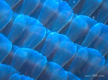 Image credit right. H. Frederick Nijhout Ph. D., Duke University. (Resized from master on www.nisenet.org)
The scales show similar features of the test scales—expanding sides and more translucent thus aiding the study and resolution of ridges and cross ribbing under brightfield illumination using the optical microscope of the late 1820s onwards. Such overlapping scales seem to preclude the possibility of cover scales.
Is any reader able to identify this species?
One further oddity which may be relevant is that from studying the slides owned by myself, Howard Lynk and Brian Stevenson there was a trend in the later 19th century for slides labelled Morpho menelaus to be of the lower wing scales only, i.e. the dense distinctly toothed scales, although this may have had an economic reason (see main article).
The authors would be very interested to hear from readers who have scale strews of the Morpho genus that match the test scale morphology.
Given the many years that have passed it may not be possible to resolve the puzzle but for the microscopist interested in the history of test objects, it certainly seems an intriguing one.
The author David Walker and colleague Howard Lynk are microscopy enthusiasts and don't have expertise in the Morpho or other genera and we welcome any comments / corrections.
Acknowledgements:
Thank you to Howard Lynk for photographing the early rare slides in his collection, for the splendid photomicrography and for providing information on the slide provenance. His imagery and knowledge of early slides was vital to help reveal the historical puzzle in combination with my own researches.
Thank you to Brian Stevenson for sharing images of slides in the main article.
Thank you to Professor H. Frederick Nijhout, Duke University for permission to show the www.nisenet.org hosted image and for helpful discussions.
Any errors in the article are solely that by one of the authors, David Walker.
Howard Lynk maintains the splendid resource 'A Cabinet of Curiosities' at www.victorianmicroscopeslides.com.
Brain Stevenson maintains the splendid resource 'Historical makers of microscopes and microscope slides' at microscopist.net.
References:
Many of the older journals and books are available on www.archive.org.
1.
C R Goring, 'On
Mr. Tulley's thick Aplanatic Object Glasses, for Diverging Rays; with an Account of a few Microscopic Test Objects', Quarterly Journal
of Science, Literature and the Arts, 1827, XXII, 265-284. (Link
is to Google Books beginning of paper.)
2. C R Goring, 'On
Achromatic Microscopes, with a description of certain Objects for
trying their Defining and Penetrating Power', Quarterly Journal
of Science, Literature and Art, 1827, 23, 410-434. (Link
is to Google Books beginning of paper.)
3. S. Bradbury, 'The Evolution of the Microscope', Pergamon Press, 1967. This book provides a detailed but very accessible discussion; chapter 5, 'The Development of the Achromatic Microscope'.
4. G. L'E. Turner, 'The Microscope as a Technical Frontier in Science', in ''Historical Aspects of Microscopy, Eds. S. Bradbury and G. L'E. Turner, The Royal Microscopical Society, 1962, pp.175-199. Figs. 9-11 plot the increase of numerical aperture and resolution of test objects with year showing the particularly rapid improvement from 1830s to ca. 1860.
5. A Pritchard and C R Goring, 'The Microscopic Cabinet ...', 1832, London.
6. J Quekett, 'Practical Treatise on the Use of the Microscope', 1848, London.
Note on the butterfly specimens. All recent specimens were sourced from an accredited European Lepidoptera dealer, imported under EU regulations from captive breeding programmes in S. America, run by and for the local people.
©
Microscopy UK or their contributors.
Published
in the March 2015 edition of Micscape.
Please
report any Web problems or offer general comments to the
Micscape
Editor .
Micscape
is the on-line monthly magazine of the Microscopy UK web site at
Microscopy-UK
© Onview.net
Ltd, Microscopy-UK, and all contributors 1995 onwards. All rights
reserved.
Main site is at www.microscopy-uk.org.uk
with full mirror at
www.microscopy-uk.net
.