|
A virtual tour of the 4026 form arranged diatom slide prepared by J D M—ller: by Jef Schoors, Belgium Credits and acknowledgements:
Slide access, microscopy and photomicrography by Jef Schoors using the facilities of |
Micscape introduction: Readers familiar with the diatom arrangements of Johann Diedrich M—ller (1844 - 1907) may be aware that his most famous arrangement was his "Universum Diatomacearum M—llerianum". Very few people have had the privilege of exploring this irreplaceable slide under the microscope first hand. But we were delighted to hear from a Belgian microscopy enthusiast Jef Schoors who was able to recently study and photograph the slide under the microscope and shares an article below on this slide and on aspects of M—ller's work. A virtual tour to species level of this slide using Jef's splendid stitched images is also provided.
When I took up microscopy again some ten years ago and joined the Royal Antwerp Microscopical Society I read about Johann Diedrich M—ller and his work in the German magazine Mikrokosmos. I was fascinated by this famous maker of diatom arrangements. And then I saw a picture of the Universum Diatomacearum M—llerianum! Placing a few of these glass skeletons in a certain layout on a cover slip already demands extreme precision and patience. But 4026 different species of diatoms meticulously arranged on one single slide is absolutely an incredible achievement! It took M—ller 4 years of preparation and no less than 40 days to mount it himself in 1890. The diatoms are arranged in an array of nine rectangles which occupy an area of 6 x 6,7 mm. The medium M—ller used was a mixture of Canada balsam and mono bromonaphthalene. The famous slide was, together with a number of other M—ller Typenplatten, on display at various exhibitions, such as in Antwerp.
Prof. em. Karel Van Camp, a member of our Microscopical Society, told me that the Universum slide is kept safely in the research department of the Botanic Garden Meise (Belgium). Prof. Van Camp has a nice collection of arranged diatom slides and he knows how keen I am on photographing them. Thanks to his connections with one of his former students, Prof. Dr. Bart Van de Vijver, algologist and researcher of the Botanic Garden, we had the opportunity to get a glimpse of the extensive Henri Van Heurck collection. Van Heurck, a Belgian industrial entrepreneur and famous diatomist, bought the 'Universum' slide from M—ller in the year 1900 for an estimated price of 6000 D Mark, the equivalent of three family houses!
In 1905 Van Heurck noticed that a number of diatoms on the slide became detached from the adhesive layer of the cover slip, and drifted from their original position on the slide. The reason was that the mono bromonaphthalene mixture never completely hardened and after 15 years started to dissolve the adhesive layer on which the diatoms were attached. Because of his age, M—ller was not able to rearrange the diatoms or replace the mounting medium. Therefore Van Heurck, who was familiar with the mounting procedure of M—ller took a risky decision. He opened the slide and replaced the medium with styrax, a mounting medium he developed. Unfortunately, some detached diatoms were not mounted again but removed together with the old medium. In the slide however we can still see some shadows of diatoms that drifted away.
Fortunately Prof. Van de Vijver granted us the permission to photograph the slide. However, because the slide can't leave the Botanic Garden Meise, photography had to be done in the offices of the research department. The microscope we used was an Olympus BX53 series with DIC and the microscope camera was an Olympus UC30 connected to the program CellSens Standard. Photo's were made in brightfield using an Olympus 4x objective. The 36 images were stitched together manually in Photoshop and uneven lighting was adjusted.
Of course I would have loved to photograph many of the specimens in detail, but time was limited. It was also the first time I used this microscope and this camera program so I had to learn fast.
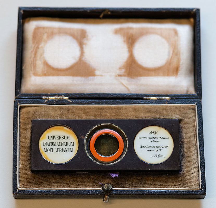
The small wooden box with the most precious diatom arrangement from J. D. M—ller, the Universum Diatomacearum Moellerianum.
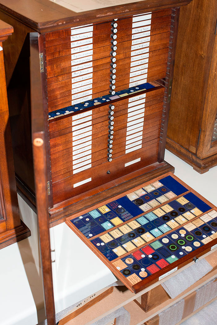
In the Botanic Garden Meise a large number of mahogany cabinets containing the M—ller collection are carefully preserved.
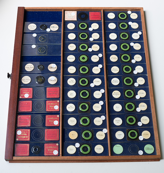
One of the many drawers with arranged diatom slides from J. D. M—ller.
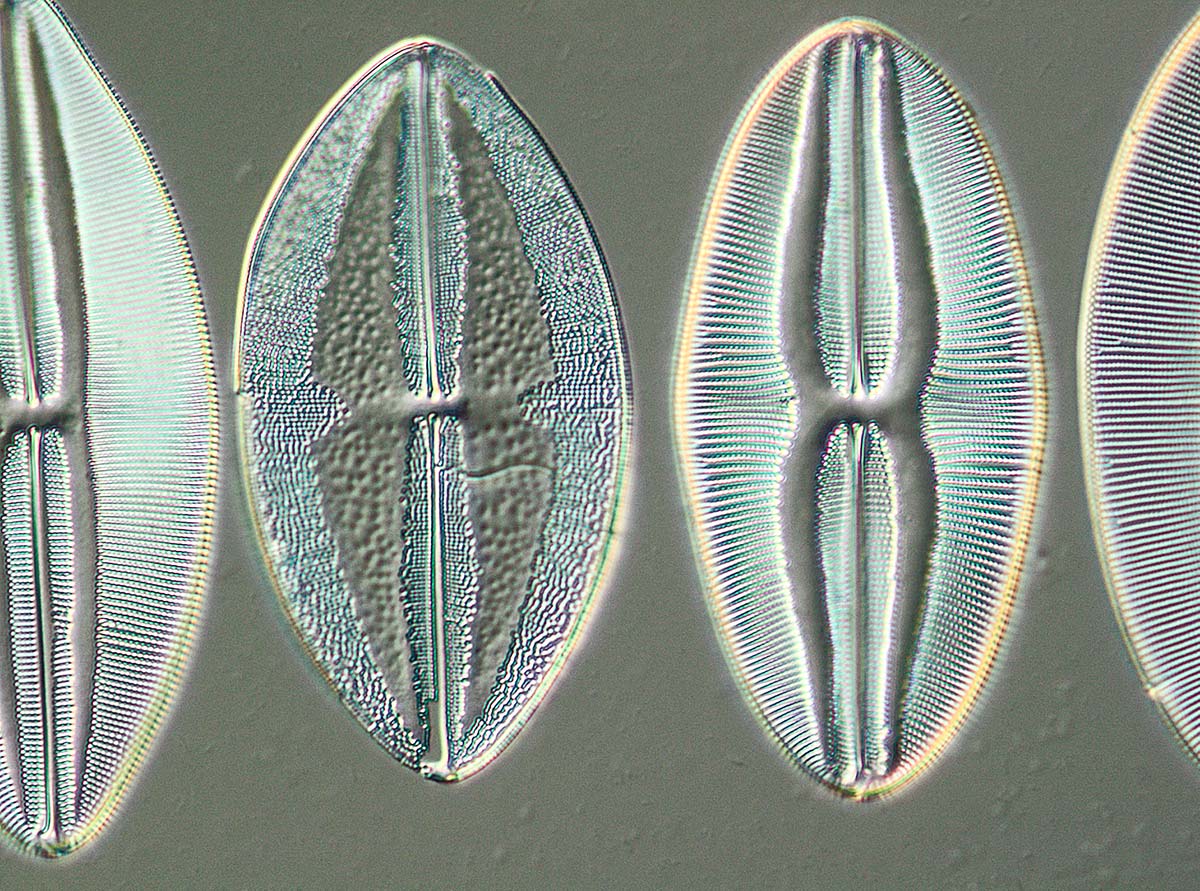
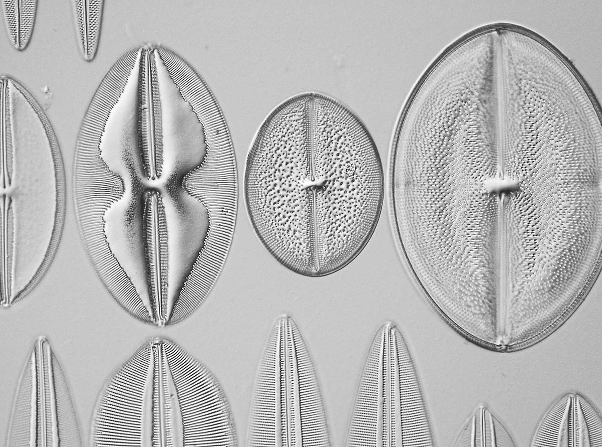
The two images above were photographed in DIC with a 40x objective. They clearly demonstrate the incredible richness in detail of each diatom. In fact, each of the many species in this fantastic slide mount deserves a separate photo, although this could take a while.
Comments to the author Jef Schoors are welcomed.
The images above of the slides at the Botanic Garden Meise are by Jef Schoors. For further image use please contact the author in the first instance.
Species list for the slide (German) in Acrobat pdf format, published 1892, 44 pages. Includes a ten page introduction by M—ller and the species list. Species are listed for each of the nine rectangular blocks from left to right and line by line, left to right.
Copied by Jef Schoors from a copy owned by and with the courtesy of the Botanic Garden Meise.
Micscape notes: Touring the slide. The master jpeg image, 7138 x 8098 pixels, 36.6 Mbytes in size was converted by Maurice Smith the Microscopy UK founder into a format that allows the detailed navigation below using 'Zoomify' script. The image can be zoomed in to any desired level by either clicking on it with the mouse or by using the zoom bar in the menu below it. The arrows in the menu bar allow the reader to scroll round and explore this marvellous slide. The small complete image top left has a blue rectangle showing where on the slide the zoom is currently set. Pressing the far right button on the menu bar resets the image.
Selected Resources:
1) The Van Heurck Collection: A phoenix rises from its ashes., by Bart Van de Vijver, Christine Cocquyt & Jan Rammeloo. Acrobat pdf, copy courtesy of Jozef Schoors.
2) Johann Diedrich M—ller, 1844 - 1907. Die Kunst, Diatomeen zu legen. Ausstellung im Zoologischen und Botanischen Museum der Universitðt Hamburg 15. November 2007 - 15. April 2008. An attractively illustrated article in German on an exhibition in 2008 of his life and work. Acrobat pdf, copy courtesy of Jozef Schoors.
3) Diatomeen im 19. Jahrhundert. Typenplatten und Salonpraparate von Johann Diedrich M—ller by Helene Kranz. Published by Goecke and Evers, 2009. A beautifully presented book in German with stunning photomicrography of many of M—ller's slides by Matthias Burba. Link is to a German shop which sells it for 15 Euros. A selection of pages can be seen here. Update August 2019. Formerly supplied by some dealers but no supplier was now found either new or used.
Related Micscape articles:
- Enjoying a M—ller 80 form diatom type-slide with a microphotograph setting by David Walker, Micscape, January 2009. Also has additional references.
- The Micscape Library has extensive resources on many aspects of diatoms by our contributors:
For a splendid introduction see Those Who Live in Glass Houses by Bill Amos, illustrated by Wim van Egmond.
The diatom forms presented on microscope slides are usually the silica frustules (the 'shells'). To admire a selection of live forms visit the 'Micropolitan Museum' by Wim van Egmond and follow the The Marine Collection / Diatom Display.
Published in the March 2015 edition of Micscape.
Please report any Web problems or offer general comments to the Micscape Editor .
Micscape is the on-line monthly magazine of the Microscopy UK web site at Microscopy-UK
ˋ
Onview.net Ltd, Microscopy-UK, and all contributors 1995
onwards. All rights reserved.
Main site is at
www.microscopy-uk.org.uk
with full mirror
at
www.microscopy-uk.net
.