|
Recruiting Amateur Microscopists And Natural Historians by Richard L. Howey, Wyoming, USA |
Since we live in a relentlessly technological age, I suppose the first step to luring people, especially young tech-savvy savants into the wonders of the natural world, would be to create an APP for identifying common protozoa and algae. However, since I am strictly a low-tech person I’ll have to leave that for others. However, I suspect that one major concession to technology will become inevitable and that is a camera that displays the image of the specimen under the microscope either on a computer screen or on a separate monitor. I have both but, I still much prefer looking through the eyepieces directly. Psychologically, this process creates for me a greater sense of immediacy and contact with what I am viewing. Furthermore, all but the very high-end and highly expensive systems produce a less satisfying image in terms of resolution, contrast and color balance. However, when you’re sharing specimens with another person, or several, having a display which allows all of you to observe, comment, and say “WOW!” at the same time is a distinct advantage.
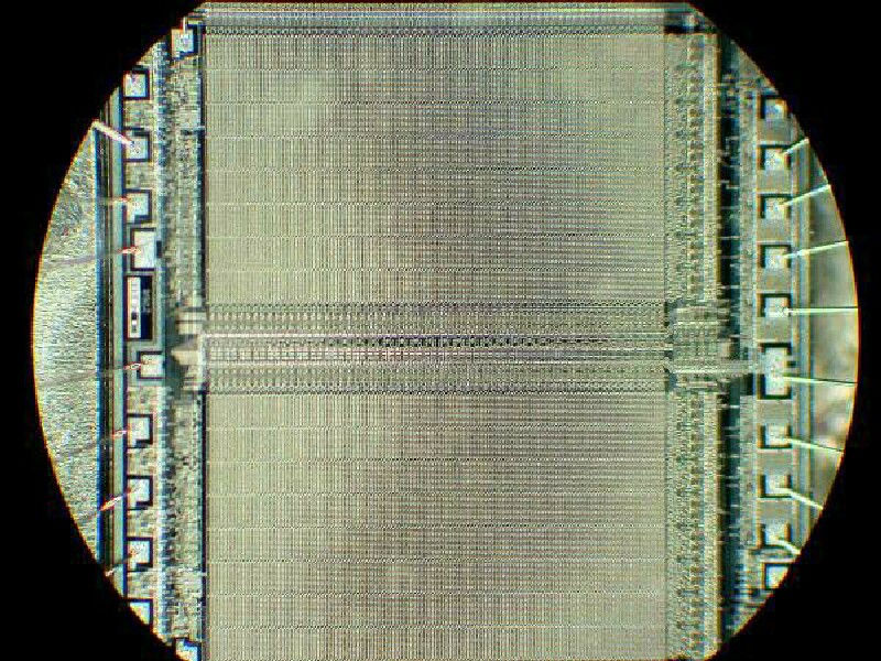
Nonetheless, to lure an innocent into the marvels of micro-worlds, it is very helpful to know something about his or her interests and use those as a basis for leading them into extended realms. Suppose our “victim” is involved with computers and the ways they perform, but has never seen a computer chip except to install one–in other words, has never seen the incredibly intricate circuitry of such a chip. Most of us use computers, but few of us have much of an idea of their internal structure or how they work. My attitude is like the one I have toward my car; I just want it to work properly, get me where I want to go and do so with the least possible hassle. If there are problems with the car, I take it to a mechanic (good ones are hard to find and are undervalued) and if I have problems with my computer, I take it to a specialist and again, good ones are hard to find and unfortunately often tend to overvalue themselves. A colleague of mine in the Physics department who is a computer aficionado (that’s a polite word for “nerd”) knows thoroughly the ins and out of these maddening, frustrating, hellishly wonderful machines and over the years, he has accumulated a drawer full of old chips. He was kind enough to give me 5 or 6 of them and I mounted 3 of them on slides. These are old, “primitive” chips compared to those being produced these days but, nonetheless, the intricacy of the circuits produces an aesthetically pleasing view.
So, here is a good lead-in to microscopy for a computer techie who doesn’t know about the architecture of chips. This is a point of departure and what I recommend is creating a moderate-sized collection of slides and specimens with considerable variety for tempting the unsuspecting into the wonderfully curious realms of the microscopic.
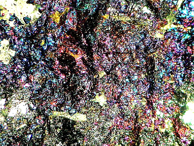
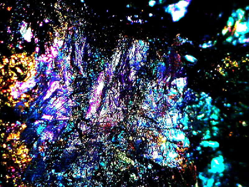
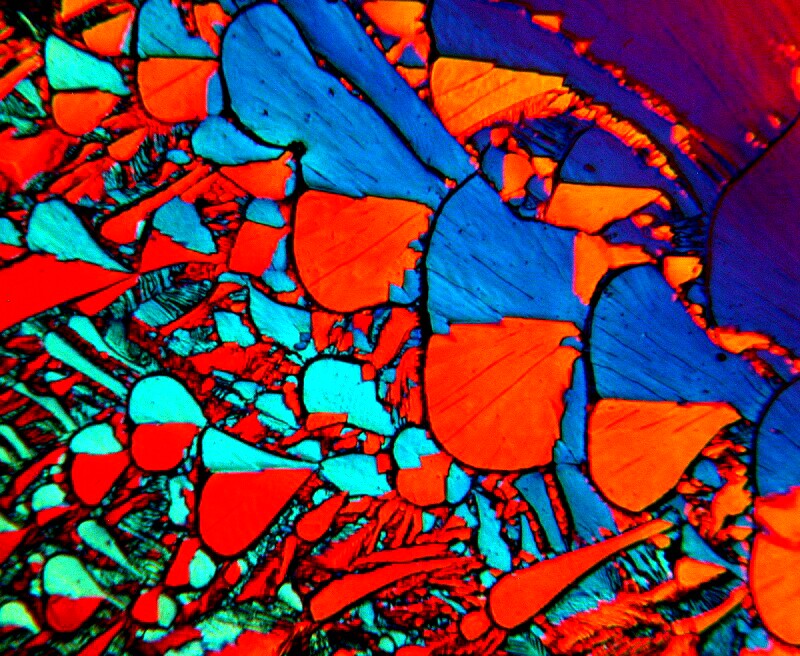
It is wise to have 2 basic groups of materials; a collection for the compound microscope and a second one for the stereo microscope. The image of the computer chips was taken with a stereo. I think it is almost always advisable to begin any “show and tell” with a stereo microscope and with lots of “show” and relatively little tell”–that is until your friend or relative is well and truly hooked. The obvious great advantage of the stereo microscope is the large range in terms of size of specimens which can be observed. Anyone with an interest in rocks and minerals is an easy target. A dazzling array of colors are readily available in a piece of the copper ore, Bornite, also known as Peacock Rock. Here are a couple of enticing images.
Furthermore, If you have a mineral micromount or two, these are almost always captivating. The specimen is usually mounted in a small plastic box to protect it and often the bottom of the specimen is glued to a piece of styrofoam cut to fit snugly in the box. Micromounts allow selecting a cluster of near perfect crystals that display a geometric purity rarely found in large specimens. Being able to prepare fine micromounts is a challenging art that requires considerable practice and skill but the rewards are significant.
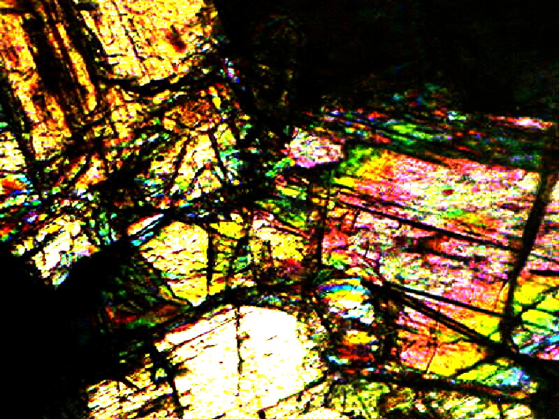
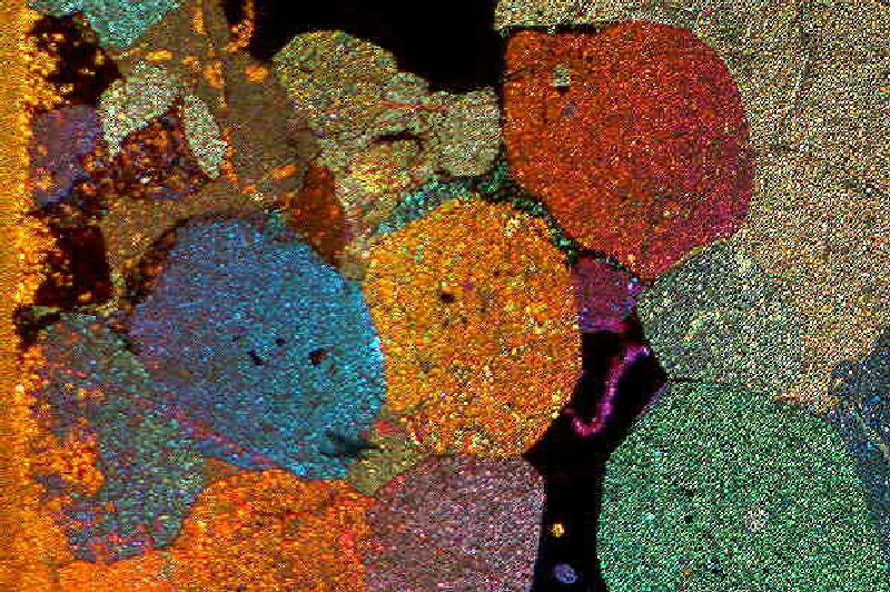
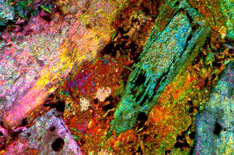
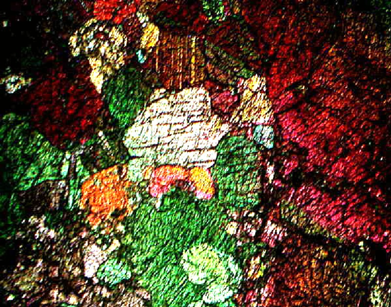
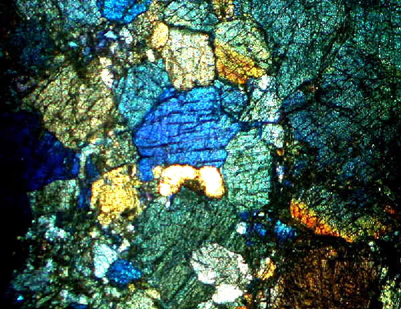
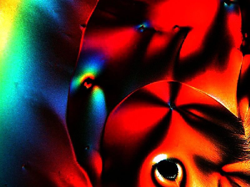
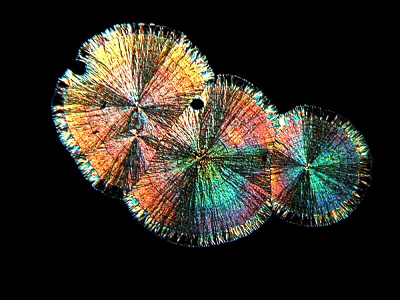
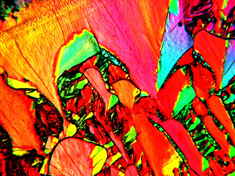
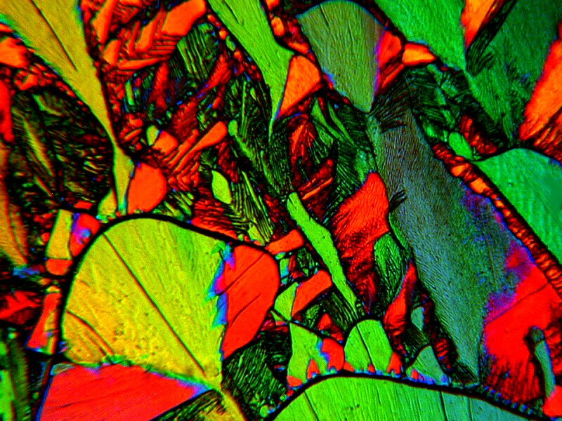
For an individual with a strong interest in things geological, a glimpse of mineral thin sections under polarized light can prove irresistible. Your demonstration kit should have 2 or 3 nice examples which I strongly recommend you purchase and regard as an investment in preserving your sanity. Well-made preparations are about 30 microns thick and can be used either with a compound microscope which has polarizing filters or with a stage polarizer on a stereo microscope. To cut such sections requires special, expensive equipment including diamond saw blades and special grinding and polishing tools. A good slide of this type can be purchased for about $20 and can be seen as a saving both in terms of your mental health and your wallet.
Below are 5 examples of such sections under polarized light.
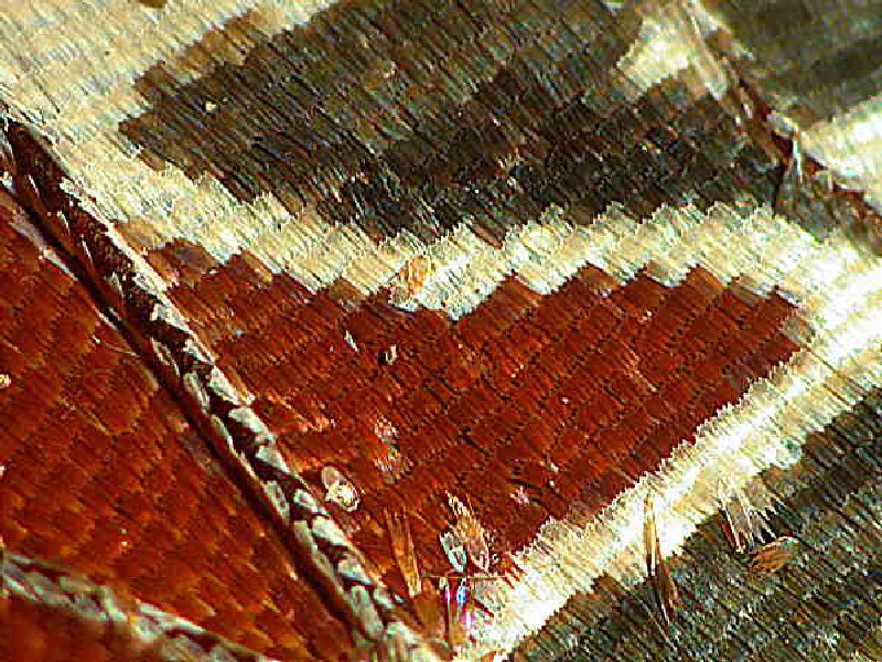
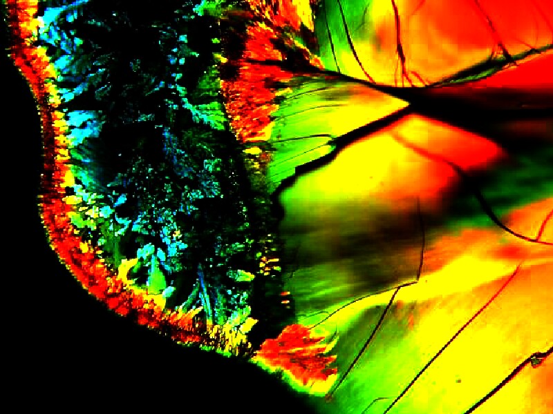
Another basic addition to your demonstration collections should be butterflies and moths. If you aren’t up to romping through meadows with a butterfly net (perhaps being chased by men in white coats with very large butterfly nets), then you might make some selective package purchases on eBay and/or check the grill of your car or truck from time to time. The scales on butterfly wings are quite amazing structures and the way in which they are attached to the wing is extraordinary.
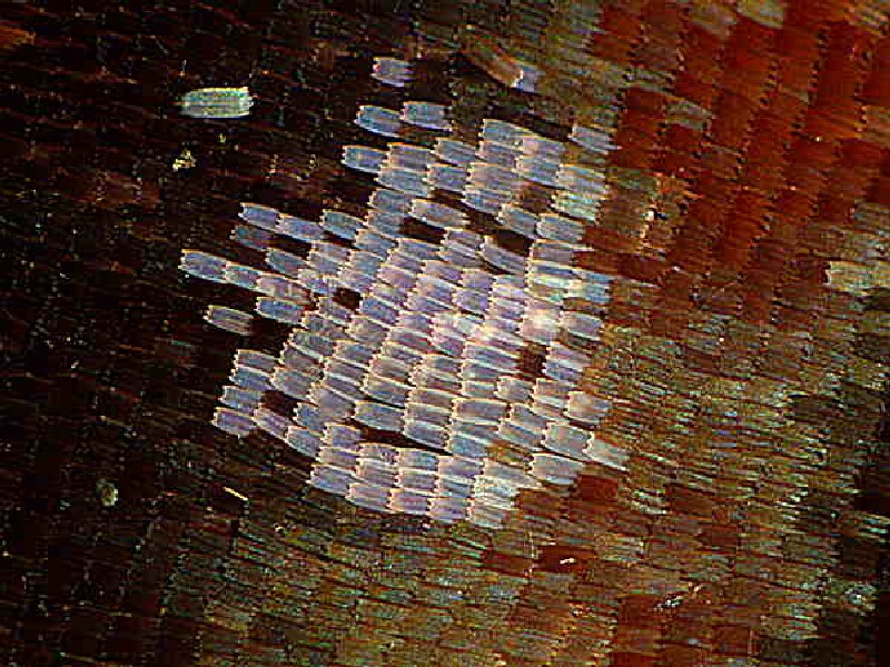
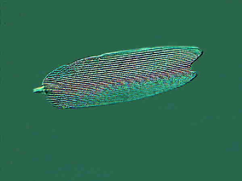
First, I’ll show you some portions of wings with some different sorts of scales. The first two are from an African Butterfly called Charax brutus. The second one shows some scales which are opalescent and the third shows a single scale.
Now, here are some images to show you the ingenious manner in which the scales are attached to the wing and then a single scale removed from that wing.
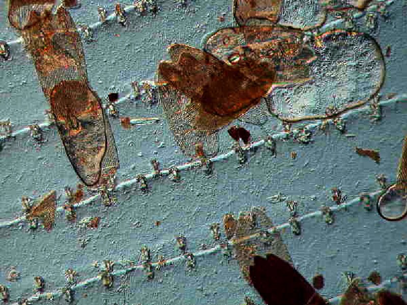
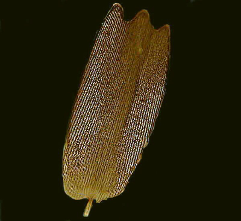
As you can see the minute “pin” tip at the base of the scale slides into a tiny pouch and there are a series of such pouches along the raised ribs which extend along the wings. The colors of the scales are of 2 basic types: 1) pigmented or 2) structural. If you hunt carefully and examine closely, you will find examples of this latter type which don’t contain a pigment, but achieve their color by refraction due to structure. So, butterflies and moths are sure winners unless you’re dealing with someone with a heart of stone.
Some people find all these “static” kinds of samples dull, because “they don’t do anything.” They want some “action”, some rock and roll, “something happenin’, dude.” It as this point that I become an advocate for retroactive abortion but, sometimes tolerance is the better part of the malodorous, so here’s where pond samples come in. If you’re going to convert the “unwashed”, then you must always have a goodly supply of pond scum on hand for their intellectual baptism.
Few can resist the charms of the busyness of a slide thick with Paramecia which fortunately are easy to obtain and culture readily. If you’re lucky, you may even find a specimen or two dividing and if you’re especially lucky you might find a conjugating pair (and, of course, everybody’s interested in sex). And, if you’re super lucky, you might happen across a large amoeba like A. proteus which are always a delight to observe when they are active. Then, if you’re supercalifragilisticexpialidociousally lucky and find some Volvox, then you’re home free, because only a troglodyte could resist the spectacle of the these green spheres rotating through the water with their indescribable elegance.
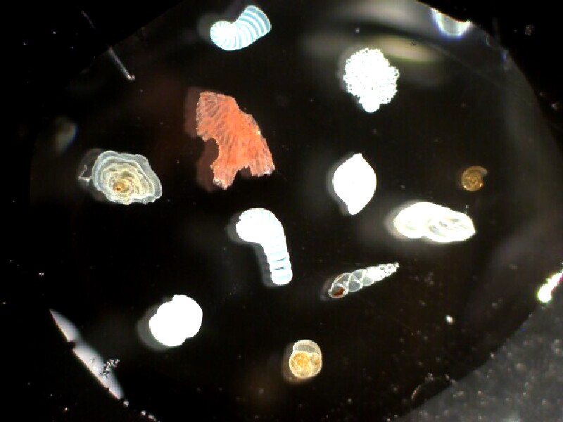
If your potential convert is a beach comber and collects shells, then you have an opportunity to show him or her a wide variety of micro-shells from forams to radiolaria to fossil bryozoa to tiny, colorful gastropod and bivalved shells. Slides of radiolaria are almost always impressive and there are usually at least a few specimens on a slide that look like miniature space vehicles.
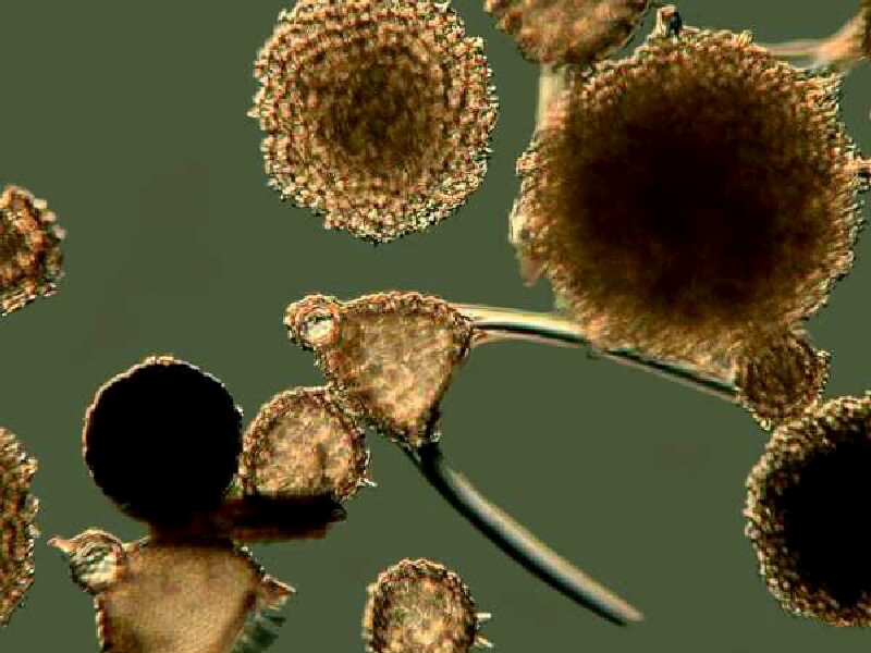
(Please forgive the quality of my image. Radiolaria are difficult to photograph, but show up very nicely when observed with the microscope and the judicious use of the fine focus).
Forams can be mounted on paleo-slides against a black background for observation with a stereo microscope. This image was taken from a slide prepared by Brian Darnton.
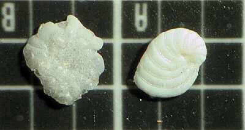
However, some forams mount up very nicely on glass slides, especially in a drop or two of immersion oil and can be observed with a compound microscope.
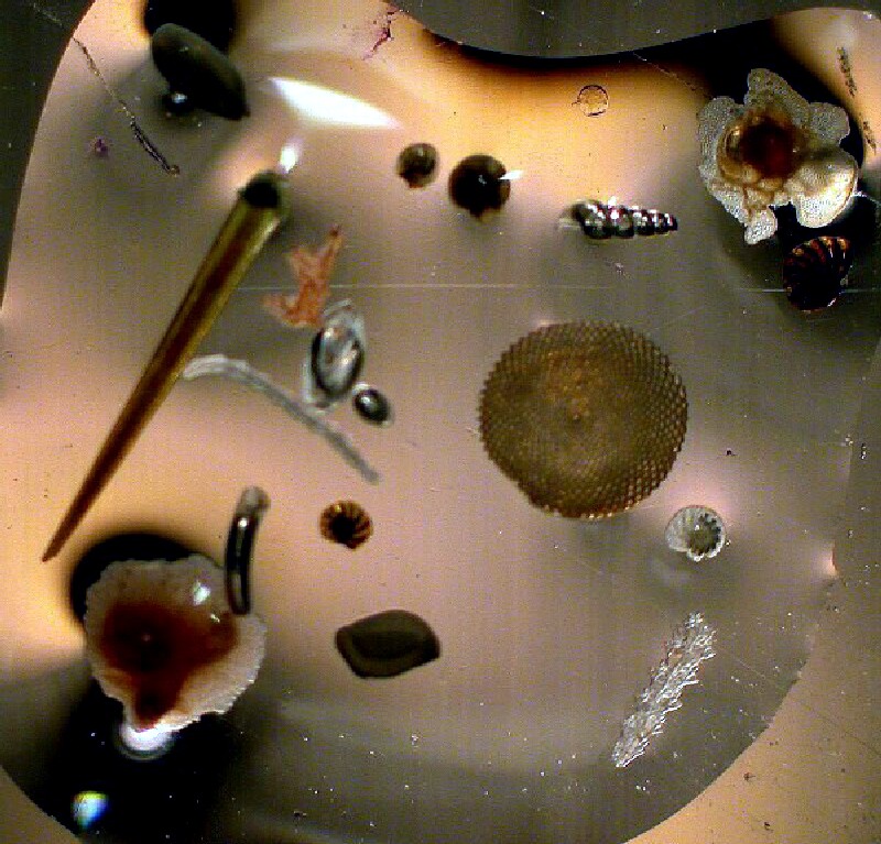
In sand samples, one can find not only small shells and fragments thereof, but spines of sea urchins and sand dollars, and spicules of sponges and soft corals.
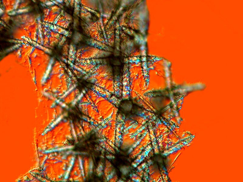
These can be mounted on slides either dry or in a resinous mountant.
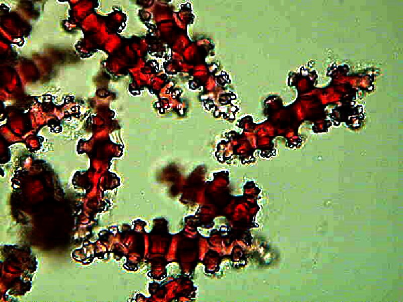
One of the most effective and spectacular bits of bait is slides of birefringent crystalline chemical compounds viewed under polarized light. Often common household items can provide impressive material but, in other cases, certain exotic mixtures can produce stunning results. Ascorbic acid (you can substitute scrapings from a Vitamin C tablet) produces very pleasing results and when mixed with certain other chemicals can generate a garden of colorful wonders.
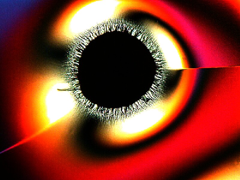
Some chemicals are especially desirable to work with since they can produce an extraordinary variety from a single slide. Below I am going to present you with a variety of images from a single slide containing a mixture of Nickel sulfate and Urea.
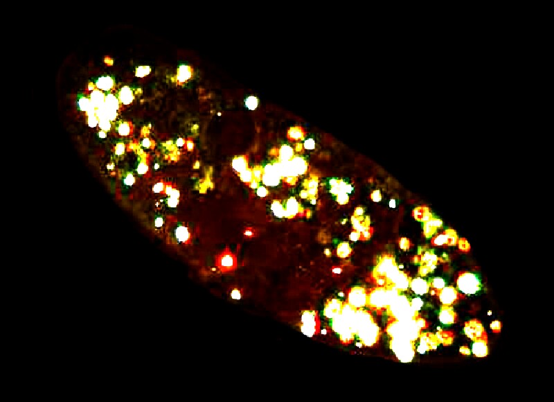
Virtually everyone enjoys a magic light show and I am going to suggest that you try 2 very different sorts. The first involves living specimens of Paramecia. Place a drop from a rich culture on a slide, put on a cover glass, and then set it up for brightfield viewing with a compound microscope. You can have the analyzer and polarizer in the light path, but set them so that they’re not crossed and you have basically a brightfield view. Let your friend or relative observe them quietly for a minute or two and then cross the polars. Paramecia accumulate calcium oxalate crystals which are birefringent, so against the black background of the crossed polars, you have micro-Christmas tree ornaments actively swimming around.
The second demonstration involves crystals again. Ideally, select a substance that is colorfully birefringent and soluble in 70% isopropyl (rubbing) alcohol. My substance of choice is the biological stain Orange G. If you pick a chemical that is only soluble in water, then you are liable to quickly lose your audience, for it can take a considerable, boring time (except to a Zen master) for the crystals to start appearing. The idea is to set everything up so that your “victim” can watch the crystals begin to appear against the blackness of the background as the alcohol evaporates. I have done this many times and still find it a delight, which almost always, the other observer shares.
With a stereo-microscope, one can display larger specimens and/or parts thereof. Small flowers can take on a whole new character and reveal structures that are unknown to most people.
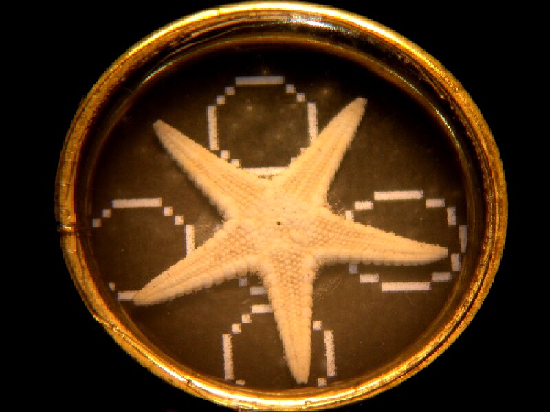
At craft shops, one can find starfish and sand dollars that are less than an inch in diameter and these can easily be turned into pleasing dry mount slides.
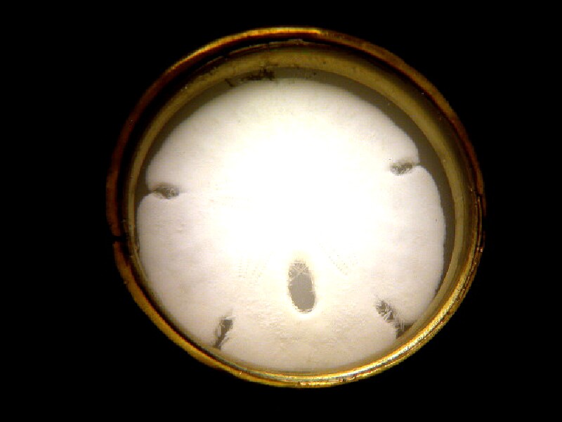
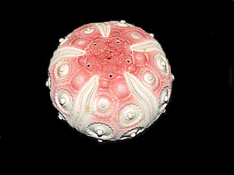
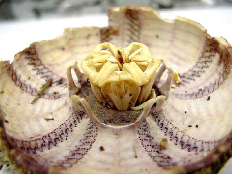
Seeing parts of the plates that comprise the “shell” or test of a sea urchin or examining its teeth which constitute that complex and intricate structure known as Aristotle’s lantern can reveal wonders never dreamed of.
What you display to entice someone toward microscopy and natural history will, of course, depend, in part, on his or her general interests, but also on your own specific interests as well. In any case, success does in large part depend upon being well prepared with a variety of specimens for both types of microscopes and you may end up thereby giving someone a special gift that will last a lifetime.
All comments to the author Richard Howey are welcomed.
Editor's note: Visit Richard Howey's new website at http://rhowey.googlepages.com/home where he plans to share aspects of his wide interests.
Microscopy UK Front
Page
Micscape
Magazine
Article
Library
Published in the May 2013 edition of Micscape Magazine.
Please report any Web problems or offer general comments to the Micscape Editor .
Micscape is the on-line monthly magazine of the Microscopy UK website at Microscopy-UK .
©
Onview.net Ltd, Microscopy-UK, and all contributors 1995
onwards. All rights reserved.
Main site is at
www.microscopy-uk.org.uk .