|
In
the first part of this work I
sought to
establish the position
of stentors within the extensive framework of the ciliate taxonomy.
Preparing now the key of the Stentoridae
family, I allow myself some freedom for the benefit of the amateur
microscopist, such as
including without ambiguity the genus Parastentor
* within
the family.
As I say below I
do not have any data for the
species Stentoropsis, so I do not
include it.
Moreover for the
reasons which I give below I consider it premature to
include Heterostentor.
* Parastentor is an
interesting
European species, which needs confirmation, and which perhaps
could be found and be redefined by some microscopist out of its
standard
locality in a Hungarian lake.
SUMMARY
OF THE FAMILY
STENTORIDAE
List of genera and
its type species
(generally this
is the first species assigned to the
genus)
Genus
Stentor Oken,
1815
Type species Stentor muelleri (Bory
of St. Vincent, 1824) Ehrenberg, 1831
Genus Maristentor, Lobban et al. 2002
Type species Maristentor
dinoferus Lobban et
al. 2002
Incertae sedis (genera
or
species for which there are doubts about its correct situation for
lack of
adequate data,)
Dennys Lynn
in her taxonomic list (to genus inclusively)
considers the
following genera of doubtful
position in the family:
Genus Stentoropsis Dogiel
and Bychowsky 1934,
Type species: S. barbi Dogiel and Bychowsky 1934
(I could not find neither a
description nor a bibliographical quotation for this genus and species,
accepted
as good by J. O. Corliss 1961 and by D. Lynn 2002. It does not appear
in any of
the consulted papers nor in any of the specialized sites on the Web.
Almost certainly the article which describes it is written in German.
Having no data
on this species I could not include it in the key of genera.)
Genus Parastentor Vuxanovici
1961,
Type species P. tentaculatus Vuxanovici 1961,
The species has two
retractile side tentacles covered in longer cilia than those of the
body. It
was observed twice, in a Hungarian Lake.
Genus Heterostentor, Song
and Wilbert 2002
Type species: Heterostentor coeruleus Song and Wilbert, 2002
Only one species known,
with intense blue pigment, and observed in the Antarctic Ocean.
Situation of
Heterostentor
In reality Heterostentor
resembles less of the features of Stentoridae than Condylostoma
auriculata does.
Its only character of Stentoridae is to have a peristomial bottom
surrounded by
a AZM similar to that of Stentor,
without undulating membrane, and the
ectodermic blue pigment. Its form is that of an ellipsoid very widened
with
one anterior contraction where the AZM is placed, and without
adhesive
foot. It is described without symbiotic algae, without cytopharynx or
visible cytostome
and its small peristomial bottom does not have cilia on its surface. It
is
a free
living protozoon never adhering to substrate as Stentor, Maristentor, Parastentor
and surely Stentoropsis does. In
my opinion so many differences would justify to separate it into a
different
family which would have to be named Heterostentoridae.
Although I give this description and its
iconography, I do not include it
in the key.
|
|
|
Heterostentor
coeruleus -
from Song & Wilbert 2002
|
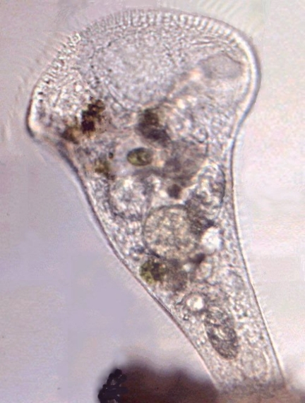
Stentor,
Maristentor and Parastentor have
the shape of a reversed cone, with an
adhesive foot, a definite pharynx and a cytostome.
|
LIST OF
VALID SPECIES OF THE GENUS STENTOR
Arranged
according to the date of its first description
|
|
01
|
St. polymorphus
|
(O F. Müller, 1773)
Ehrenberg, 1838
|
Cosmopolitan
|
|
02
|
St. Niger
|
(OF. Müller, 1773) Ehrenberg
1831
|
Europe
|
|
03
|
St. multiformis
|
(O F. Mueller, 1786) Ehrenberg
1838
|
Marine populations?
Cosmopolitan?
|
|
04
|
St. muelleri
|
(Bory St.
Vincent, 1824) Ehrenberg, 1838
|
Cosmopolitan **
|
|
05
|
St. coeruleus
|
Ehrenberg, 1830
|
Cosmopolitan **
|
|
06
|
St. roeselii
|
Ehrenberg, 1835
|
Cosmopolitan **
|
|
07
|
St. igneus
|
Ehrenberg, 1838
|
Cosmopolitan
|
|
08
|
St. barretti
|
Barrett 1870
|
Europe
|
|
09
|
St. elegans
|
Fromentel 1876
|
Central and
Eastern Europe **
|
|
10
|
St. amethystinus
|
Leidy, 1880
|
Cosmopolitan? **
|
|
11
|
St. fuliginosus
|
Forbes 1891
|
Cosmopolitan?
|
|
12
|
St. pyriformis
|
Johnson, 1893
|
North America and
Mexico
|
|
13
|
St. baicalius
|
(Swarczewsky 1929)
Foissner 1994
|
Lake Baikal
|
|
14
|
St. loricatus
|
Barry 1950
|
News Zealand
|
|
15
|
St. introversus
|
Tartar, 1958
|
North America
|
|
16
|
St. caudatus
|
Dragesco 1970
|
Africa
|
|
17
|
St.
multimicronucleatus
|
Dragesco 1970
|
Tropical Africa
|
|
18
|
St. katashimai
|
Kumazawa 1973
|
Japan
|
|
19
|
St. tartari
|
Narayana Murthy
and
Kasturi Bai 1974
|
described from Bangalore (India), and Kenya (Africa)
|
|
20
|
St. araucanus
|
Foissner and
Wölfl 1994
|
Andean lakes of
southern South America
|
|
21
|
St. cornutus
|
Kumazawa, 2002
|
Japan
|
|
** The species
marked by 2 asterisks are illustrated
in the key.
IDENTIFICATION
KEY
Abbreviations: Mn -
macronucleus, Mic. -
micronucleus, µ - microns, AZM Adoral Zone of Membranelles
For a more
convenient (and different) presentation, we will use here numbers
instead of letters.
1 (2)
|
Marine species. Shaped as very
wide trumpets, with a vast peristomial
bottom abruptly widened. Peristome divided by one deep central notch,
which
makes it appear bilobate. With many bands of cortical pigment and
abundant zooxanthella
(symbiotic algae derived from dinoflagelates).
With many cilia dispersed
in the vast
peristomial bottom. AZM similar to that of Stentor |
Maristentor
|
|
Only one species with a rounded
macronucleus located close to the anterior
end. Has a length of a
around 900
microns. It was
identified until now only on the island
of Guam. |
M.
dinoferus Lobban
and all, 2002 |
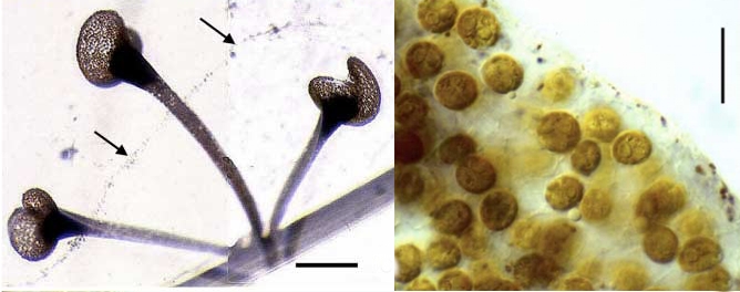
|
|
Maristentor
dinoferus - the second photo show the dinoflagellate related
zooxanthelles. from Lobban et
al. 2002
|
| 2(1) |
Freshwater species |
3 |
| 3(4)
|
with
retractile
and ciliated side
tentacles |
Parastentor |
|
Only
one species (Hungary)
|
P. tentaculatus Vuxanovichi
(1961) |
| 4(3)
|
with circular or reniforme
peristome. Cilia of the peristomial bottom
laid out in regular lines
which follow the contour of the peristome. Body
covered with longitudinal lines of cilia, which can contain Stereocils
("rigid sensory
hairs") of a size larger than the cilia, with
intermediate ectoplasmic bands and pigmented granules or not. Without
side tentacles |
5 |
5
|
we
have arrived at the genus
Stentor |
go to 6
|
|
The genus is
known
by the majority of microscopists, the most frequent species,
which normally have an average size (from 1 to 1.5 mm) have the
definite
shape of
a widened "trumpet". However some species of the
genus vary a lot, and
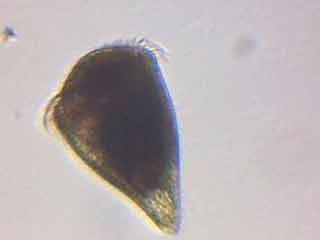
there are some conical, short forms (less than 200 microns), with a
very
wide peristomial
bottom, which gives them the shape of a reversed bell. When
contracted all the
forms adopt an ovoid or pear shaped aspect,
truncated by the AZM in the anterior end.
The number of
lines
of cilia of the peristomial bottom, like those of
the body,
is an important secondary specific character. Some species of Stentor
can
have symbiotic algae called zoochlorelles.
After this first
basic
dichotomy, the following options require more space to
continue with a
rigorous description
of only two options. Thus, and having shown to the beginners the
principle
of a dichotomous taxonomic key, to save space I give up it for a simple
table
with multiple choices and very easy to follow.
|
|
| 6
(7) |
With
zoochlorelles |
|
|
a)
Nucleus vermiforme,
blue
pigment...............................................................
|
araucanus |
|
b)
Nucleus
moniliform........................................................................................
|
polymorphus |
|
c) Nucleus
multiglobular....................................................................................
|
|
|
c1)
2 or 3 Mn, red pigment
(the
population
must be searched with care because of the possibility of the
subject being a rare multiglobular specimen of St. fuliginosus)
|
tartari |
|
c2)
2 (still 5) macronucleus.
Colourless.......................................................
|
pyriformis |
|
d) One single
and
globular macronucleus
|
|
|
d.1
Pigment crimson,
which can appear brown or even black, by effect of the zoochlorelles.
Mic.
surrounded by pigment granules
(see fuliginosus).................................................................................................
|
amethystinus |
|
|
|
Fig. 10 - Stentor
amethystinum, specimen
anaesthetized with potassium iodide. Picture by Christian
Colin. The combination of
the
pigment color and the zoochlorelles produces the
opaque and characteristic dark color.
|
|
d.2
maroon pigment or
reddish. Mic. not surrounded by granules of pigment (see amethystinus)........................................................................
|
fuliginosus |
|
According to
Foissner and Wölfl, 1994. It could be a
synonym of amethistinum. In
the two species the combination of zoochlorelles with the pigment tends
to mask the color, which appears almost black.
|
|
| 7
(6) |
Without
zoochlorelles |
|
|
a)
Nucleus vermiform
|
|
|
a1)
colourless*, a gelatinous
lorica.........................................................
|
roeselii |
|
a2)
green pigment, with
lorica.................................................................
|
loricatus |
|
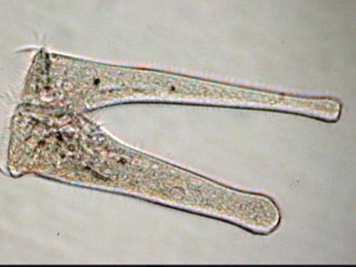
b)
Nucleus nodular,
peristomial bottom bilobate, long and thin cytoplasmic filament
connecting it to
the substratum, with lorica.......................................
|
barretti |
|
|
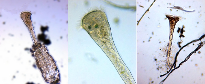
Stentor
roeselii - At
left a typical specimen with
consolidated lorica.
In the center the vermiform macronucleus characteristic of the species.
On
the right-hand side a specimen with a loose lorica, probably in
construction.
The
three pictures from Dominique Voisin
|
|
c) Nucleus moniliform
|
|
|
c1) bluish color
|
|
|
c1.1)
With buccal
pouch..........................................................................
|
coeruleus |
|
c1.2)
Without buccal
pouch, retractile peristomial edge.......................
|
introversus |
|
(in specimens
retracted of introversus, the peristomial edge form one
large pad
around the peristomial bottom, assimilating
almost completely the AZM)
|
|
|
|
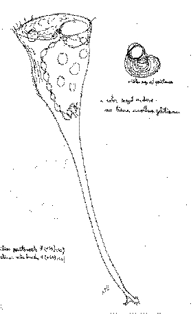
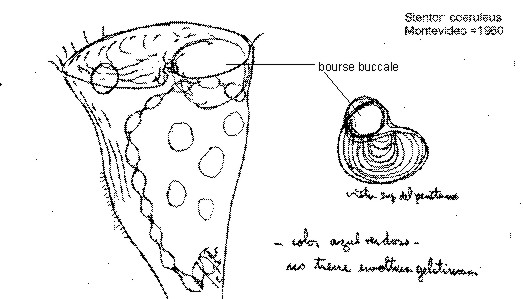
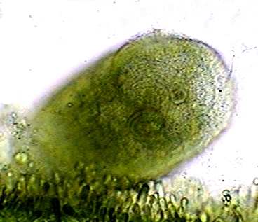
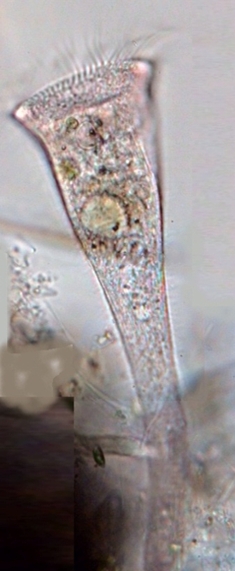
| St. coeruleus - picture
by Jean Marie Cavanihac |
Specimen from Montevideo (1960) personal drawing
with the assistance of an eyepiece grid |
|
The image on the
right-hand side (scanned from a 22 X 36 cm
original), is not intended to show that I found this stentor at Montevideo, but to
show that the "buccal pouch" of Kumazawa (2002) is a reality. For
better underlining this fact, I add below an augmented detail.
|
|
The
comments on the drawing read: a) upper view of the peristome.
b) bluish color, c) it does not have a
gelatinous sheath |
|
c2)
colourless
|
|
|
c2.1) Without
lorica
|
|
|
c2.1.1) Without
buccal
pouch
|
|
|
30-42
segments in Mn.
Short and squat in the shape of a bell with a lengthened
foot........................................................................
|
caudatus |
|
7-18
segments in Mn, in
the shape of long
trumpet..................................................................
|
cornutus |
|
c2.12)
With buccal pouch;
small and regularly
conical.............................................................................
|
katashimai |
|
c2.2)
With lorica,
without pouch, very long and thin, with long and rigid stereocils in regular
groups.......................................................................................
|
muelleri |

|
|
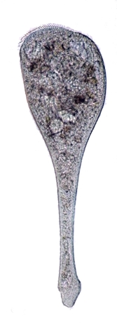
| One
colony of St, prob. muelleri on
Cladophora |
This is not a beautiful picture,
but one sees the concentric lines of
cilia in the peristomial bottom |
| Material
fixed with hot AFA, stained with Fast Green and mounted in NPM.
Cancún,
in a brackish aquarium. |
|
d)
Multiglobular nucleus
(4 globules), without zoochlorelles,
bluish............................................................................................................
|
baicalius |
|
e) Single globular
nucleus
|
|
|
e1) Colourless *
|
|
|
Macronucleus
40-62 µ,
52-142 Mic..............................
|
multimicronucleatus |
|
One
oval Macronucleus <40
µ......................................
|
elegans |
|
NOTE: * Colourless
: they can be transparent, but according to their size and the opacity
of the
cytoplasm they can appear yellowish, but their ectoplasmic granules are
not
pigmented.
|
|
|
|
|
| Two
images of a probable St. elegans from
Jean Marie
Cavanihac. It is a very rare species. The second photo shows what
appears to be
a "buccal pouch", a detail which is not described in this species.
However in the illustration presented by Foissner and Wölfl 1994,
and in spite
of its small size, one distinguishes an insinuation of the pouch. One
must pay
attention to this detail if the species is found again. |
|
e2) Coloured
|
|
|
Shades
of red........................................................
|
igneus |
|
Blue,
with a small lorica.......................................
|
multiformis |
|
Dark
coffee...........................................................
|
niger |
APPENDIX
Stentor
reproduction
Stentor
is a ciliate and like all those, it has two different reproduction
methods. One (which is primarily a cloning) consists of transverse
division of
an individual which, by doing this, distributes in equivalent manner
the
nuclear
contents between the two sister cells, without the phenomenon of
chromosomes reduction.
This phenomenon was carefully documented by Christian Colin in a
specimen which probably is Stentor
coeruleus.
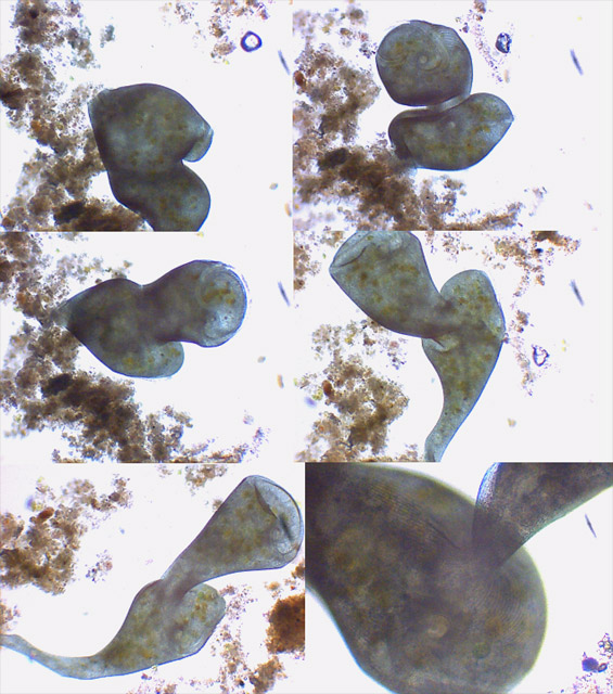
The other method implies sexual activity, and the
intervention of two individuals who, linked by their oral end, destroy
their
macronucleus, and through the established cytoplasmic bridge exchange
the
micronucleus which has already undergone the process called chromatic
reduction. Finally
they rebuild their macronucleus.
An infrequent
image of this phenomenon was provided
by Jean Marie Cavanihac and reproduced below.
| Note:
I suggest that those who find some stentors and who wishes to
use the key, use the “cut and paste” facility to extract the key and to
copy it
in a text processor for an easier consultation. |
|
REFERENCES
Colin,
Christian,
2003: Mon
Stentor-demon .Magazine
MicrOscOpies on - line
CORLISS,
John O., 1961: The
Ciliate
Protozoa. 310 pgs. XXII
plates. Pergamon Press
FOISSNER
W. & WÖLFL S.,
1994: Revision of
the
genus Stentor OKEN (Protozoa, Ciliophora) and description of S.
araucanus Nov.. spec. from South American lakes. – Journal of Plankton
Research - Vol.16 - pp. 255-289.
KAHL,
A., 1935.- Wimpertiere
oder ciliata.
on line: HTTP://ameba.Ihosei.ac.jp/DIB/Kahl/
KUMAZAWA
H, 2002.- Notes
on the
taxonomy of Stentor Oken (Protozoa, Ciliophora) and a description of a
new
species. Journal of
Plankton Research – Vol. 24 No.1 - pp. 69-75
LOBBAN,
C.S., SCHEFTER, M, SIMPSON,
A.G.B., LADLE, X.,
PAWLOWSKI, J., AND FOISSNER, W., 2002 - Maristentor
dinoferus N. gen., N. sp., a
giant heterotrich ciliate
(Spirotrichea: Heterotrichida) with zooxanthellae, from coral reefs of Guam, Marian Islands. –Marine
Biology, 2002. Vol. 140: pp 411-423
HTTP://www.uog.edu/dns/Maristentor_article.Pdf
DENNIS H. LYNN
(on
line, unpublished) June 14, 2002 - Classification of
the Phylum Ciliophora
HTTP://www.uoguelph.will
ca/~ciliates/classification/genera.HTML
For the most
recent, published revision by D.H. Lynn
see:
LYNN, D.H.
AND E.B. SMALL. 1997. Revised
Classification of the Phylum Ciliophora Doflein, 1901. Rev. Soc.
Mex. Hist. Nat. Vol.47: pp 65-78.
SONG,
Weibo and WILBERT, Norbert.
2002 - Faunistic
Studies on Marine Ciliates from the Antarctic Benthic Area,
Including Descriptions of One Epizoic Form, 6 New Species, and 2 New
Genera
(Protozoa: Ciliophora) Acta
Protozool. Vol.41: 23 - 61.
HTTP://www.nencki.gov.pl/pdf/ap/ap583.pdf
Fauré-Fremiet, et. 1936 - Condylostoma
(Stentor) auriculatus (Gruber) Bull. Soc. Zool.
France 61: 511-519.
(I
was able to consult this last work by courtesy of Prof. Daniel Nardin.)
Acknowledgements
I
thank in particular Doctor Foissner
and Doctor
Kumazawa, who made possible this
article by kindly sending their papers to me, and to all those
authors who liberally
shared their research on the Web. And especially to Domenique Voisin
and Jean
Marie Cavanihac, who allowed the free use of their pictures to
illustrate this
article. It is at the same time a Homage to Christian Colin, and a
recognition of the important labour of research and documentation made
in the past
few years by the participants in the French Forums of Microscopy.
|
|