Image
gallery: 'Big' slides
Images
of large microscope slide subjects created using a Nikon Coolscan IV ED 35mm
film scanner.
by David Walker, UK
In a recent article I shared my experiences of imaging prepared microscope slide subjects using a 35 mm film scanner, a Nikon Coolscan IV ED (2900 dpi). Although a technique new to me as a microscopy hobbyist, the practice is apparently very well established with professionals e.g. for imaging large histological sections and Nikon make an adaptor for their Coolscan scanners to scan microscope slides, although I used a homemade one.
From my own trials I was impressed with the ease of imaging very large subjects up to 35x24mm with even lighting to give good quality results, so I have delved into my modest microscope slide collection and have scanned some of the largest subjects I have, a selection are below. Some were so large they still required two image merges, although this is far fewer than the stitching needed if a low power microscope objective was used.
The imaging technique can be extended to cross polarisation just by temporarily mounting a polarising filter on top of and below the microscope slide. Use of a quarter or full wave retardation plate with the crossed polars is also possible as there's a generous height in the Coolscan slide holder. Thus a film scanner can act as a quick and easy to use digital macroscope with polarisation facilities for imaging e.g. thin rock sections and crystal slides with a field of view not readily possible with a typical compound microscope.
As remarked in the earlier article, a film scanner isn't needed for this sort of imaging but is very convenient if one is available. A modest consumer digicam (ca. >3 megapixel) with macro can often match if not outperform a film scanner as the digicam can zoom in to fill the frame whereas the scanner can't. Even lighting can be achieved with a digicam by e.g. using a 35mm photo slide light box which has daylight balanced lighting. Digicams have the advantage in that they are less likely to show any debris in the slide mount with diffuse light sources, film scanners with non-diffuse light sources can be very unforgiving in this respect.
Comments to the author David Walker are welcomed.
Update: The performance of the Coolscan IV is compared with that of a Minolta Scan Elite 5400 in a May 2005 article.
Image note: The film scanner can potentially create up to 10.1 megapixel images for subjects that fill the 35 mm film area so the extensive resizing below inevitably loses detail and sharpness. Some resharpening has been added to compensate. A couple of images are clickable to view the cropped masters.
INSECTS
These were slides prepared by the author using material provided by and with the guidance of the sadly missed Eric Marson (Northern Biological Supplies) at one of his Belstead House courses.
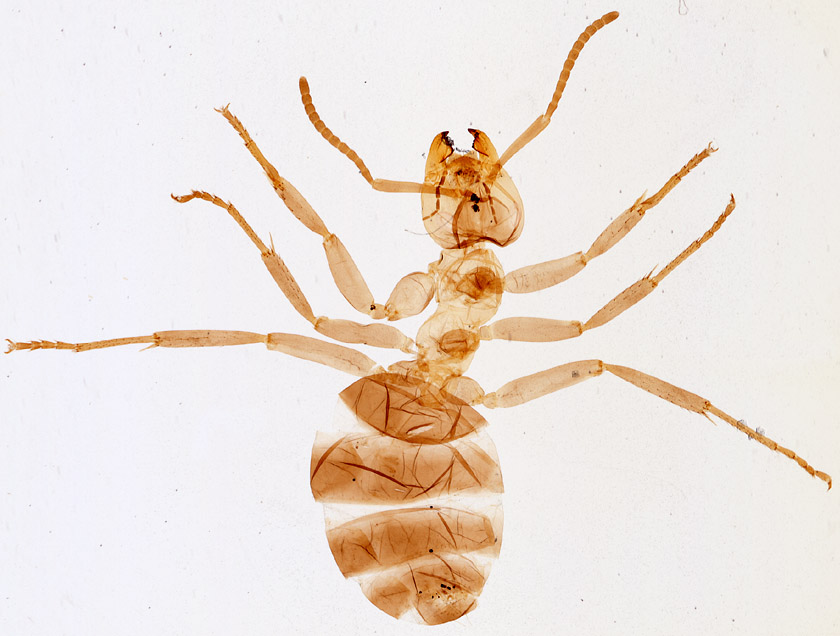
Wood ant, Formica, worker. 13 x 16 mm.
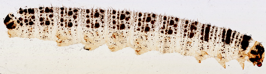
Large
white, Pieris, butterfly larva, 37 x 6 mm. Two scans stitched using the automatic
'Photomerge' command
in Photoshop Elements®.
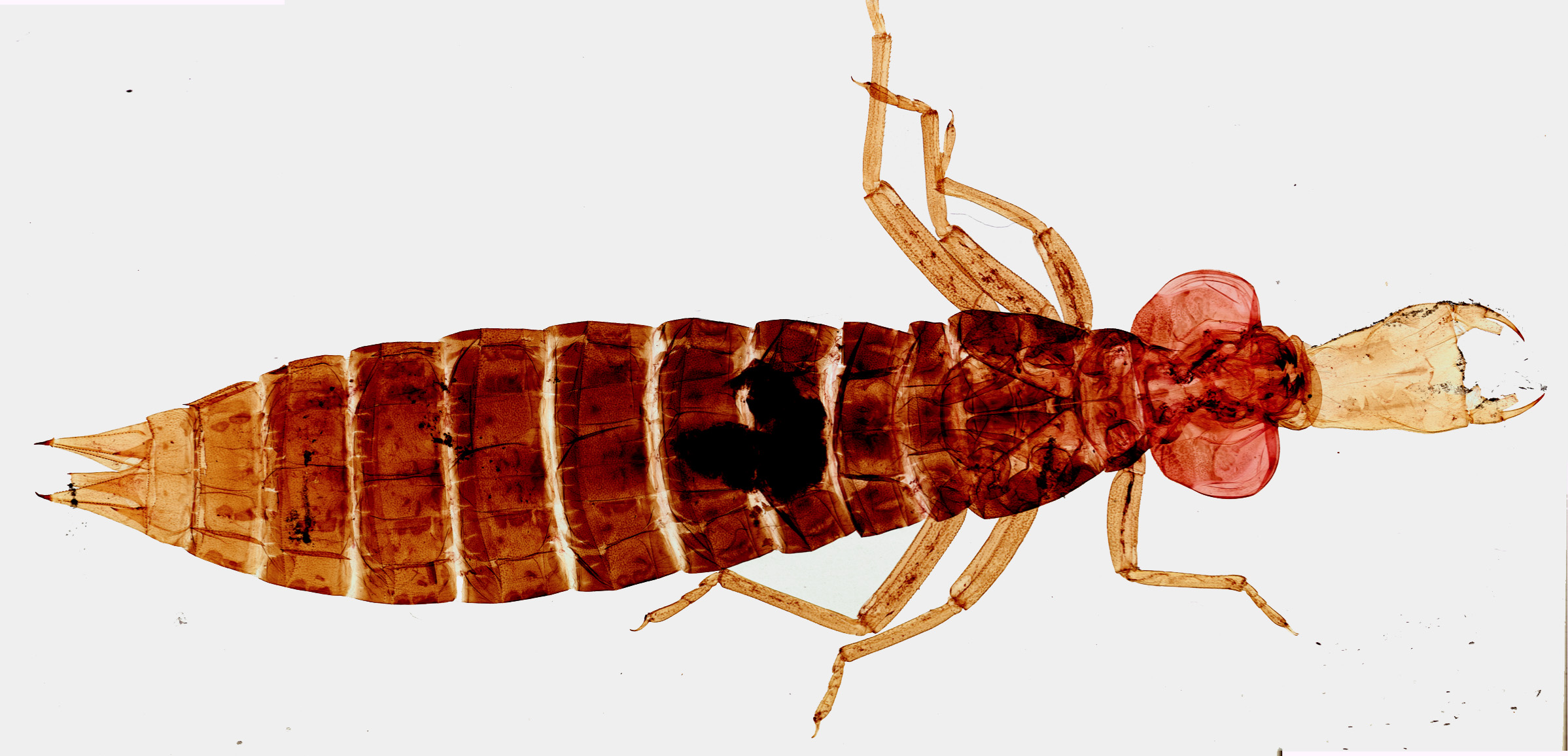
Aeshna dragonfly nymph. 41 x 18 mm. The largest subject in my slide collection. It could be imaged with two scans which were stitched. When the outline of the subject is simple, as here, the 'bucket fill' command in image editing software can be used to cleanly remove slide debris. Click image to view master.
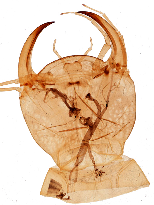
Head
of water beetle larvae, Dytiscus, 12 x 7 mm.
'Bucket fill' of background.
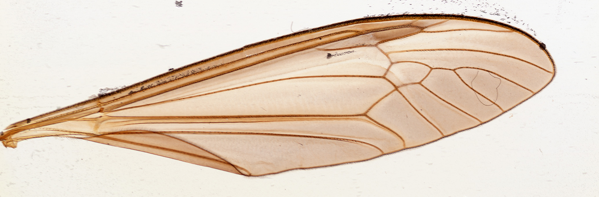
Crane fly, Tipula wing, 18 x 4.5 mm. Click image to view master.
CROSSED POLARISATION FILTERS
In the earlier trials I used two odd bits of polaroid filter to hand, but have since made two 25mm x 25mm squares (i.e. the width of a standard slide) of polaroid, each with the plane of polarisation parallel to an edge, which allows quick and precise extinction alignment. Small quantities of easy to cut plastic polarisation / retardations sheets are cheaply available from a variety of sources. e.g. Knight Optical in the UK. The field of view in the two examples below were each ca. 13 x 13 mm and limited by the size of retardation filter to hand. Up to 35 x 24 mm can be achieved i.e. the size of the 35 mm film frame if filters of the appropriate size were used.
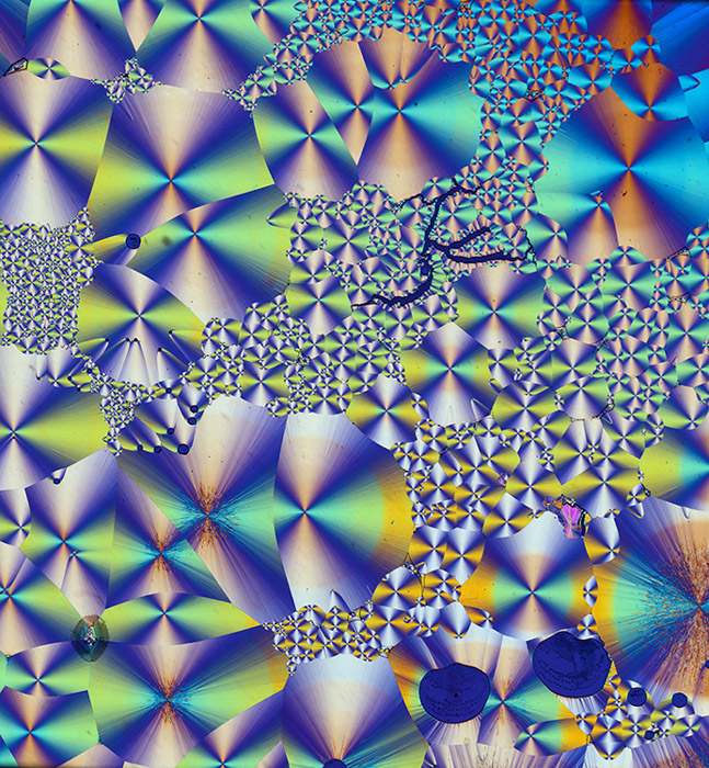
Cholesterol
acetate, crossed polars and lambda retardation plate. Biosil slide.
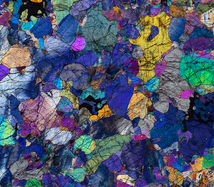
Peridotite,
Italy. Thin
rock section. Slide C, Open University (UK) set S260.
Crossed polars and lambda retardation plate.
Please report any Web problems or offer general comments to the Micscape Editor .
Micscape is the on-line monthly magazine of the Microscopy UK web site at Microscopy-UK