Trials on scanning microscope slides of large subjects with a 35 mm Nikon Coolscan IV ED slide scanner
by David Walker, UK
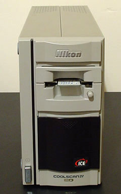
I recently treated myself to a secondhand 35 mm slide scanner, a Nikon Coolscan IV ED with 2900 dpi optical resolution. This was bought to rejuvenate slides and negatives which I'd built up over many years of casual 'snapping'. A few years ago good quality slide scanners attracted high prices and were well beyond my budget, but prices of new models have been steadily dropping and correspondingly, the previous to current models are becoming good value when bought secondhand.
Nikon sell an attachment for most of their Coolscan range to allow microscope slides to be scanned, which prompted my interest as a microscopy enthusiast as it seems to offer a route for easily imaging slide subjects up to 35 x 23 mm (i.e. the 35 mm film scanning aperture) with even illumination. The attachment is called the 'FH-G1 Medical holder' (see e.g. Nikon Singapore site for image and downloadable pdf document) and supports a microscope slide in the film plane of the 35 mm slide holder. The item is expensive (street price typically US $270) so was interested in making a homemade holder. An inspection of the chamber where the photo film attachments are inserted suggests that the mechanics and optics keep well away from the film plane during scanning, so a home made support should be as effective, costs nothing and most importantly wouldn't harm the scanner. Nikon's downloadable documentation for the FH-G1 recommends that microscope slides are inserted with coverslip uppermost and the total thickness of microscope slide / coverslip / cement can be up to 2 mm. So as long as a homemade holder places the microscope slide within 1 mm or so of the normal film plane, the scanner's autofocus or manual focus override hopefully should cope.
The slot into which the 35 mm photo slide is inserted is a generous height so I made a simple modification to an empty plastic 35mm slide mount; the top half of the mount was removed and replaced with two cardboard pieces to guide and loosely hold the microscope slide in place. A subsequent Google search showed that a similar approach has been adopted by some professional users as well, suggesting this is a successful and safe design. (See disclaimer.)
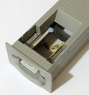
Left: The homemade slide mount. Two blobs of Blu-Tack® is extra security to ensure the slide doesn't fall out in the scanner. Middle: The slide holder in the 35 mm photo slide holder. The plastic mount is gently pushed into the holder not the slide. The button right safely pushes out the slide mount after scanning. Right: The Coolscan IV must be used vertically not horizontally for microscope slide scanning with a homemade slide holder.
Results
I was unable to find on the Web actual examples of such scans with the FH-G1 / homemade equivalents or how a film scanner performed in microscopy terms so wasn't sure what sort of quality to expect. So the easiest way to answer how a film scanner performed was 'try it and see' .....
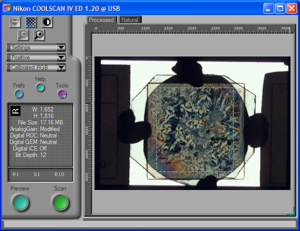
The Nikon Scan software supplied allows cropping and many image adjustments on the preview before full scan. The default settings were fine for many slides, the pre-scan histograms showed good capture of highlight, midtone and shadow detail. For lighter slides the brightness was decreased, for the crossed polar studies (example shown above) the Analogue Gain was set to maximum (2). No sharpening at all was needed before scans except a little post processing on smaller images to correct for softening caused by resizing.
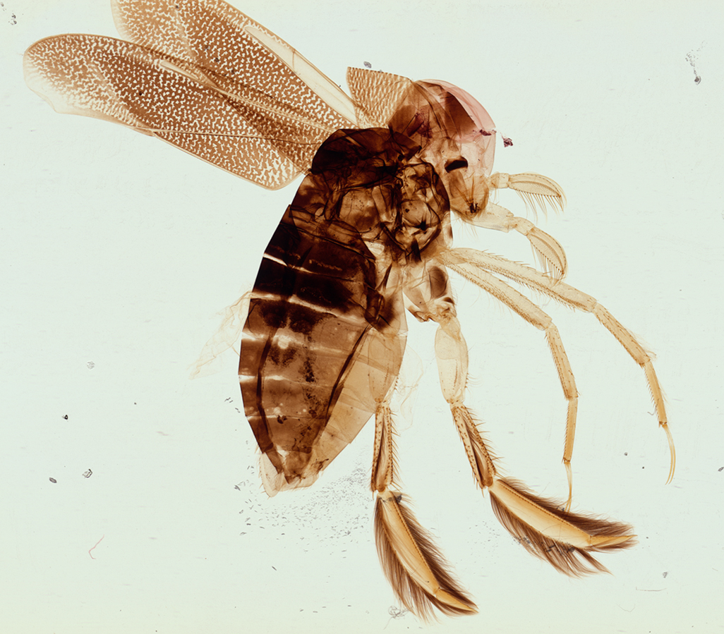
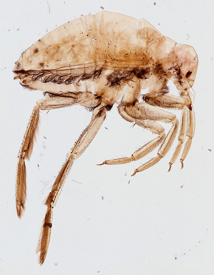
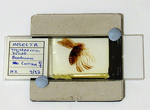
Water boatman, Corixa, female (NBS slide), subject 22x22 mm. The scanner gives good tone and detail. The scans are sharp suggesting the home made slide holder positions the slide well enough for the autofocus. Click image to view master.
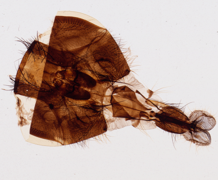
Blowfly head and tongue, Calliphora (NBS slide), subject 6x4 mm. The exoskeleton is quite dense in parts but default settings give detail in all areas. Click image to view master.
Stained octopus tentacle with suckers, (Biosil slide), subject 10x5.5 mm. Large scale features are shown in the master image with the Coolscan IV 2900 dpi scanner. As a hobbyist I'm uncertain of the professional applications of microscope slide scanning with the Coolscan range e.g. with Nikon's 'Medical holder' or homemade equivalent; possibly studies of gross structures of histology sections. I'd appreciate feedback from readers doing these sort of scans as to what the medical uses of such slide scans are. (Update August 26th 2004 - see footnote on resources providing an insight into this, kindly provided by George McNamara).
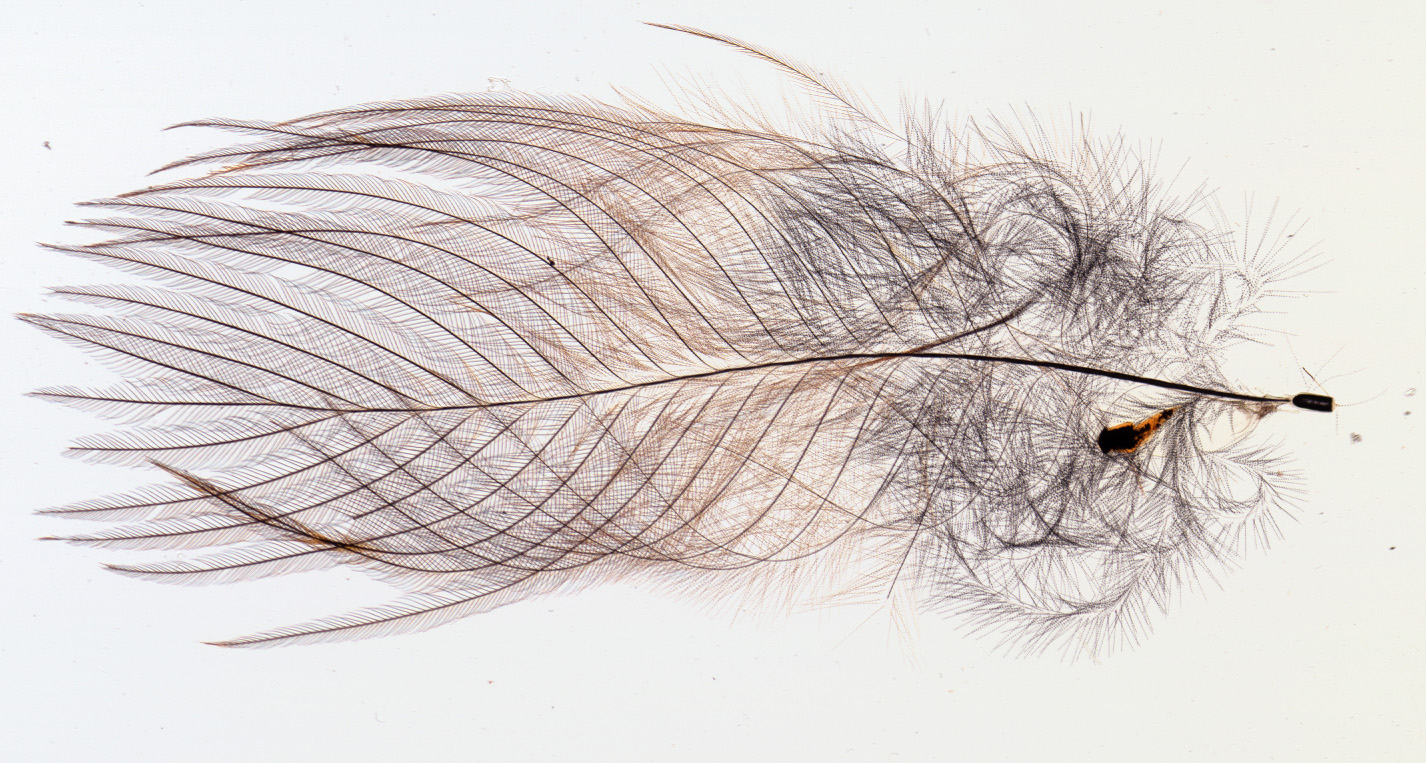
Robin's breast feather, (Biosil slide), unresized detail from full scan. Field of full scan 10x5 mm. Digital cameras can start to show aliasing, 'jaggies' and other artefacts with this sort of fine line detail. The scanner seems to do a reasonable job, keeping smooth linear detail and records the genuine dotted effect in the finer strands on the right. Click image to view full master scan.

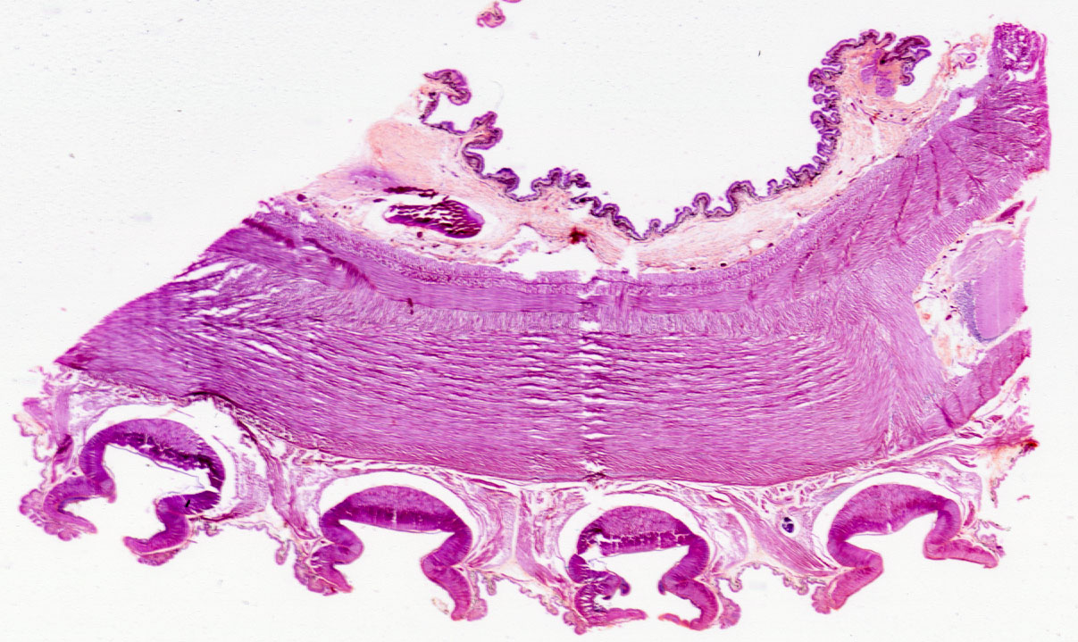
Pumpkin, stained T/S and L/S section, (Biosil slide), field width 24 mm. The large scan area is useful for multi section mounts like this and for showing the overall structure and larger features of botanical sections. In the master image, the smallest features just distinguished are ca. 20 microns across. To put this resolution in a microscopy perspective, a specialist 1.25x NA0.04 planapo compound microscope objective has a theoretical resolution of ca. 7 microns. A more typical 3.5x NA0.10 achromatic objective has a resolution of 2.75 microns - both for green light 550 nm. Click image to view master.
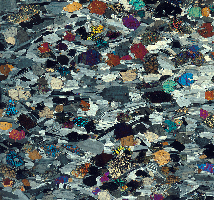
Gabbro, thin rock section between crossed polars, field ca. 15x15 mm. Open University slide K in set S260. I tried attaching a Polaroid filter above and below suitable slide with a little Blu-Tack® which still allowed the slide to be safely inserted without jamming. With default settings, scans were dark although images could be pulled back in software. But turning the Analogue Gain of the scanner to max did give good straight scans. The Coolscan V has a better dynamic range than the Coolscan IV (for better shadow detail in dense 35 mm slides) so should cope even better. Click image to view master. As a novice at interpreting these sort of slides, the ability to view in a single wide field image the types, features and relative amounts of the component minerals was very useful, cf visual scanning of a much smaller field on a microscope slide.
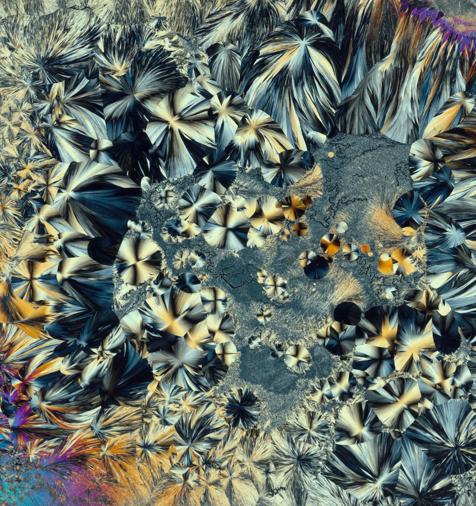
Santonin (NBS slide), field 15x15 mm, this is almost the full coverslip view and limited only by the size of the Polaroid pieces to hand (see software capture image above). The typical field of view of the author's 3.5x LOMO objective with 7x eyepiece and 1.5x binocular head is 3.5 mm, so it's one way of enjoying such slides in the 'bigger picture'. Click image to view master.
Comparison with a digital camera
A convenient way of imaging large microscope subjects is with a consumer digicam in macro mode, with or without macro extenders. The Sony S75 3.3 megapixel camera I use can achieve a similar field of view to the scanner with a reversed 50mm SLR lens attached albeit with vignetting. Digicams which excel at macro e.g. the Coolpix 4500 or 9xx series could also do a comparable job with a better ability than the S75 to zoom in on detail and are good value secondhand. Vishnu Reddy in a Micscape article has shared results from a digital macroscope built around a Coolpix 9xx for copying microscope slides .
In principle the 10.1 megapixel full 35 mm frame image scan from the Coolscan IV should exceed the performance of the S75 3.3 megapixel digicam, but in practice many microscope slide subjects occupy a smaller field than the 35 mm frame on the scanner with no ability to zoom in, whereas a digicam has the ability to zoom in to fill the frame.
|
Sony S75 with reversed Nikkor 50 mm f1.4 lens attached.
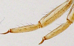
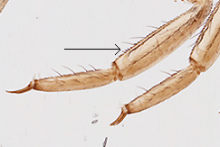
Leg detail from water boatman resized to match the image right. The whole insect filled ca. 70% of the camera's field of view. Even tones like the background when viewed at full resolution can be noisy on digicams. Update Aug. 25th. I've recently been trying the very powerful noise reduction software called Neat Image, which has ready made 'Device Noise Profiles' for most consumer digicams. It does an excellent job of removing the even tone camera noise without affecting detail. Click here to see master of above image treated with Neat Image. Compare background of treated with master clickable untreated image above. The digicam image above was taken using a diffuse light box for viewing 35 mm slides and does not emphasise the slide debris / dust. |
Coolscan IV
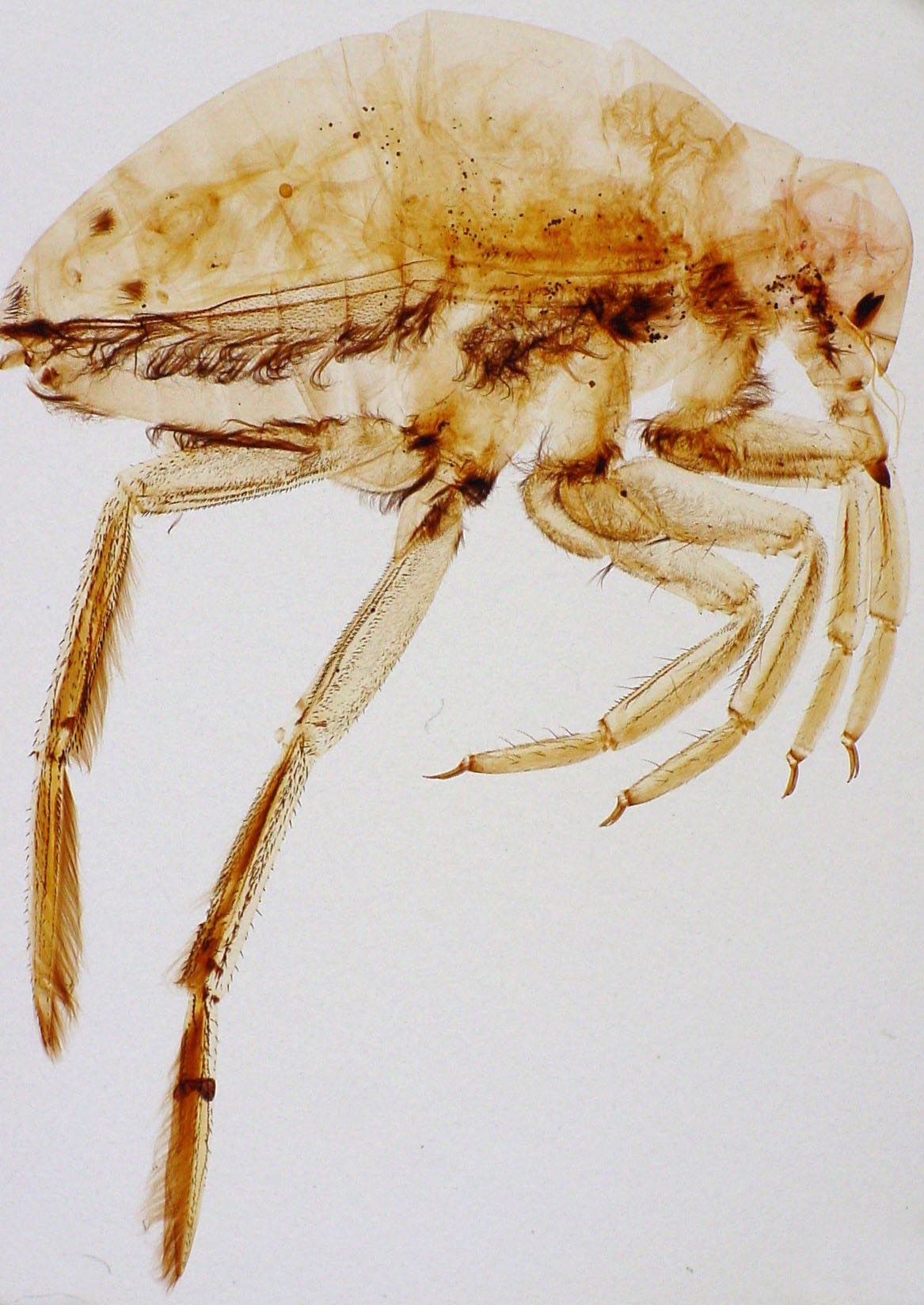
The same subject only occupied about 60% of the full imaging field. Resolution seems comparable to the Sony S75 but less noisy; both were able to resolve the arrowed hairs which were measured to be 58 microns apart under a microscope. The collimated light source of the Coolscan IV right shows the slide debris more but perhaps gives better edge contrast.
|
Conclusions to date: I think the Coolscan IV 2900 dpi scanner does a good job of creating a digital image of suitable slides at low mags and certainly good enough for screen / web use and small prints; the current model the Coolscan V with 4000 optical dpi should do better. The scanner for me offers most potential when the subject fills the 35 mm frame area to exploit the full pixel count. I've started to scan some large subjects in my microscope slide collection just to enjoy viewing the mount in its entirety, in addition to visual scan viewing with the low power objective; it should be stressed that even the most modest 3.5x objective will resolve far more detail visually.
I don't think purchasing a scanner by the hobbyist would be justified for microscope slide scanning alone, for many subjects a consumer digicam in macro mode would also give good results (including for the large field crossed polar images), but if a scanner is owned, the Coolscan IV or similar models offer a useful way of quickly imaging larger microscope slides.
Comments to the author David Walker are welcomed.
Updates: A further image gallery of scanned large slide subjects was shared in the October 2004 Micscape.
The performance of the Coolscan IV is compared with that of a Minolta Scan Elite 5400 in a May 2005 article.
(Disclaimer: Using a film scanner with homemade attachments is at the owner's risk, the author accepts no liability for damage to scanner or owner. If the scanner is damaged it would invalidate the maker's guarantee. The author believes the method to be safe when used with care for a Coolscan IV used in its vertical position but may not be for other makes or models of a different design.)
Footnote 1: Thank you to George McNamara (Childrens Hospital Los Angeles - Image Core) who kindly emailed and provided a fascinating insight into the professional use of 35 mm slide scanners for digitising microscope slides. Some of George's notes and the resources he mentioned are below, shared with his permission:
1) http://home.earthlink.net/~tiki_goddess/ - a stunning image of a microscope slide of a stained whole mouse embryo by George presented on his home pages. The page also provides information on the 4000 dpi scanner he used with links to manufacturers' resources.
2) http://www.jhc.org/cgi/content/full/51/2/151 - Computerized Quantification of Tissue Vascularization Using High-resolution Slide Scanning of Whole Tumor Sections by Christophe F. Chantrain, Yves A. DeClerck, Susan Groshen, and George McNamara.
3) Montague PR, Meyer M, Folberg R (1995) Technique for the digital imaging of histopathologic preparations of eyes for research and publication. Ophthalmology. 1995 Aug;102(8): 1248-51.
George remarks that ref. 3 is 'the earliest paper I've seen on using a 35 mm scanner for microscope' and he would like to know of anyone who used one before 1995. (Please contact George McNamara via the email contact on his web page at http://home.earthlink.net/~tiki_goddess/ ).
4) Flatbed scanners: George notes that 'some flatbed scanners are getting into the price range and resolution to make them worth considering. There are a number of papers on using flatbed scanners to digitize 2D and 3D specimens (Beer 1999 Amer J Surgical Pathology, for example) and one by Krout et al 2002, J Neuroscience Methods on using the (expensive) Creo Scitech high resolution scanner with ~2 um pixel size.'
Footnote
2: March 2005 update: Thank you also to Paulette Herlin who kindly supplied the following
refs. of her professional work with slide scanning. She writes: We are
currently using this acquisition mode for getting 2700 to 4000dpi virtual
slides from histological sections of tumors and quantifying tissue and cell
markers at the François Baclesse Cancer Centre in Caen (France). May I give
you some references:
1 - Nga Tran Kim, Nicolas Elie, Benoit
Plancoulaine, Paulette Herlin, Michel Coster. An original approach for
quantification of blood vessels on the whole tumour section. "Analytical
Cellular Pathology" 25 (2003) 63-75.
2 - Nicolas Elie, Benoit
Plancoulaine, Jehan-Pierre Signolle, Paulette Herlin. A simple Way of
Quantifying Immunostained Cell Nuclei on the whole histologic Section.
"Cytometry" Part A 56A:37-45 (2003).
3 - Nicolas Elie. Contribution à
létude du stroma des tumeurs ovariennes humaines par traitement et analyse
dimages numériques. Thèse de doctorat de lUniversité de Caen
Basse-Normandie, 16 décembre 2003.
4 - Ronan Francoise, Jean Jacques
Michels, Benoit Plancoulaine, Paulette Herlin. Optimal Resolution for
automatic quantification of blood vessels on digitized images of the whole
cancer section. "Image Analysis and Stereology" (2005) 24:1-9.
5 -
Nicolas Elie, Alexandre Labiche, Jean Jacques Michels, Paulette Herlin, Low
resolution evaluation of ovarian tumor stromal compartment. Accepted for
publication in "Image Analysis and Stereology", mars 2005.
Please report any Web problems or offer general comments to the Micscape Editor .
Micscape is the on-line monthly magazine of the Microscopy UK web site at Microscopy-UK