 |
PART 4 INTESTINAL OXYUROIDS,
INHABITANTS OF PERIPLANETA
AMERICANA FROM DURANGO AND CANCÚN, MÉXICO
|
|
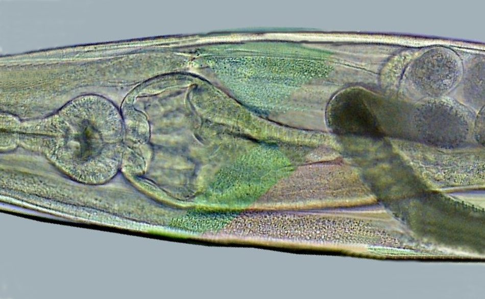
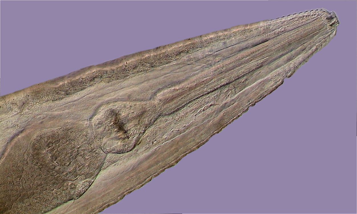
The image is a picture
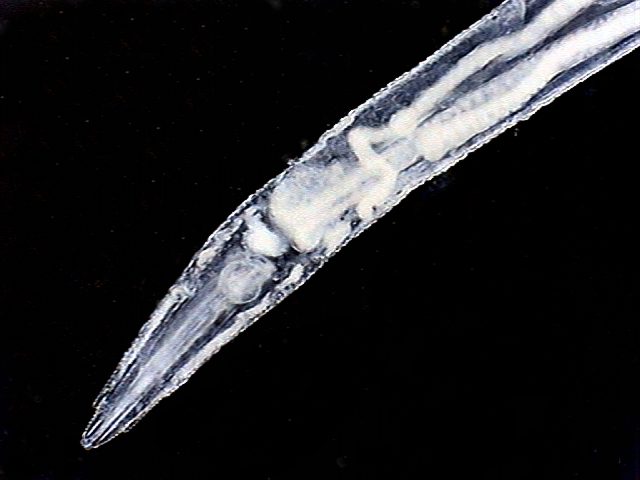
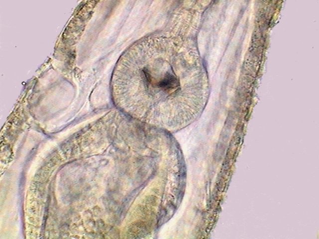
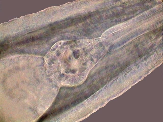
at the level of the bulb in the digestive system of a thelastomatid
nematode of the genus Thelastoma.
At right there is the long and thin
pharynx, conected to the bulb via a tight sphincter. The bulb itself
connects to the intestine at left through a muscular valve.
Materials absorbed through the pharynx are ground in the bulb and
passed to the intestine to be digested. Image taken with the 10x
objective, with the aid of a COL-J contrast disc. Except where stated
otherwise, the included images are personal pictures obtained with a
digital camera of 0.4 Mpx. integrated in my American Optical DC3-163-P
microscope equipped with planachromatic optics (Ocular 10x, Objectives:
x4 (NA 0.10), x10 (NA 0.25), x40 (NA 0.65) and x 100 HI (NA 1.25)). The
original ones have been captured at 640 x 480 px. and reduced or
trimmed as was necessary to include them in this work. An entire
image’s work-up, including mosaics of several images was
made in Photo Paint. In the image legends the objective
with which it was taken is indicated, just as a suggestion of the power
used because of the different sizes of each image. A number of
contrast devices (Rheinberg discs, darkfield discs, COL discs, and
the Mathias arrow), had been used to impart color, or relief to the
images. |
|
INTRODUCTION
The parasitic nematodes of cockroaches are near relatives of the well-known Enterobius vermicularis, a parasite of the intestine of young children. They belong to an order of parasitic Nematodes (OXYURIDA) with numerous species, that parasitize vertebrates and invertebrates, mainly arthropods. In English the oxyurida used to be called "pin worms" because of their thin shape and because the body generally had one long sharp pointed tail. The order is normally divided into two Superfamilies, due to the affinity of each one to vertebrate hosts (Superfamily OXYUROIDEA) or invertebrate ones (Superfamily THELASTOMOIDEA). Each superfamily has a long list of subordinated families, and each one of these a long list of genera and species. They are normally assigned to the Order OXYURIDA with about 850 species. Note: those who
need to
refresh their taxonomy knowledge can review: The cockroaches being arthropods, it's expected that their parasites belong to the second superfamily, which is integrated into five families. The species found in Periplaneta
americana belong to the type family (the first
described family) of the Superfamily: THELASTOMATIDAE. The
family has at least 9 genera accepted as valid, but in the
Periplaneta americana from North America, there are only
three species described of three genera
Never, better that
in this case can it be said that an image is worth a thousand words. We
will add commentaries only when the labels applied cannot be fully
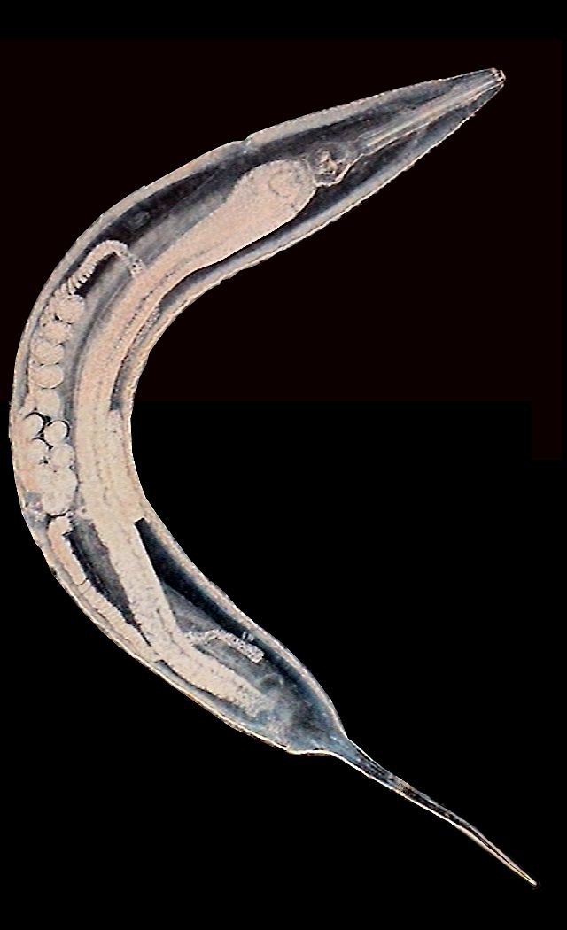
understood. Thelastoma bulhoësi (female)
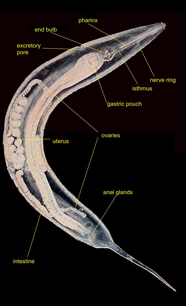
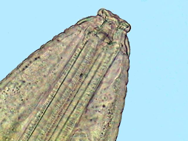
This female is only a little shorter that the one of Hammerschmidtiella. cuticle appears clearly ridged in cross-section. The anterior end is provided with eight lips easily visible and a second circle of muscular papillae that can be folded backwards. A short single anterior segment can be telescoped within the body, and is more mobile and independent than the rest of the body. After the anus, the body narrows rapidly where the long thin and pointed tail starts.
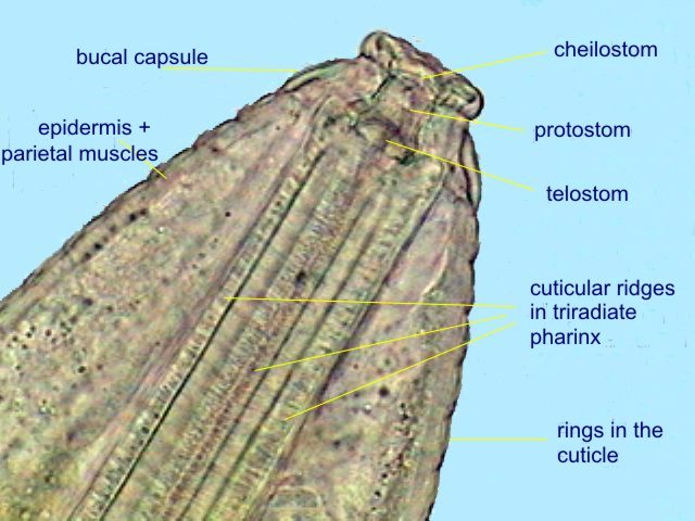
The mouth gives
entrance to one short and cylindrical buccal
capsule
in which can be clearly distinguished a cylindrical (almost annular)
anterior
portion, the cheilostom or vestibule, a
medium and
longerportion, named protostom
and finally another small basal
portion
with its inferior angles rounded, the telostom,
that opens in the pharynx or esophagus.
This
is long, straight and slightly conical, without a corpus or a
pseudobulb.
(The “corpus” is a widened portion of the pharynx anterior to the
pseudobulb). The
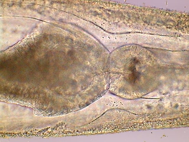
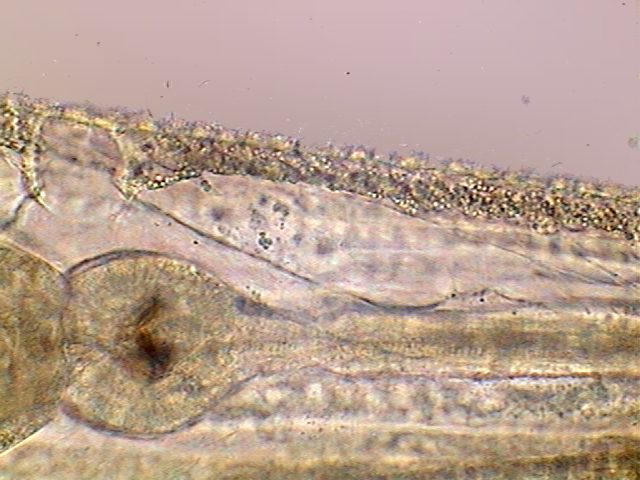
access to the bulb (a
muscular and spherical expansion at the end of the pharynx) is
controlled by a very
evident sphincter. You can see these
structures in the title image.
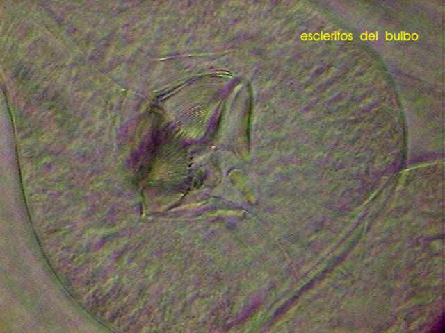
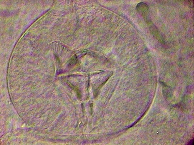
The bulb is spherical, muscular and provided with 3 valves and extensive cuticle, triangular, with its base anterior, with an indented free edge. It is evident that the bulb is a grinding tool.
In
other nematodes the excretory system has associated cells, visible
and relatively large called "renettes".
But in the oxyuroidea these do not exist. In addition the walls of the
excretory
tubes do not show cellular nuclei.
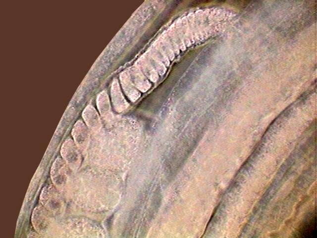
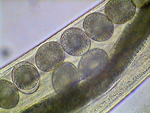
The
species is didelphic, i.e. it has two ovaries, an
anterior one and another posterior, that are continued with a unique
large uterus
normally full of eggs in different stages of development.
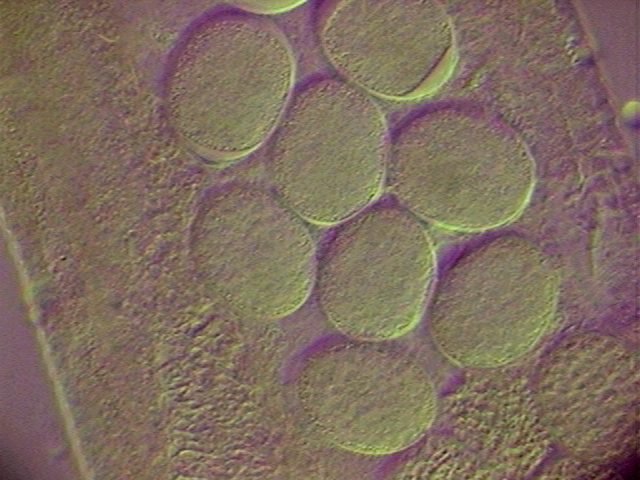
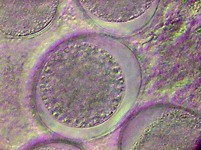
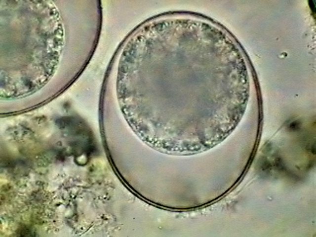
Each ripe ovule shows its large clear nucleus which soon becomes enveloped by the shell of the egg.
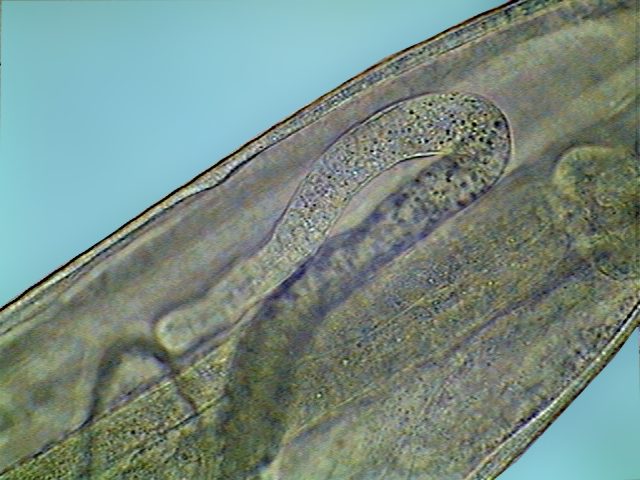
In fig. 19, apparently corresponding to a young female, the end of the vagina, full of sperm, is very visible, suggesting a previous copulation. This sperm must travel to the seminal vesicles at the start of the ovaries to be stored in the seminal vesicles to fertilize the maturing eggs.
A
spermatic receptacle full of sperm (each one of the little dots inside the receptacle)
is seen at
a greater magnification in the fig. 20. Hymann says that in the early
days of nematology
this large receptacle, which does not have a flagellum as a tail, were
confused with
nematode larvae, due to their location.
To be continued. |
