by David Walker,
UK
|
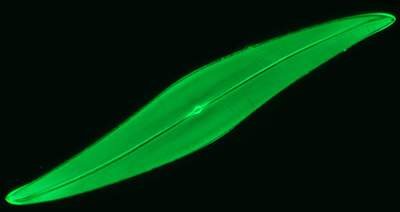
Diatom Pleurosigma
angulatum - a versatile 'demonstration' subject.
by David Walker,
UK |
|
Prepared slides of selected test diatom frustules are in many microscopy hobbyist's slide collections, including the authors. Old mounts of test slides e.g by Watson and NBS are often seen in eBay auctions or from dealers but test slides are still available.
Although the toughest test diatoms like Amphipleura pellucida can be fun (or should that be frustrating) to try, the species that I seem to find most often under a microscope objective is Pleurosigma angulatum. Not so much deciding what is real or an artifact at the very highest powers with an optical microscope which seems to have prompted much discussion in the literature, but simple studies at lower powers when the punctae become just resolved.
 The
species is very useful
for assessing various aspects of objectives used frequently i.e the 20x to 40x
range and also for demonstrating some
microscopic principles. Below are some simple demos which may be of
interest using a selection of modest to higher spec objectives the author has
access to. They are from the perspective of the author's casual interest in
diatoms and limited knowledge of microscope practice rather than as an afficionado
of either, comments on any errors in interpretation are welcomed.
The
species is very useful
for assessing various aspects of objectives used frequently i.e the 20x to 40x
range and also for demonstrating some
microscopic principles. Below are some simple demos which may be of
interest using a selection of modest to higher spec objectives the author has
access to. They are from the perspective of the author's casual interest in
diatoms and limited knowledge of microscope practice rather than as an afficionado
of either, comments on any errors in interpretation are welcomed.
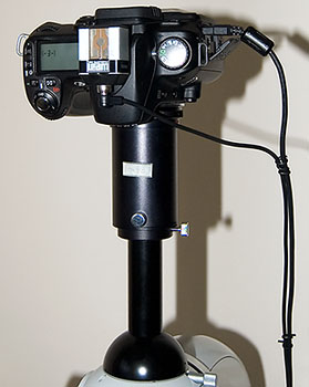
Note on camera setup: A Nikon D300 DSLR body was used on a Zeiss monocular tube on top photo port of a Zeiss Photomicroscope III with Optovar at 1.25x (shown right). A lensless third party 35 mm film microscope adaptor was used. A Zeiss Kpl W 10X eyepiece was used as the projection lens on a 12 mm collar to create a real projected image rather than virtual. This fills the frame of the APS sensor but does give about a 2/3rd crop of visual field. This sensor crop is actually a benefit for studying diatoms. Camera controlled by remote Nikon Control Pro software. Exposures ca 1 second to bring the exposures out of the known vibration problem settings of this setup.
The importance of an objective's numerical aperture (NA) for resolving detail
The NA of a typical 20x achromatic objective is ca. 0.40 - 0.45, e.g. the LOMO pre-DIN 20X NA0.40 or the Zeiss DIN 25X NA0.45. Trying to resolve the punctae of P. angulatum with such an objective in white or green light is unlikely no matter whether contrast enhancement is tried. Using a higher mag eyepiece than normal will also have no benefit.
The punctae on the P. angulatum test slide I used (Klaus Kemp's 8 form slide) were 0.64 microns apart (see footnote 1 on taking measurements) and by using one form of the resolution equation R = 0.61 x l / NA (see footnote 2), this suggests an NA of ca. 0.52 is theoretically required for green light of 550 nm, i.e. somewhat higher NA than 0.40.
This can be demonstrated in practice if a higher spec 20-25x objective is available with an NA of 0.60- 0.65, such as the LOMO 20X NA0.65 apo or Zeiss 25X NA0.60 Neofluar; the punctae can be readily seen in brightfield (a little oblique may help contrast).
I find this type of objective also a good test of a photomicro' setup, as the punctae at this lowish mag can border on visual acuity so if the camera is faithfully recording the fine punctae it suggests it is well setup.
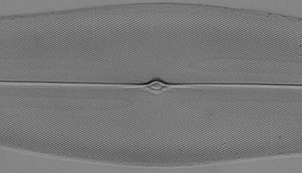
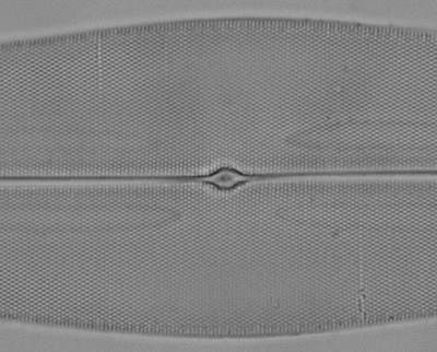
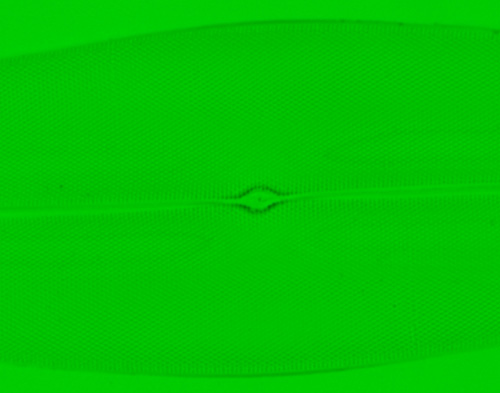
Zeiss
25X NA0.45 planachromat, green light, slight oblique. The punctae are not resolved.
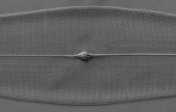
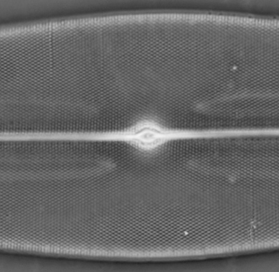
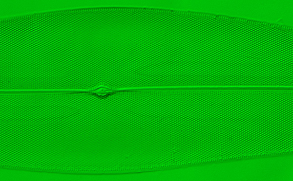
Zeiss
25X NA0.60 (phase) Neofluar, brightfield green light, slight oblique. The punctae
are resolved.
Compare image from the same objective in phase mode below.
Decreasing the wavelength of light increases resolution of a given objective
The
resolution equation also indicates that resolution is directly proportional
to wavelength. So what wavelength of light is theoretically required to resolve
P. angulatum with an NA0.45 objective? The equation gives 470 nm which
is a deep blue.
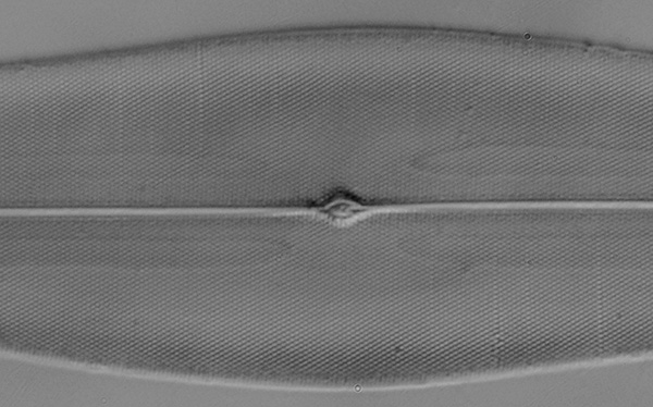
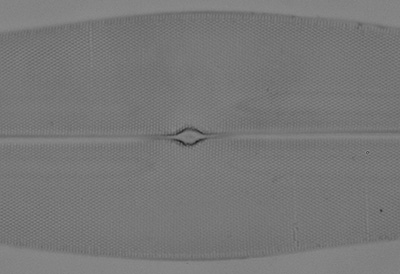
Zeiss 25X NA0.45 planachromat, Wratten 47B gelatine filter, 50% oblique. This typical deep blue filter has a broad spectral transmission, in this case ca. 380 - 500 nm peaking at 430 nm (from 'Kodak Photographic Filters Handbook'). The punctae are beginning to be evident but not very well.
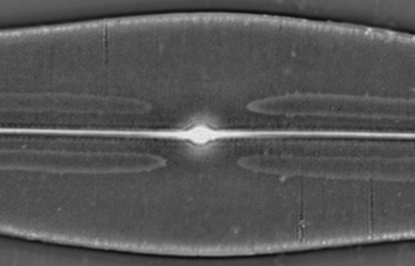
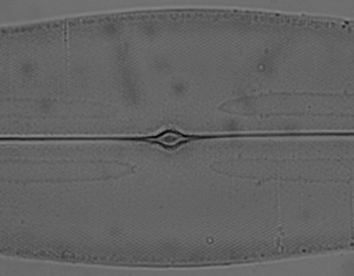
Zeiss 25X NA0.45 planachromat, 400 nm interference filter on lamphouse, slight oblique. This filter has a much narrower 40 nm half bandwidth and shorter wavelength than the Wratten 47B above. The punctae are better resolved with less oblique.
Although an interesting demonstration, it's arguable whether a modest NA objective would be used to increase resolution by decreasing wavelength; the objective may not be well corrrected for spherical aberration at shorter wavelengths. Lower wavelengths tend to be more useful with high NA objectives in photomicrography to maximise the resolution for subjects such as diatoms with fine frustule detail.
The loss of resolution in phase contrast
Phase contrast is a valuable contrast enhancement technique but the objective's phase plate is smaller (often considerably so) than its full aperture and reduces the effective NA of the objective when used in phase rather than brightfield. Frithjof Sterrenburg makes some interesting comments on this in his Microscopy Primer. The resolution loss may not be critical for subjects where the contrast benefit is most prized but isn't the best technique if optimum resolution needs to be maintained; oblique or DIC (if available) may be better.
For example, the Zeiss 25X NA0.60 phase Neofluar readily resolves the punctae in brightfield with green light, but does not in phase. See below.
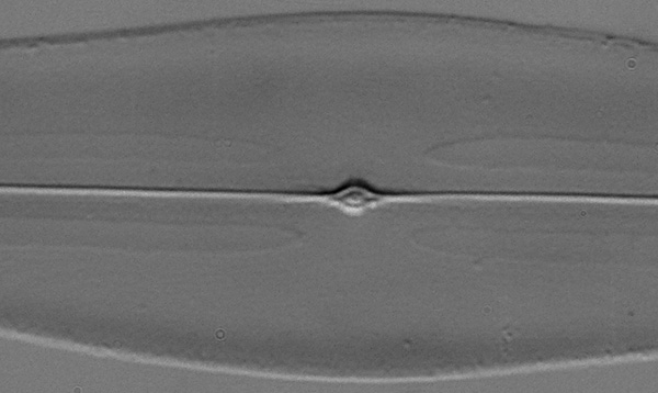
Zeiss 25X NA0.60 Neofluar, green light, brightfield.
Zeiss 25X NA0.60 Neofluar, green light, phase. The punctae resolution is lost and for this sort of subject, the image adopts a rather 'muddy' look cf. brightfield.
Phase objectives with higher NA can compensate to some extent. For example, a Zeiss 45X NA0.75 Neofluar is able to resolve the punctae in phase (NA0.65 is more typical for a 40X achromatic phase objective). This extra resolution with phase can be useful when studying fine features of e.g. pond life.
Left:
Zeiss 40X NA0.75 phase Neofluar, brightfield with green light slight
oblique. Punctae are resolved.
Right: Same objective in phase, green
light. Punctae are still resolved as the phase ring hasn't reduced the
NA below that required to resolve them, unlike the 25X NA0.65 objective
above.
Query: Although the loss of resolution in phase is well commented on, I don't recall seeing a reference that quantifies the loss; any references welcomed. If it is directly related to the condenser phase ring size, then for the Zeiss 40X NA0.75 Neofluar, the outer boundary of condenser phase annulus occupies 40% of the objective's back focal plane (measured accurately with condenser iris graduation). Does it then follow that its effective NA would decrease to ca. 0.30 (i.e. 0.40 x NA0.75 = NA0.30)? If so, the objective shouldn't be able to resolve the punctae of P. angulatum in green light with phase, but it clearly does with ease and from earlier, suggests that the NA is at least 0.52 in phase to resolve this diatom.
The effect of condenser iris setting on resolution and image quality
The microscopy textbooks and makers' manuals discuss the importance of correctly setting the condenser iris for each objective. Setting the iris to ca. 2/3rds or 4/5ths of full aperture by inspecting the back focal plane of the objective are typical guidelines although the makers of some modern objectives state that the designs can work well near full aperture. Although it's often best to study the image critically with a given objective while adjusting the iris to see what is most suited, to balance effective NA, image contrast and depth of field. P. angulatum is a useful subject to study the effect of iris setting.
Practical hint: Many condenser iris are graduated either numbered or unnumbered to allow reproducible iris settings.
Zeiss 40x NA0.75 Neofluar, brightfield green light - effect of condenser iris setting:
Left:
Iris at full aperture of objective. The full NA of the objective is
being used and punctae are resolved, but image is of low contrast.
Right:
Iris 4/5th of full aperture. This about the optimum for this subject,
punctae are still resolved with improved contrast.
Above: Iris 1/4 of full aperture. The objective's effective NA has been dropped below that required to resolve the punctae well. Contrast has increased although notice how out of plane debris in the optical train become evident.
Off-axis oblique for contrast enhancement without resolution loss
Of the 160mm tube length Zeiss objectives I own, even the higher spec ones seem to perform best with condenser iris stopped down to ca. 70%, especially oil immersion optics. But a modest amount of oblique with objective at full aperture can give good contrasty images without resolution loss. Extreme oblique needs to be used with care in case artifacts are generated. A phase condenser is a ready source for off-axis oblique, simply by moving the brightfield port off-centre. See the Micscape Library Oblique section for articles on methods of creating variations of oblique both off-axis and circular oblique.
Zeiss 40X NA1.0 oil immersion apo at 90% of full aperture, brightfield. Achromatic-aplanatic condenser oil immersed. From the author's examples at least (no delamination) they don't perform well near full aperture giving low contrast images of an already low contrast subject, obscuring resolved detail. Zeiss, in their manuals (ca. 1970s) for 160mm objectives recommend ca. 2/3rds of full aperture is set with condenser iris.
Zeiss
40X NA1.0 apo at full aperture with 50% oblique using an offset brightfield
port in phase condenser. Oblique 45° to long axis. Contrast is markedly
improved using the full aperture of the objective.
Experimenting with the direction of the oblique relative to the diatom long axis can be explored to give the best results. The older textbooks discuss the importance of oblique orientation (practice and theory) in some detail for resolving the punctae of diatoms such as A. pellucida. e.g. Spitta's 'Microscopy' 1909; his photomicrographs presented in this volume of test diatoms are some of the finest ever taken with an optical microscope.
********
An optical microscope can only 'interpret' the actual structure of a diatom, especially if the finer detail is near or beyond the resolution of the microscope. To see what the punctae of P. angulatum really look like, Frithjof Sterrenburg illustrates SEMs of this species and remarks on 'true images' of diatoms in his Micscape article 'A second look at some well known test diatoms'.
The classic modern textbook 'The Diatoms. Biology and Morphology of the Genera' by Round, Crawford and Mann also has a wealth of SEMs of diatoms that include many of the optical microscope test species and is relatively affordable (ca. Ł43) now that a 'digitally printed' paperback version was published in 2007.
Comments to the author David Walker are welcomed.
Footnotes:
1) Measuring the punctae separation: For accurate measurements an objective such as a 90x achromatic oil immersion is ideal to give well separated punctae. An eyepiece reticle could be calibrated for counting punctae visually using a good 0.01 mm micrometer slide. I much prefer to print out a full camera field image of the well resolved diatom and then an image of the micrometer slide, (or alternatively on computer screen measurements using the calibration / measurement feature found in most microscopy imaging software). This gives an accurate measurement of the horizontal and vertical field widths, to allow a good number of punctae to be counted and separation averaged.
|
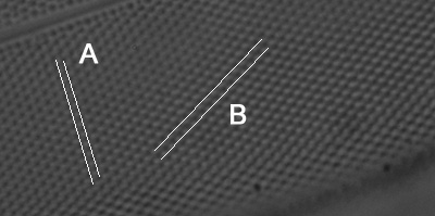
Right: Crop from an achromatic 90x NA.125 oil immersion resolves the punctae well (a little oblique helps contrast). A printout allows accurate measurements of punctae separation over a long distance and averaged, once field width has been calibrated with a similar print-out of a micrometer under exactly the same conditions. For this article and comments on resolution, punctae separation along a line B of 0.64 µm was used, (separation was 0.65 µm along a line A). Some older literature measures the number of 'lines per inch' separation although its not always defined what orientation of lines is being used. The separation between lines of orientation 'A' was 0.56 µm for this specimen, but lines if seen e.g in oblique, are an optical microscope's interpretation of incomplete punctae resolution.
|
|
2)
The textbooks discuss various forms of the equation linking resolution R (nm
or µm)
to wavelength l (nm
or µm)
and NA of objective.
The equation R = 0.61 x l /
NA was used.
Acknowledgement: The author used the invaluable 'Test plate 8 forms' (Hyrax mount) which includes the species P. angulatum, made by Klaus Kemp.
Published in the September 2008 edition of Micscape.
Please report any Web problems or offer general comments to the Micscape Editor .
Micscape is the on-line monthly magazine of the Microscopy UK web site at Microscopy-UK
© Onview.net Ltd, Microscopy-UK, and all contributors 1995
onwards. All rights reserved.
Main site is
at www.microscopy-uk.org.uk
with full mirror
at www.microscopy-uk.net
.