Victorian Rambles - Part I
by David Walker, UK
Over the years my brother and I have collected a modest selection of old microscope slides. When the budget and occasion permits we treat ourselves to interesting single old slides or small collections of up to a dozen or so; larger collections can be prohibitive for us in price nowadays, especially if they contain examples by the famous mounters. Old slides can be enjoyed on many levels, and this occasional series is a random dip into our collection to illustrate some hopefully interesting slide subjects. Different lighting techniques are explored to show their relative merits where appropriate.
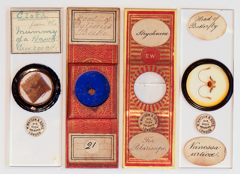
The four Victorian slides discussed.
'Cloth from the Mummy of a Hawk, Circa 200BC'
It is not always the subject itself that may be of particular interest under the microscope; sometimes it can be the evocative context. This slide is a good example. A textile expert may be able to deduce far more from it than I can, but does offer to the casual observer a hands-on contact to a great culture of over two millennia ago which is rather appealing. I wasn't aware that dead birds were mummified but a number of fascinating resources were found online.
The 'Object Lessons' website shows a photograph of a mummified hawk and an X-ray with a short summary of the hawk's importance and appeal to the ancient Egyptians.
The BBC h2g2 website has an overview of 'Animal Mummification' in ancient Egypt with an entry on the hawk.
The research of a subject that would not normally be undertaken is another aspect of microscope slides that can be of interest, this is greatly helped nowadays with ready access to Internet resources.
The slide has the typical Watson address label for the years 1882-1908 (ref. 1) and an old handwritten label in green ink. This assumes that the description is correct of course, how many could distinguish the real thing from a tatty piece of Victorian cloth? The cloth piece has what looks like a simple warp and weft weave.
As mentioned in my 'Forays into Fluorescence - 2' article, since setting up a simple epi-fluorescent system using the normal 100W quartz tungsten microscope lamp, I find that checking a dry mounted old slide subject for autofluorescence can be worthwhile, especially with deep blue excitation, as it can often show an interesting aspect of the subject. The visual image may be of low intensity without a mercury arc lamp and the colours unclear, but a modern budget digital SLR like the author's Nikon D5000 can often take a competent image.
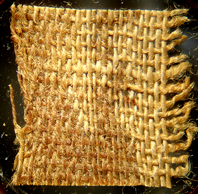
Image taken using top lighting on a Meiji EMZ1 stereo, 15x to fill field of Sony P200.
The cloth piece is ca. 11 x 10 mm in size.
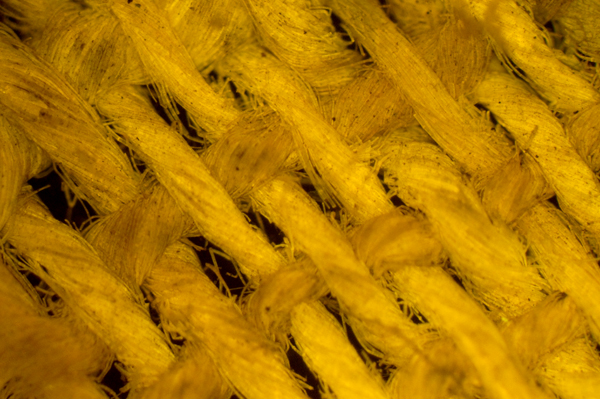
Epi-fluorescence image using blue-violet excitation with 100W quartz halogen lamp. Zeiss 2.5/0.08 objective.
Nikon D5000 DSLR, 10 seconds ISO 800. It may be the cloth treatments that fluoresce.
'Foot of Dytiscus Beetle'
Despite being a keen pond dipper, I've never come across a live adult great diving beetle Dytiscus marginalis which can grow up to 24-38 mm with larvae up to 60 mm (ref. 2). They must be magnificent to see.
'Natural England' shows a photograph of both the adult and larva. I like the description in Fitter and Manuel of the larvae (ref. 2) who describe them as 'formidable beasts' and who note that 'fingers of careless collectors' may be on the menu if not handled with care.
The male leg is a classic subject for the microscope and described and illustrated in for example, 'Carpenter' as shown below. The complex features are described by Carpenter as 'a very remarkable apparatus of suckers' and are associated with the male attaching itself to the female during mating. Some of the older books on freshwater life have splendid extended observations of the typical behaviour of Dytiscus adult beetle and larvae, e.g. W Furneaux's 'Life in Ponds and Streams', 1919. The author's slide is for top light only but the leg can form an excellent subject with transmitted light, especially with crossed polars, as Dr Stephen Lowry strikingly shows in his Nikon Small World 2007 'Image of Distinction'.
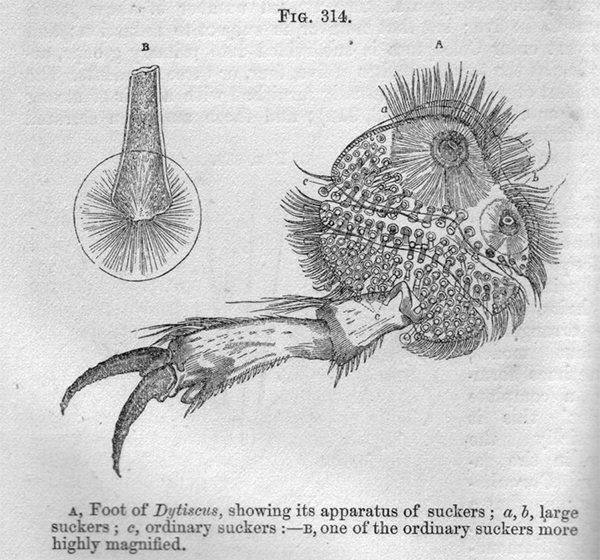
Figure from 'The Microscope and its Revelations' by the Rev. W B Carpenter, 1857, second edition.
A comparison of off-axis top lighting and epi-fluorescence are shown below.
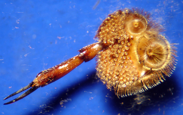
Image taken using Meiji EMZ1 stereo using its built-in top light and Sony P200 digicam on eyepiece. As with many old dry mounts, it suffers from fungal growth, both under the coverslip and on the subject itself. I'm not so sure the blue background suits this type of subject, black may have been better. The leg is ca. 6 mm long and first joint of the tarsi ca. 3 mm wide.
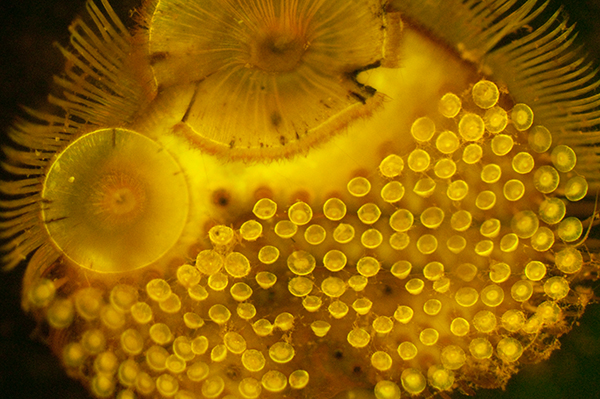
Epi-fluorescence image using blue-violet excitation with 100W quartz halogen lamp. Zeiss 2.5/0.08 objective. Nikon D5000 DSLR, exposure 15 seconds at ISO 800.
The fungal growth does not fluoresce giving a 'cleaner' image of the subject.
Closer look with autofluorescence. Zeiss 6.3/0.16 objective. Exposure 5 secs at ISO 800.
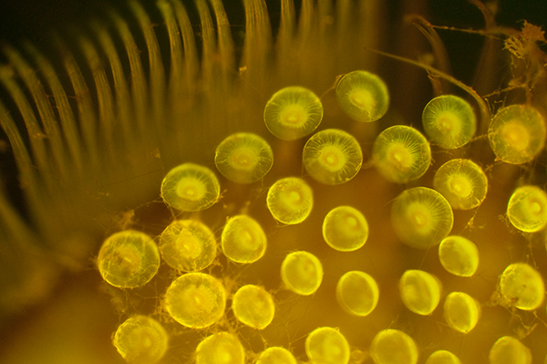
'Strychnine'
This chemical seems to have a particular macabre appeal to study, perhaps because of its many associations with poisoning, both real and fictional. Wikipedia has both a page on the compound Strychnine and a separate page devoted to 'Strychnine poisoning', the latter which is a fascinating but at times unpleasant read.
The author's example of the chemical is safely immobilised in this attractively papered slide with the 'EW' monogram of the famous mounter Edmund Wheeler. Curiously the slide itself rather than the cover slip has broken some time in its life but the paper and coverslip hold it together. The damage doesn't unduly affect the study of the attractive formations under polarised light in particular.
It always surprises me how remarkably robust the paper and adhesives used were on some Victorian papered slides, considering how many times slides of this age may have been handled during their lives. There's often barely a scuff mark or peeling of the paper, even at the sharp edges or corners of the slide.
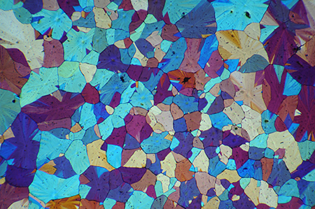
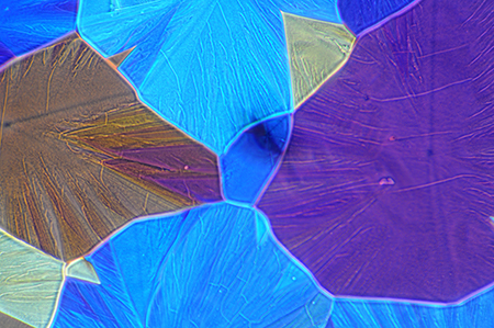
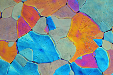
The strychnine crystals were large interlocking flat plates with fine detail, which suited exploration under different lighting conditions, as shown below.
|
Zeiss 2.5/0.08 objective. Crossed polars with lambda tint plate. |
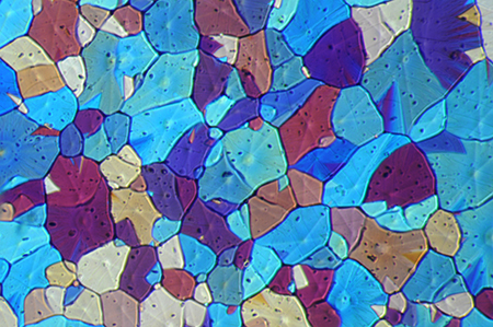
Zeiss 6.3/0.16 objective. Crossed polars with lambda tint plate. |
|
Zeiss 6.3/0.16 objective. Crossed polars with lambda tint plate. |
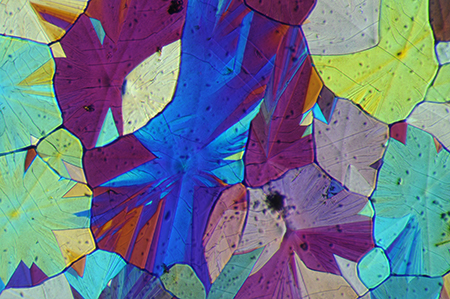
Zeiss 6.3/0.16 objective. Darkfield with crossed polars with lambda tint plate gives an attractive stained glass window effect. |
|
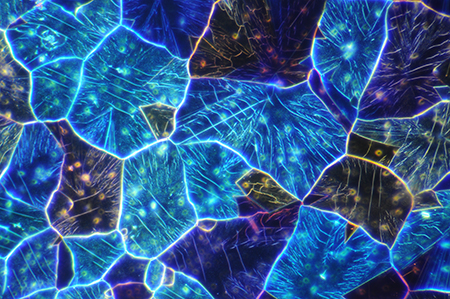
Zeiss 16/0.40 Neofluar objective. Phase with crossed polars and lambda tint plate. the phase brings out the fine structure of the crystal plates. |
Zeiss 16/0.35 objective. Nomarski DIC with lambda tint plate. DIC gives some depth as well as bringing out fine structure in the plates. The optical sectioning effect of DIC, useful for microorganisms, is less useful here as depth of field is lost. Stacking images may recover this. |
'Head of Butterfly Vanessa urticae'
Although the Victorian papered slides can be very attractive, my favourite slides are the later unpapered examples by Watson; I like their elegant simplicity with the typical printed oval labels with the subject title, subtitle and 'maker's' label. The laid out parts of larger subjects like insects can be admired with hand lens or low power stereo, as in this example of a beautifully presented head of the small tortoiseshell butterfly. Photographing these sort of subjects well in their entirety can be an enjoyable challenge as the relative merits of different image capture routes can be explored, e.g. using camera and macro lens, low power stereo microscope, stitching images with low power compound microscope objective or other methods such as using a 35 mm slide film scanner.
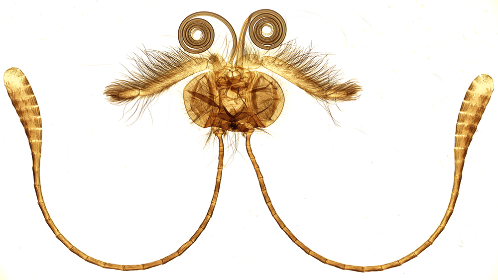
As displayed: Image using 35mm film scanner (Minolta Dimage 5400 Elite), 5400 dpi scan resized for web page.
Use mouse rollover to see equivalent stereo microscope image. The horizontal field width is ca. 16 mm.
Although the scanner's spatial resolution can barely match the lowest power compound microscope objective (discussed in this article), its ability to scan subjects up to the 35x24 mm film frame size in one scan, its speed and guaranteed even lighting make it a favourite route of the author for web imagery of large subjects.
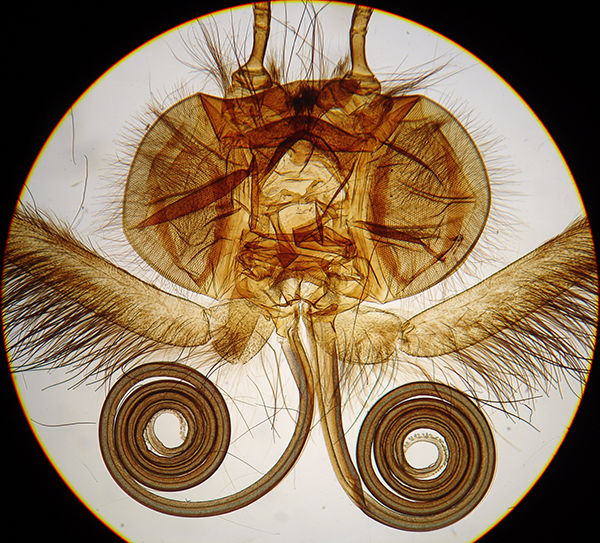
A Meiji EMZ1 stereo microscope was also used at 15X (see mouse rollover) with Sony P200 digital camera on eyepiece and optical zoom to fill field. The built in transmitted lighting of many stereos is basic and not always possible to achieve even lighting. Despite tonal levels adjustment it's hard to match the bright white background that the scanner achieves. The collimated beam of the scanner also gives contrasty edge detail cf the softer image that the back lit diffuser gives on the typical light base of a stereo.
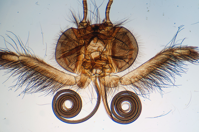
Zeiss 2.5/0.08 objective and capturing the full field of the 10x eyepiece with Sony P200 digicam supported over the eyepiece. This illustrates the problems of imaging large subjects with a compound microscope. Using a 35 mm film frame crop or in author's case the smaller APS sensor crop on a Nikon D5000 DSLR further limits the field area captured.
Zeiss 1/0.04 objective. The author possesses one of these cumbersome none parfocal objectives, but it's the one I like / use least in the Zeiss optical armoury. The visual images of large subjects are useful as the eye tolerates any slight uneven lighting, but a camera is unforgiving; the Zeiss Photomicroscope III's otherwise excellent lighting struggles to illuminate the background evenly to within the fraction of a stop which a camera demands. The numerical aperture of 0.04 is also only half that of a typical stereo (the author's Meiji is ca. 0.075), and this is evident in photography where the images lacks crispness no matter how carefully I focus with Live View on the camera. Blue aberration on contrasty edges of the subject is also seen.
Comments to the author are welcomed.
References
1) B. Bracegirdle, 'Notes on Modern Microscope Manufacturers', Quekett Microscopical Club, London, 1996.
2) R. Fitter and R. Manuel, 'Collins Field Guide to Freshwater Life', 1986.
Published in the September 2010 edition of Micscape.
Please report any Web problems or offer general comments to the Micscape Editor .
Micscape is the on-line monthly magazine of the Microscopy UK web site at Microscopy-UK
©
Onview.net Ltd, Microscopy-UK, and all contributors 1995
onwards. All rights reserved.
Main site is at
www.microscopy-uk.org.uk.